Product Specification
| Host |
Rabbit |
| Antigen |
SOX-10 |
| Synonyms |
/ |
| Immunogen |
Synthetic Peptide |
| Location |
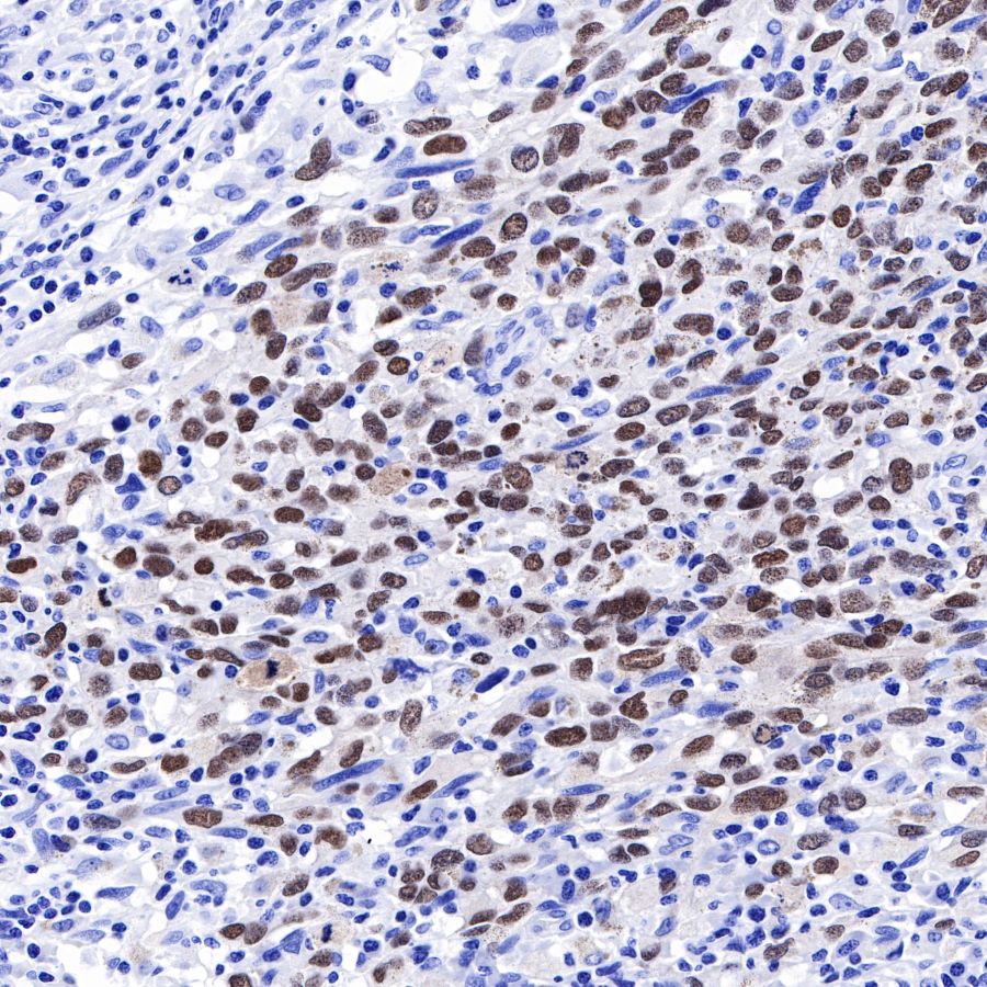
Cytoplasm, Nucleus |
| Accession |
P56693 |
| Clone Number |
SDT-136-59 |
| Antibody Type |
Rabbit mAb |
| Application |
WB, IHC-P, ICC, ICFCM |
| Reactivity |
Hu, Ms, Rt |
| Predicted Reactivity |
Pg, Av |
| Purification |
Protein A |
| Concentration |
0.25 mg/ml |
| Physical Appearance |
Liquid |
| Storage Buffer |
PBS, 40% Glycerol, 0.05%BSA, 0.03% Proclin 300 |
| Stability & Storage |
12 months from date of receipt / reconstitution, -20 °C as supplied |
Dilution
| application |
dilution |
species |
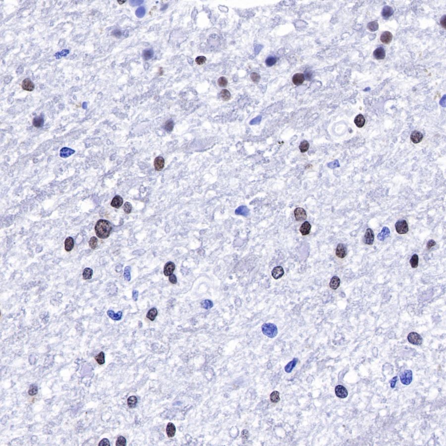
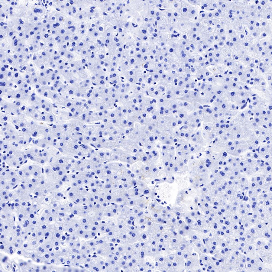
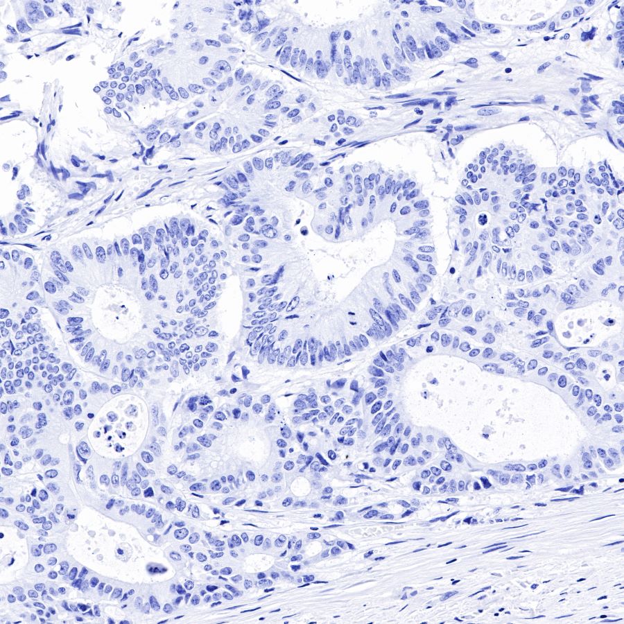
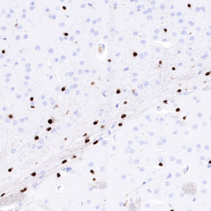
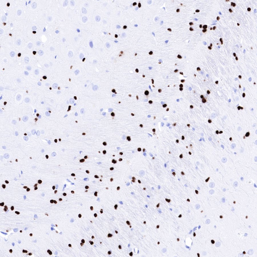
| IHC-P |
1:1000 |
|
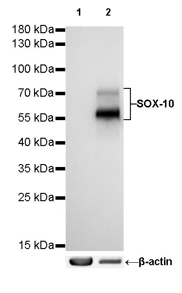
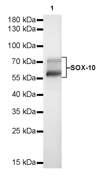
| WB |
1:500 |
|
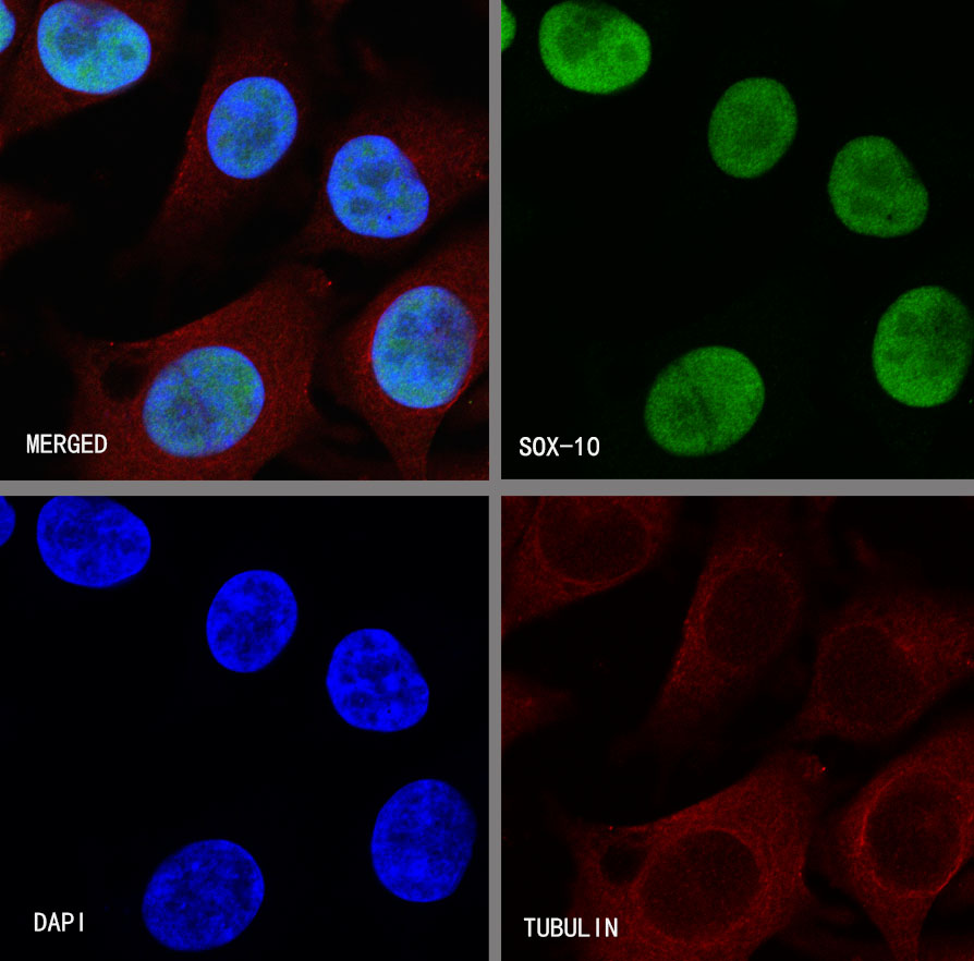
| ICC |
1:250 |
|
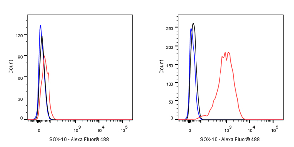
| ICFCM |
1:250 |
|
Background
Transcription factor SOX-10 is a protein that in humans is encoded by the SOX10 gene. This gene encodes a member of the SOX (SRY-related HMG-box) family of transcription factors involved in the regulation of embryonic development and determination of cell fate. The encoded protein acts as a transcriptional activator after forming a protein complex with other proteins. This protein acts as a nucleocytoplasmic shuttle protein and is important for neural crest and peripheral nervous system development.