WB result of Annexin A2 Rabbit mAb Primary antibody: Annexin A2 Rabbit mAb at 1/5000 dilution Lane 1: LNCaP whole cell lysate 20 µg Lane 2: HeLa whole cell lysate 20 µg Lane 3: HepG2 whole cell lysate 20 µg Lane 4: K-562 whole cell lysate 20 µg Negative control: LNCaP whole cell lysate Secondary antibody: Goat Anti-Rabbit IgG, (H+L), HRP conjugated at 1/10000 dilution Predicted MW: 39 kDa Observed MW: 35 kDa
S-RMab®Annexin A2 Recombinant Rabbit mAb (SDT-444-67)
S-RMab®Annexin A2 Recombinant Rabbit mAb (SDT-444-67)
Price:
Regular price
$59.00 SGD
Regular price
Sale price
$59.00 SGD
Unit price
per
For shipping services or bulk orders, you may request a quotation.
Secure checkout with

View full details
Product Details
Product Details
Product Specification
| Host | Rabbit |
| Antigen | Annexin A2 |
| Synonyms | Annexin II, Annexin-2, ANXA2, Calpactin I heavy chain, Calpactin-1 heavy chain, Chromobindin-8, Lipocortin II, Placental anticoagulant protein IV, PAP-IV, Protein I, p36 |
| Immunogen | Recombinant Protein |
| Location | Secreted |
| Accession | P07355 |
| Clone Number | SDT-444-67 |
| Antibody Type | Recombinant mAb |
| Application | WB, IHC-P, ICC |
| Reactivity | Hu |
| Predicted Reactivity | Ms, Pg, Dg |
| Purification | Protein A |
| Concentration | 0.5 mg/ml |
| Conjugation | Unconjugated |
| Physical Appearance | Liquid |
| Storage Buffer | PBS, 40% Glycerol, 0.05% BSA, 0.03% Proclin 300 |
| Stability & Storage | 12 months from date of receipt / reconstitution, -20 °C as supplied |
Dilution
| application | dilution | species |
| WB | 1:5000 | |
| ICC | 1:50 |
Background
Annexin A2 is a membrane scaffolding and binding protein, which mediated various cellular events. Its functions are generally affected by cellular localization. In the cytoplasm, they interacted with different phospholipid membranes in Ca2+ -dependent manner and play vital roles including actin binding, remodeling and dynamics, cytoskeletal rearrangement, and lipid-raft microdomain formation. However, upon cell exposure to certain stimuli, annexin A2 translocates to the external leaflets of the plasma membrane where annexin A2 was recently reported to serve as a virus receptor, play an important role in the formation of virus replication complex, or implicated in virus assembly and budding [PMID: 30746844].
Picture
Picture
Western Blot
WB result of Annexin A2 Rabbit mAb Primary antibody: Annexin A2 Rabbit mAb at 1/5000 dilution Lane 1: mouse heart lysate 20 µg Secondary antibody: Goat Anti-Rabbit IgG, (H+L), HRP conjugated at 1/10000 dilution Predicted MW: 39 kDa Observed MW: 35 kDa
WB result of Annexin A2 Rabbit mAb Primary antibody: Annexin A2 Rabbit mAb at 1/5000 dilution Lane 1: rat heart lysate 20 µg Secondary antibody: Goat Anti-Rabbit IgG, (H+L), HRP conjugated at 1/10000 dilution Predicted MW: 39 kDa Observed MW: 35 kDa
Immunohistochemistry
IHC shows positive staining in paraffin-embedded human liver. Anti-Annexin A2 antibody was used at 1/500 dilution, followed by a HRP Polymer for Mouse & Rabbit IgG (ready to use). Counterstained with hematoxylin. Heat mediated antigen retrieval with Tris/EDTA buffer pH9.0 was performed before commencing with IHC staining protocol.
IHC shows positive staining in paraffin-embedded human placenta. Anti-Annexin A2 antibody was used at 1/500 dilution, followed by a HRP Polymer for Mouse & Rabbit IgG (ready to use). Counterstained with hematoxylin. Heat mediated antigen retrieval with Tris/EDTA buffer pH9.0 was performed before commencing with IHC staining protocol.
IHC shows positive staining in paraffin-embedded human stomach. Anti-Annexin A2 antibody was used at 1/500 dilution, followed by a HRP Polymer for Mouse & Rabbit IgG (ready to use). Counterstained with hematoxylin. Heat mediated antigen retrieval with Tris/EDTA buffer pH9.0 was performed before commencing with IHC staining protocol.
IHC shows positive staining in paraffin-embedded human prostate. Anti-Annexin A2 antibody was used at 1/500 dilution, followed by a HRP Polymer for Mouse & Rabbit IgG (ready to use). Counterstained with hematoxylin. Heat mediated antigen retrieval with Tris/EDTA buffer pH9.0 was performed before commencing with IHC staining protocol.
IHC shows positive staining in paraffin-embedded human lung squamous cell carcinoma. Anti-Annexin A2 antibody was used at 1/500 dilution, followed by a HRP Polymer for Mouse & Rabbit IgG (ready to use). Counterstained with hematoxylin. Heat mediated antigen retrieval with Tris/EDTA buffer pH9.0 was performed before commencing with IHC staining protocol.
IHC shows positive staining in paraffin-embedded human breast cancer. Anti-Annexin A2 antibody was used at 1/500 dilution, followed by a HRP Polymer for Mouse & Rabbit IgG (ready to use). Counterstained with hematoxylin. Heat mediated antigen retrieval with Tris/EDTA buffer pH9.0 was performed before commencing with IHC staining protocol.
IHC shows positive staining in paraffin-embedded mouse colon. Anti-Annexin A2 antibody was used at 1/500 dilution, followed by a HRP Polymer for Mouse & Rabbit IgG (ready to use). Counterstained with hematoxylin. Heat mediated antigen retrieval with Tris/EDTA buffer pH9.0 was performed before commencing with IHC staining protocol.
IHC shows positive staining in paraffin-embedded mouse kidney. Anti-Annexin A2 antibody was used at 1/500 dilution, followed by a HRP Polymer for Mouse & Rabbit IgG (ready to use). Counterstained with hematoxylin. Heat mediated antigen retrieval with Tris/EDTA buffer pH9.0 was performed before commencing with IHC staining protocol.
IHC shows positive staining in paraffin-embedded rat colon. Anti-Annexin A2 antibody was used at 1/500 dilution, followed by a HRP Polymer for Mouse & Rabbit IgG (ready to use). Counterstained with hematoxylin. Heat mediated antigen retrieval with Tris/EDTA buffer pH9.0 was performed before commencing with IHC staining protocol.
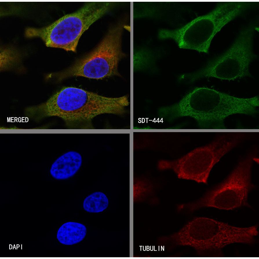
Immunocytochemistry
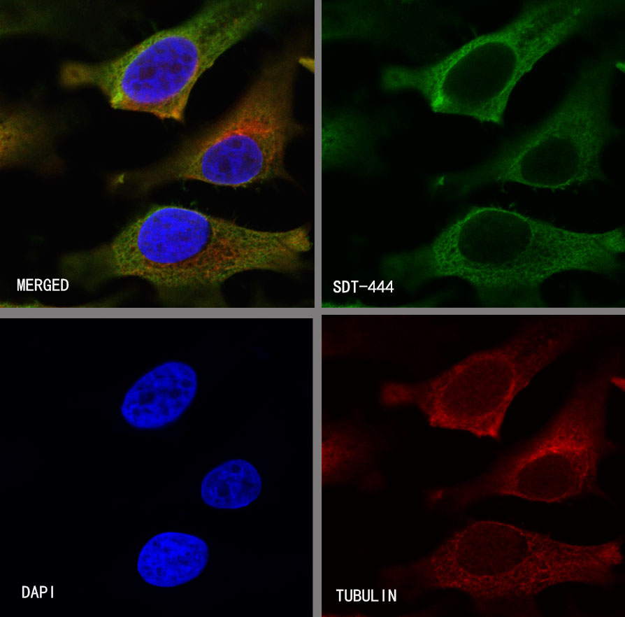
ICC shows positive staining in HeLa cells. Anti-Annexin A2 antibody was used at 1/50 dilution (Green) and incubated overnight at 4°C. Goat polyclonal Antibody to Rabbit IgG - H&L (Alexa Fluor® 488) was used as secondary antibody at 1/1000 dilution. The cells were fixed with 4%PFA and permeabilized with 0.1% PBS-Triton X-100. Nuclei were counterstained with DAPI (Blue).Counterstain with tubulin (Red).