S-RMab® PD-1 Recombinant Mouse mAb (SDT-R143)
S-RMab® PD-1 Recombinant Mouse mAb (SDT-R143)
Price:
Regular price
$131.00 SGD
Regular price
Sale price
$131.00 SGD
Unit price
per
For shipping services or bulk orders, you may request a quotation.
Secure checkout with

View full details
Product Details
Product Details
Product Specification
| Host | Mouse |
| Antigen | PD-1 |
| Synonyms | Programmed cell death protein 1, hPD-1, CD279 |
| Immunogen | N/A |
| Location | Cell membrane |
| Accession | Q15116 |
| Clone Number | SDT-R143 |
| Antibody Type | Recombinant mAb |
| Isotype | IgG1 |
| Application | WB, IHC-P, ICC, ICFCM, IP |
| Reactivity | Hu |
| Purification | Protein G |
| Concentration | 2 mg/ml |
| Tag | N/A |
| Physical Appearance | Liquid |
| Storage Buffer | PBS, 40% Glycerol, 0.05% BSA, 0.03% Proclin 300 |
| Stability & Storage | 12 months from date of receipt / reconstitution, -20 °C as supplied |
Dilution
| application | dilution | species |
| IHC-P | 1:200-1:500 | null |
| ICFCM | 1:200 | null |
| ICC | 1:100 | null |
| WB | 1:1000 | null |
| IP | 1:200 | null |
Background
Programmed cell death protein 1, also known as PD-1 and CD279 (cluster of differentiation 279), is a protein on the surface of T and B cells that has a role in regulating the immune system's response to the cells of the human body by down-regulating the immune system and promoting self-tolerance by suppressing T cell inflammatory activity. This prevents autoimmune diseases, but it can also prevent the immune system from killing cancer cells. PD-1 is an immune checkpoint and guards against autoimmunity through two mechanisms. First, it promotes apoptosis (programmed cell death) of antigen-specific T-cells in lymph nodes. Second, it reduces apoptosis in regulatory T cells (anti-inflammatory, suppressive T cells).
Picture
Picture
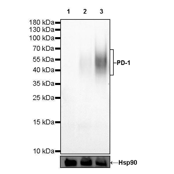
Western Blot
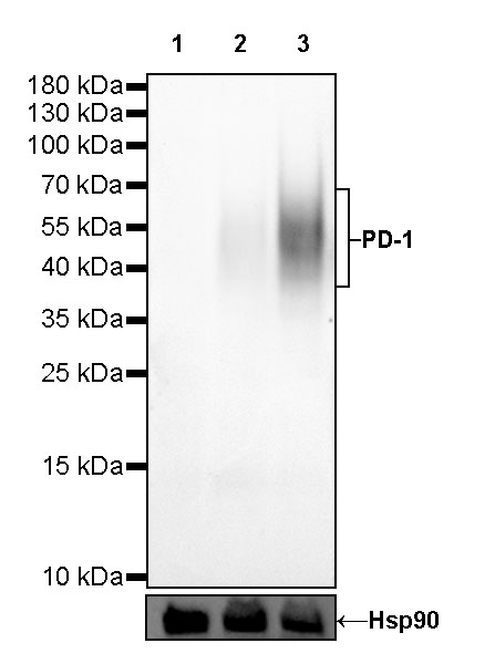
WB result of PD-1 Mouse mAb
Primary antibody: PD-1 Mouse mAb at 1/1000 dilution
Lane 1: Jurkat whole cell lysate 20 µg
Lane 2: MOLT-4 whole cell lysate 20 µg
Lane 3: MOLT-4 treated with Ionomycin (500ng/ml 24 hr) and PMA (10ng/ml 24 hr) whole cell lysate 20 µg
Negative control: Jurkat whole cell lysate
Secondary antibody: Goat Anti-mouse IgG, (H+L), HRP conjugated at 1/10000 dilution
Predicted MW: 32 kDa
Observed MW: 38~70 kDa
FC
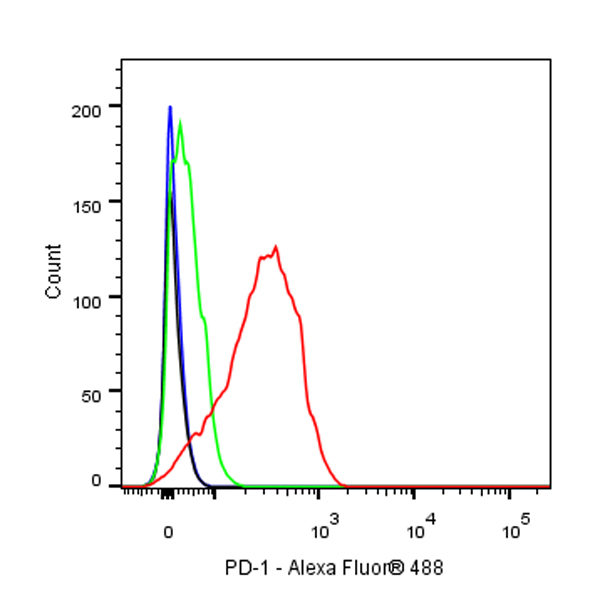
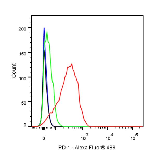
Flow cytometric analysis of Molt-4 (Human cervix adenocarcinoma epithelial cell) cells, treated with 10ng/ml PMA and 500ng/ml Ionomycin for 24h (Red) or untreated (Green), labeling PD-1 at 1/200 dilution (1 μg) compared with a Mouse monoclonal IgG isotype control (Black) and an unlabeled control (cells without incubation with primary antibody and secondary antibody) (Blue). Goat Anti - Mouse IgG Alexa Fluor® 488 was used as the secondary antibody.
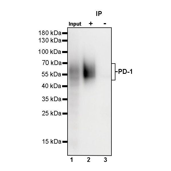
IP
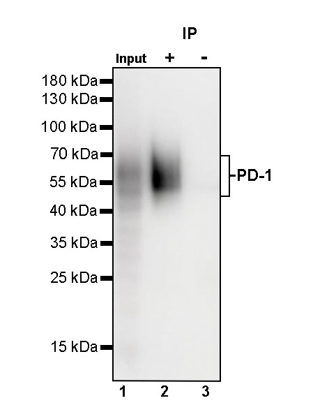
PD-1 Mouse mAb at 1/200 dilution (1 µg) immunoprecipitating PD-1 in 0.35 mg MOLT-4 treated with Ionomycin (500ng/ml 24 hr) and PMA (10ng/ml 24 hr) whole cell lysate.
Western blot was performed on the immunoprecipitate using PD-1 Mouse mAb at 1/1000 dilution.
Secondary antibody (HRP) for IP was used at 1/2000 dilution.
Lane 1: MOLT-4 treated with Ionomycin (500ng/ml 24 hr) and PMA (10ng/ml 24 hr) whole cell lysate 20 µg (Input)
Lane 2: PD-1 Mouse mAb IP in MOLT-4 treated with Ionomycin (500ng/ml 24 hr) and PMA (10ng/ml 24 hr) whole cell lysate
Lane 3: Mouse monoclonal IgG1 IP in MOLT-4 treated with Ionomycin (500ng/ml 24 hr) and PMA (10ng/ml 24 hr) whole cell lysate
Predicted MW: 32 kDa
Observed MW: 38~70 kDa
(This blot was developed with high sensitivity substrate)
Immunohistochemistry
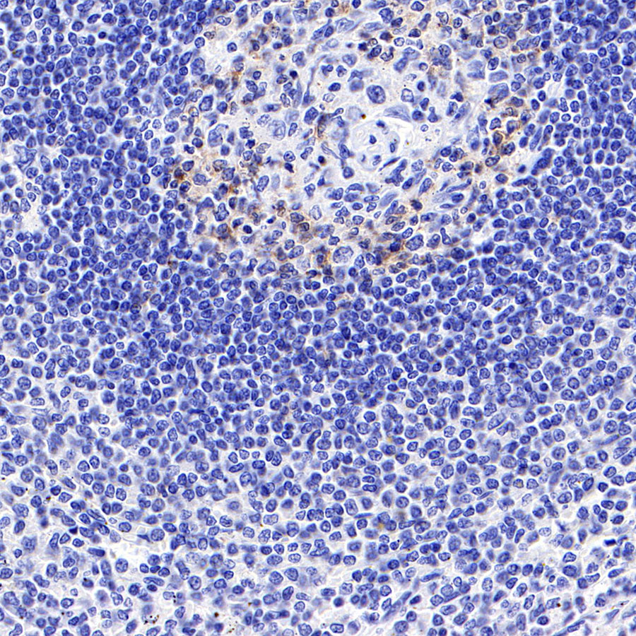
IHC shows positive staining in paraffin-embedded human tonsil. Anti-PD-1 antibody was used at 1/200 dilution, followed by a HRP Polymer for Mouse & Rabbit IgG (ready to use). Counterstained with hematoxylin. Heat mediated antigen retrieval with Tris/EDTA buffer pH9.0 was performed before commencing with IHC staining protocol.
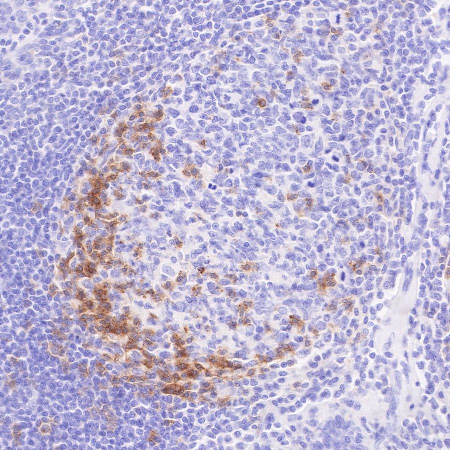
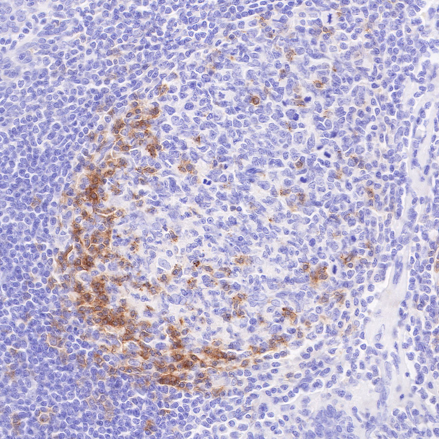
IHC shows positive staining in paraffin-embedded human spleen. Anti-PD-1 antibody was used at 1/500 dilution, followed by a HRP Polymer for Mouse & Rabbit IgG (ready to use). Counterstained with hematoxylin. Heat mediated antigen retrieval with Tris/EDTA buffer pH9.0 was performed before commencing with IHC staining protocol.
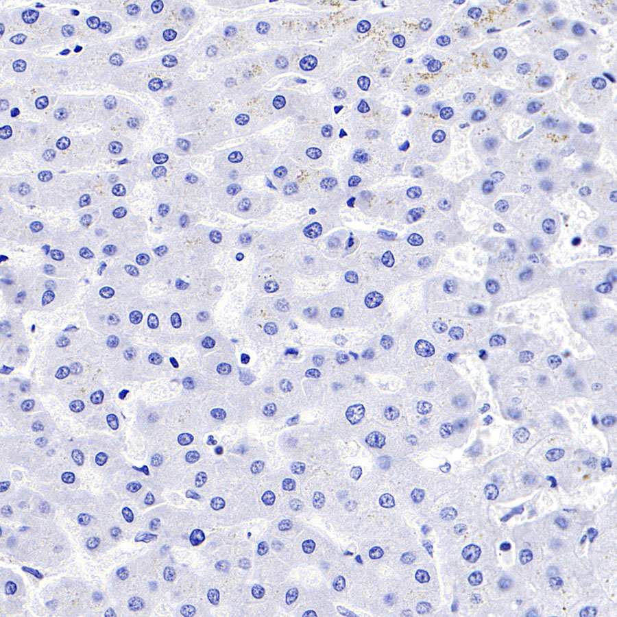
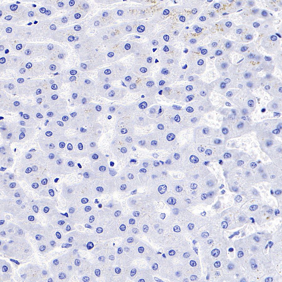
Negative control: IHC shows negative staining in paraffin-embedded human liver. Anti-PD-1 antibody was used at 1/500 dilution, followed by a HRP Polymer for Mouse & Rabbit IgG (ready to use). Counterstained with hematoxylin. Heat mediated antigen retrieval with Tris/EDTA buffer pH9.0 was performed before commencing with IHC staining protocol.
Immunocytochemistry
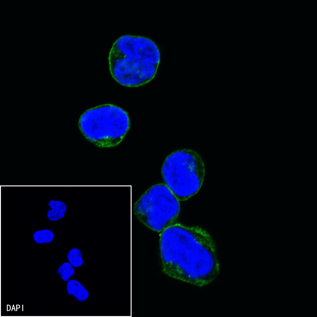
ICC shows positive staining in MOLT-4 cells treated with Ionomycin (500ng/ml,24h) + PMA (10ng/ml,24h). Anti-PD-1 antibody was used at 1/100 dilution (Green) and incubated overnight at 4°C. Goat polyclonal Antibody to mouse IgG - H&L (Alexa Fluor® 488) was used as secondary antibody at 1/1000 dilution. The cells were fixed with 4% PFA and permeabilized with 0.1% PBS-Triton X-100. Nuclei were counterstained with DAPI (Blue).
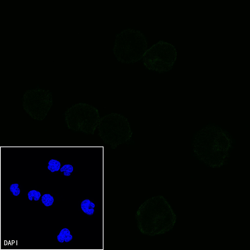
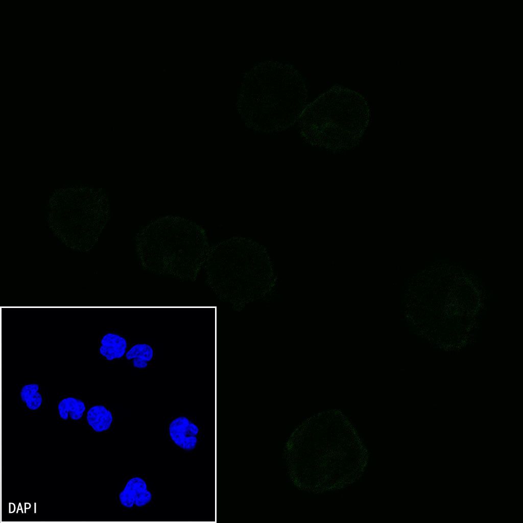
Negative control:ICC shows negative staining in MOLT-4 cells untreated with Ionomycin (500ng/ml,24h) + PMA (10ng/ml,24h). Anti-PD-1 antibody was used at 1/100 dilution and incubated overnight at 4°C. Goat polyclonal Antibody to mouse IgG - H&L (Alexa Fluor® 488) was used as secondary antibody at 1/1000 dilution. The cells were fixed with 4% PFA and permeabilized with 0.1% PBS-Triton X-100. Nuclei were counterstained with DAPI (Blue).