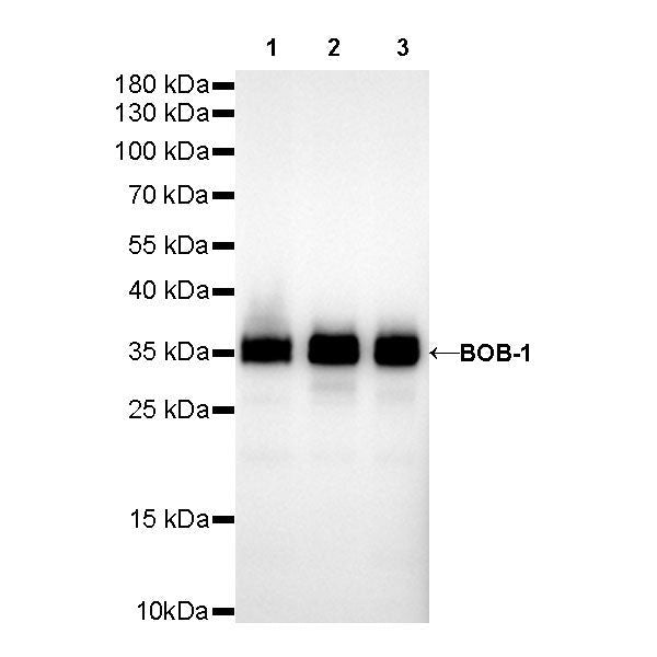
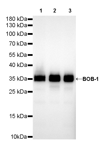
WB result of BOB-1 Rabbit mAb
Primary antibody: BOB-1 Rabbit mAb at 1/500 dilution
Lane 1: Raji whole cell lysate 20 µg
Lane 2: Ramos whole cell lysate 20 µg
Lane 3: Daudi whole cell lysate 20 µg
Secondary antibody: Goat Anti-Rabbit IgG, (H+L), HRP conjugated at 1/10000 dilution
Predicted MW: 27 kDa
Observed MW: 35 kDa
Exposure time: 40s
S-RMab® BOB-1 Recombinant Rabbit mAb (SDT-R115)
S-RMab® BOB-1 Recombinant Rabbit mAb (SDT-R115)
Price:
Regular price
$131.00 SGD
Regular price
Sale price
$131.00 SGD
Unit price
per
For shipping services or bulk orders, you may request a quotation.
Secure checkout with

View full details
Product Details
Product Details
Product Specification
| Host | Rabbit |
| Antigen | BOB-1 |
| Synonyms | OBF-1, OCA-B, OCT-binding factor 1 |
| Immunogen | N/A |
| Location | Nucleus |
| Accession | Q16633 |
| Clone Number | SDT-R115 |
| Antibody Type | Rabbit mAb |
| Application | WB, IHC-P, ICC, ICFCM, IP |
| Reactivity | Hu |
| Purification | Protein A |
| Concentration | 0.25 mg/ml |
| Physical Appearance | Liquid |
| Storage Buffer | PBS, 40% Glycerol, 0.05% BSA, 0.03% Proclin 300 |
| Stability & Storage | 12 months from date of receipt / reconstitution, -20 °C as supplied |
Dilution
| application | dilution | species |
| IHC-P | 1:1000 | null |
| WB | 1:500 | null |
| ICC | 1:250 | null |
| IP | 1:25 | null |
| ICFCM | 1:250 | null |
Background
BOB-1 interacts with the sequence-specific DNA-binding POU transcription factors (named after the founding family members PIT1, OCT1/2, and UNC86), the ubiquitously expressed OCT1 (POU2F1) and lymphoid-specific OCT2 (POU2F2) [PMID: 33864944IF: 9.2 Q1 ]. As a transcriptional co-activator, BOB-1 itself does not bind DNA but is rather recruited into transcriptional regulation via interaction with DNA-bound POU-domain transcription factors OCT1 and OCT2. The POU-domain is a unique bipartite structure allowing DNA recognition with remarkable flexibility [PMID: 12213595, PMID: 29335749].
Picture
Picture
Western Blot
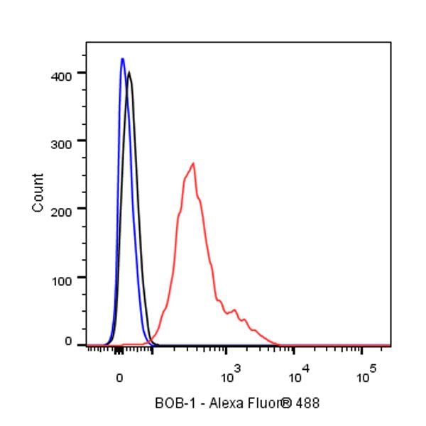
FC
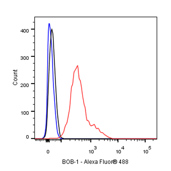
Flow cytometric analysis of 4% PFA fixed 90% methanol permeabilized Ramos (Human Burkitt's lymphoma B lymphocyte) labelling BOB-1 antibody at 1/250 dilution (0.1 μg) / (Red) compared with a Rabbit monoclonal IgG (Black) isotype control and an unlabelled control (cells without incubation with primary antibody and secondary antibody) (Blue). Goat Anti - Rabbit IgG Alexa Fluor® 488 was used as the secondary antibody.
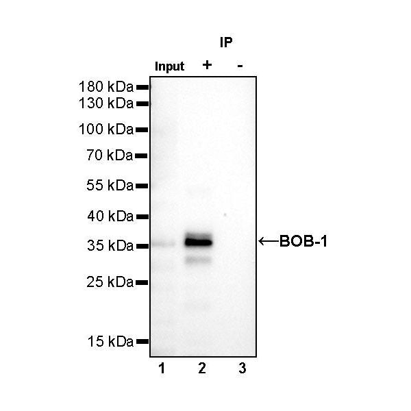
IP
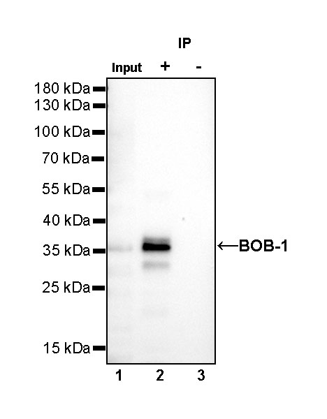
BOB-1 Rabbit mAb at 1/25 dilution (1 µg) immunoprecipitating BOB-1 in 0.4 mg Daudi whole cell lysate.
Western blot was performed on the immunoprecipitate using BOB-1 Rabbit mAb at 1/1000 dilution.
Secondary antibody (HRP) for IP was used at 1/400 dilution.
Lane 1: Daudi whole cell lysate 10 µg (Input)
Lane 2: BOB-1 Rabbit mAb IP in Daudi whole cell lysate
Lane 3: Rabbit monoclonal IgG IP in Daudi whole cell lysate
Predicted MW: 27 kDa
Observed MW: 36 kDa
Exposure time: 15 s
Immunohistochemistry
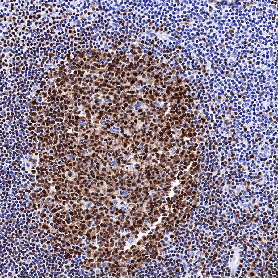
IHC shows positive staining in paraffin-embedded human tonsil. Anti-BOB-1 antibody was used at 1/1000 dilution, followed by a HRP Polymer for Mouse & Rabbit IgG (ready to use). Counterstained with hematoxylin. Heat mediated antigen retrieval with Tris/EDTA buffer pH9.0 was performed before commencing with IHC staining protocol.

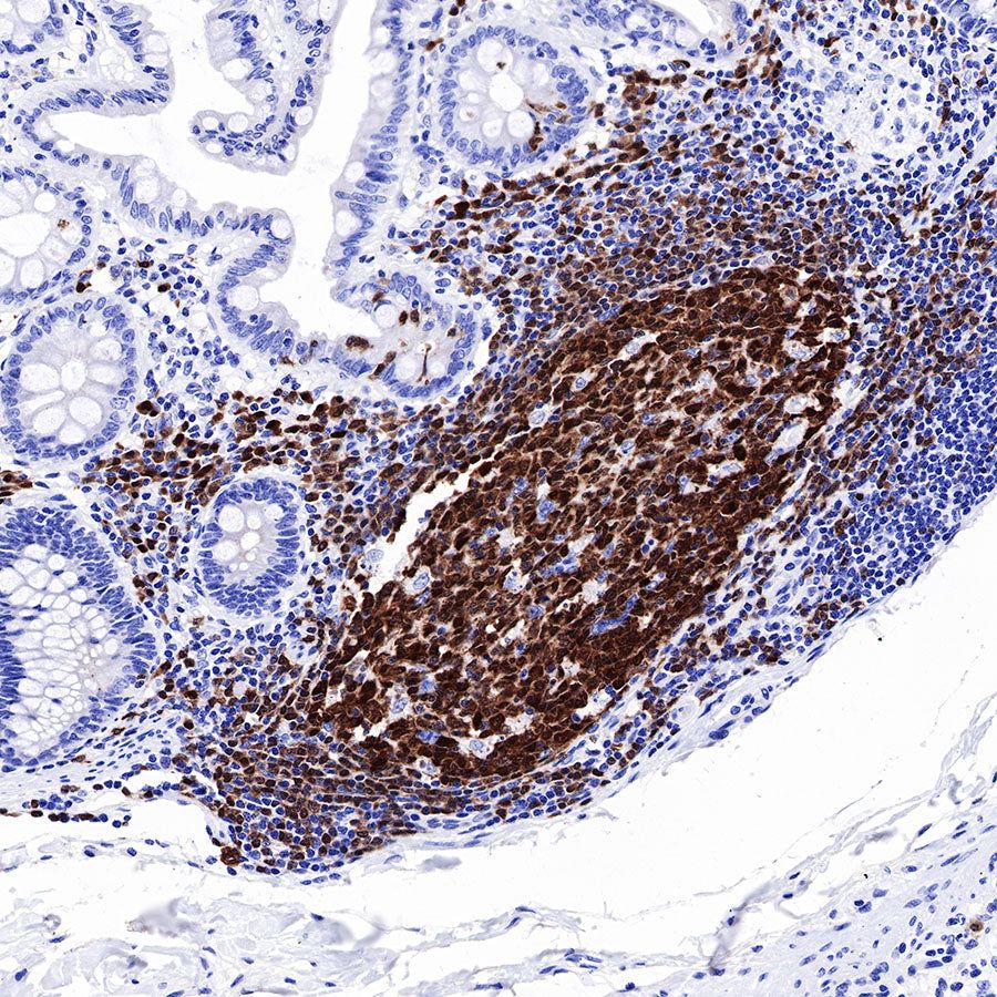
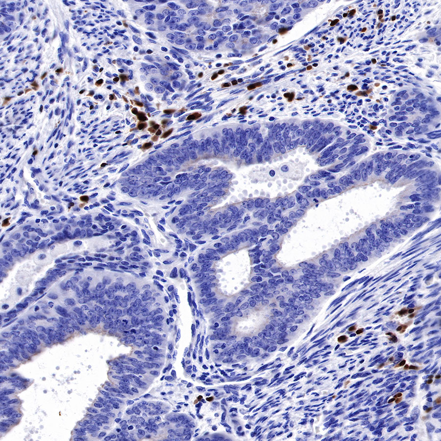
IHC shows positive staining in paraffin-embedded human colon. Anti-BOB-1 antibody was used at 1/1000 dilution, followed by a HRP Polymer for Mouse & Rabbit IgG (ready to use). Counterstained with hematoxylin. Heat mediated antigen retrieval with Tris/EDTA buffer pH9.0 was performed before commencing with IHC staining protocol.
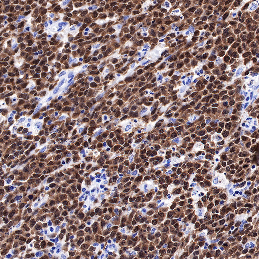
IHC shows positive staining in paraffin-embedded human diffuse large B-cell lymphoma. Anti-BOB-1 antibody was used at 1/1000 dilution, followed by a HRP Polymer for Mouse & Rabbit IgG (ready to use). Counterstained with hematoxylin. Heat mediated antigen retrieval with Tris/EDTA buffer pH9.0 was performed before commencing with IHC staining protocol.
IHC shows positive staining in paraffin-embedded human endometrial cancer. Anti-BOB-1 antibody was used at 1/1000 dilution, followed by a HRP Polymer for Mouse & Rabbit IgG (ready to use). Counterstained with hematoxylin. Heat mediated antigen retrieval with Tris/EDTA buffer pH9.0 was performed before commencing with IHC staining protocol.
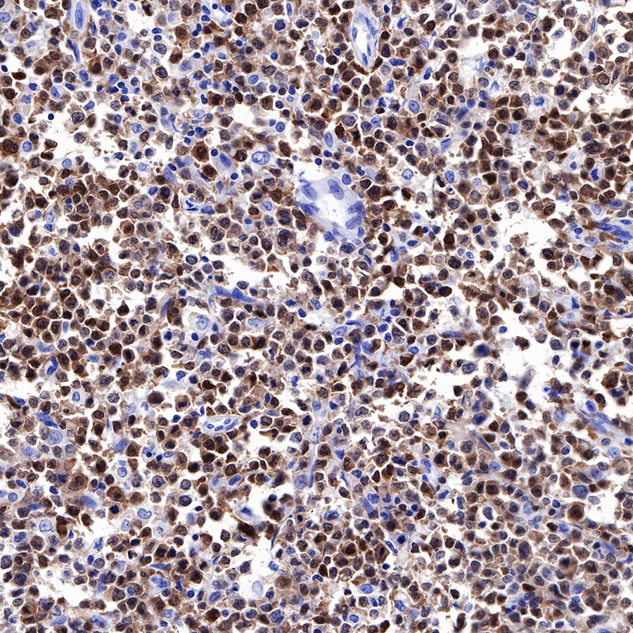
IHC shows positive staining in paraffin-embedded human diffuse large B-cell lymphoma. Anti-BOB-1 antibody was used at 1/1000 dilution, followed by a HRP Polymer for Mouse & Rabbit IgG (ready to use). Counterstained with hematoxylin. Heat mediated antigen retrieval with Tris/EDTA buffer pH9.0 was performed before commencing with IHC staining protocol.
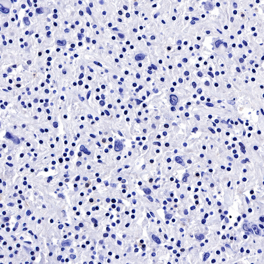
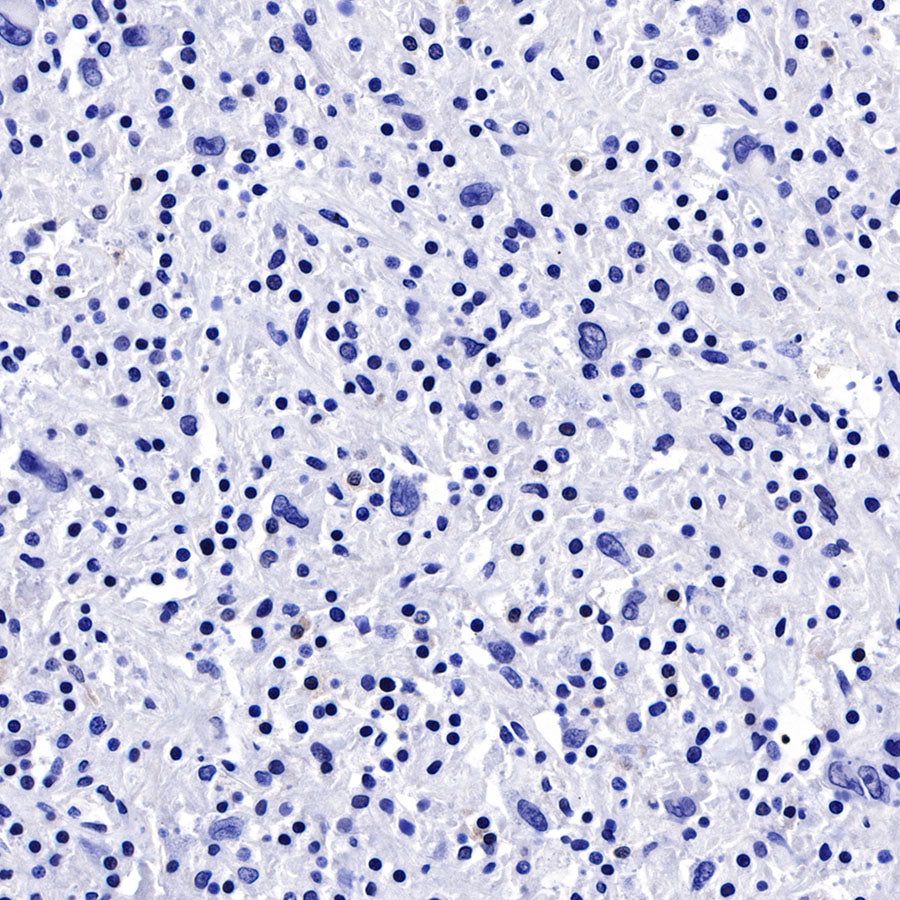
Negative control: IHC shows negative staining in paraffin-embedded human Hodgkin's lymphoma. Anti-BOB-1 antibody was used at 1/1000 dilution, followed by a HRP Polymer for Mouse & Rabbit IgG (ready to use). Counterstained with hematoxylin. Heat mediated antigen retrieval with Tris/EDTA buffer pH9.0 was performed before commencing with IHC staining protocol.
Immunocytochemistry
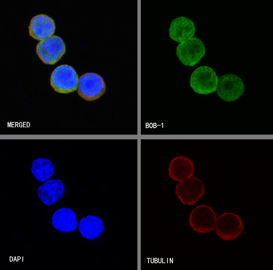
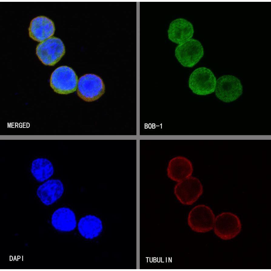
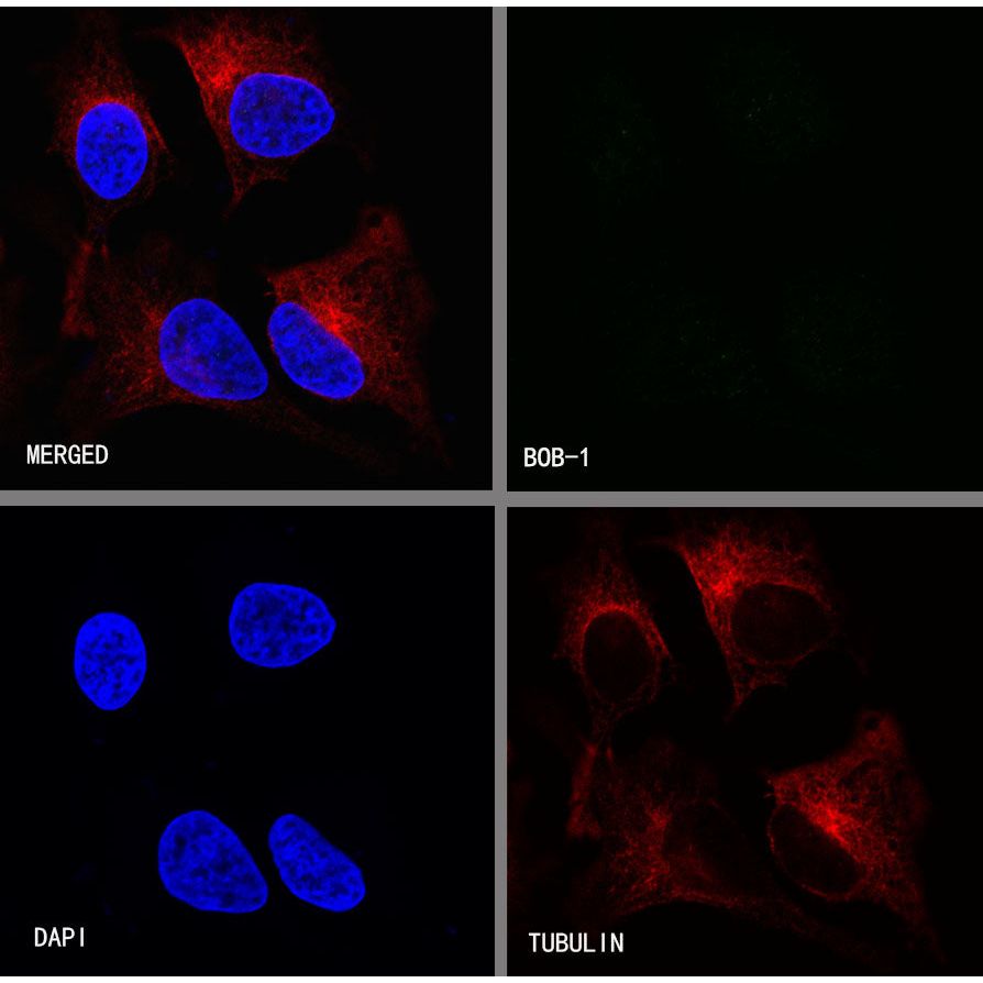
ICC shows positive staining in Ramos cells. Anti-BOB-1 antibody was used at 1/250 dilution (Green) and incubated overnight at 4°C. Goat polyclonal Antibody to Rabbit IgG - H&L (Alexa Fluor® 488) was used as secondary antibody at 1/1000 dilution. The cells were fixed with 4% PFA and permeabilized with 0.1% PBS-Triton X-100. Nuclei were counterstained with DAPI (Blue). Counterstain with tubulin (red).
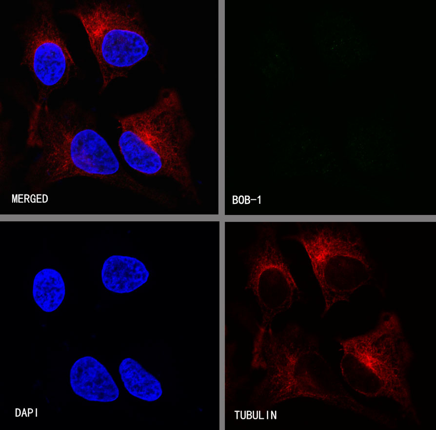
Negative control:ICC shows negative staining in HeLa cells. Anti-BOB-1 antibody was used at 1/250 dilution and incubated overnight at 4°C. Goat polyclonal Antibody to Rabbit IgG - H&L (Alexa Fluor® 488) was used as secondary antibody at 1/1000 dilution. The cells were fixed with 4% PFA and permeabilized with 0.1% PBS-Triton X-100. Nuclei were counterstained with DAPI (Blue). Counterstain with tubulin (red).