
WB result of Factor XIIIa Rabbit mAb
Primary antibody: Factor XIIIa Rabbit mAb at 1/1000 dilution
Lane 1: Human plasma lysate 5 µg
Secondary antibody: Goat Anti-Rabbit IgG, (H+L), HRP conjugated at 1/10000 dilution
Predicted MW: 68 kDa
Observed MW: 84 kDa
Factor XIIIa Recombinant Rabbit mAb (SDT-644-7)
Factor XIIIa Recombinant Rabbit mAb (SDT-644-7)
Price:
Regular price
$131.00 SGD
Regular price
Sale price
$131.00 SGD
Unit price
per
For shipping services or bulk orders, you may request a quotation.
Secure checkout with

View full details
Product Details
Product Details
Product Specification
| Host | Rabbit |
| Antigen | Factor XIIIa |
| Synonyms | Coagulation factor XIII A chain, Coagulation factor XIIIa, Protein-glutamine gamma-glutamyltransferase A chain, Transglutaminase A chain, F13A1, F13A, FXIIIA |
| Immunogen | Synthetic Peptide |
| Location | Cytoplasm, Secreted |
| Accession | P00488 |
| Clone Number | SDT-644-7 |
| Antibody Type | Recombinant mAb |
| Isotype | IgG |
| Application | WB, IHC-P, IP |
| Reactivity | Hu |
| Purification | Protein A |
| Concentration | 0.5 mg/ml |
| Conjugation | Unconjugated |
| Physical Appearance | Liquid |
| Storage Buffer | PBS, 40% Glycerol, 0.05%BSA, 0.03% Proclin 300 |
| Stability & Storage | 12 months from date of receipt / reconstitution, -20 °C as supplied |
Dilution
| application | dilution | species |
| WB | 1:1000 | |
| IHC | 1:1000 | |
| IP | 1:50 |
Background
Factor XIII is a betaglobulin found in plasma and is composed of two subunits. Factor XIIIA is the catalytic subunit. Factor XIIIa is expressed in platelets, megakaryocytes, fibroblast-like cells in the placenta, uterus, and prostate, monocytes and macrophages and dermal dendritic cells. It is most commonly recognized as a marker of fibrohistiocytic proliferations. Factor XIIIa has also been used with CD34 to differentiate between dermatofibroma and dermatofibrosarcoma protuberans.
Picture
Picture
Western Blot
IP
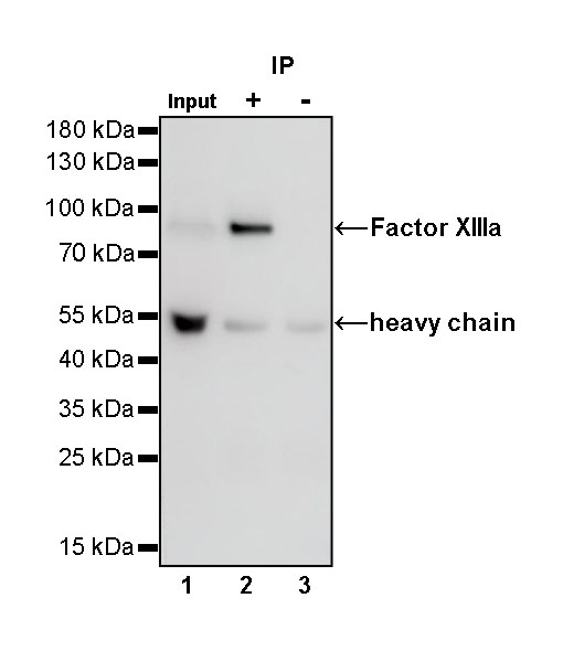
Factor XIIIa Rabbit mAb at 1/50 dilution (1 µg) immunoprecipitating Factor XIIIa in 0.4 mg human plasma lysate.
Western blot was performed on the immunoprecipitate using Factor XIIIa Rabbit mAb at 1/1000 dilution.
Secondary antibody (HRP) for IP was used at 1/400 dilution.
Lane 1: human plasma lysate 20 µg (Input)
Lane 2: Factor XIIIa Rabbit mAb IP in human plasma lysate
Lane 3: Rabbit monoclonal IgG IP in human plasma lysate
Predicted MW: 68 kDa
Observed MW: 84 kDa
Immunohistochemistry
IHC shows positive staining in paraffin-embedded human tonsil. Anti-Factor XIIIa antibody was used at 1/1000 dilution, followed by a HRP Polymer for Mouse & Rabbit IgG (ready to use). Counterstained with hematoxylin. Heat mediated antigen retrieval with Tris/EDTA buffer pH9.0 was performed before commencing with IHC staining protocol.
IHC shows positive staining in paraffin-embedded human spleen. Anti-Factor XIIIa antibody was used at 1/1000 dilution, followed by a HRP Polymer for Mouse & Rabbit IgG (ready to use). Counterstained with hematoxylin. Heat mediated antigen retrieval with Tris/EDTA buffer pH9.0 was performed before commencing with IHC staining protocol.
IHC shows positive staining in paraffin-embedded human placenta. Anti-Factor XIIIa antibody was used at 1/1000 dilution, followed by a HRP Polymer for Mouse & Rabbit IgG (ready to use). Counterstained with hematoxylin. Heat mediated antigen retrieval with Tris/EDTA buffer pH9.0 was performed before commencing with IHC staining protocol.
IHC shows positive staining in paraffin-embedded human breast cancer. Anti-Factor XIIIa antibody was used at 1/1000 dilution, followed by a HRP Polymer for Mouse & Rabbit IgG (ready to use). Counterstained with hematoxylin. Heat mediated antigen retrieval with Tris/EDTA buffer pH9.0 was performed before commencing with IHC staining protocol.
IHC shows positive staining in paraffin-embedded human fibrous histiocytoma. Anti-Factor XIIIa antibody was used at 1/1000 dilution, followed by a HRP Polymer for Mouse & Rabbit IgG (ready to use). Counterstained with hematoxylin. Heat mediated antigen retrieval with Tris/EDTA buffer pH9.0 was performed before commencing with IHC staining protocol.
IHC shows positive staining in paraffin-embedded human dermatofibrosarcoma protuberans. Anti-Factor XIIIa antibody was used at 1/1000 dilution, followed by a HRP Polymer for Mouse & Rabbit IgG (ready to use). Counterstained with hematoxylin. Heat mediated antigen retrieval with Tris/EDTA buffer pH9.0 was performed before commencing with IHC staining protocol.
