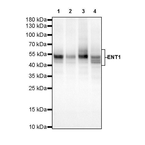
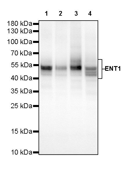
WB result of ENT1 Rabbit mAb
Primary antibody: ENT1 Rabbit mAb at 1/1000 dilution
Lane 1: HEK-293 whole cell lysate 20 µg
Lane 2: 293T whole cell lysate 20 µg
Lane 3: HeLa whole cell lysate 20 µg
Lane 4: U-87 MG whole cell lysate 20 µg
Secondary antibody: Goat Anti-Rabbit IgG, (H+L), HRP conjugated at 1/10000 dilution
Predicted MW: 50 kDa
Observed MW: 40~60 kDa
(This blot was developed with high sensitivity substrate)
ENT1 Recombinant Rabbit mAb (SDT-R182)
ENT1 Recombinant Rabbit mAb (SDT-R182)
Price:
Regular price
$131.00 SGD
Regular price
Sale price
$131.00 SGD
Unit price
per
For shipping services or bulk orders, you may request a quotation.
Secure checkout with

View full details
Product Details
Product Details
Product Specification
| Host | Rabbit |
| Antigen | ENT1 |
| Synonyms | Equilibrative nucleoside transporter 1, hENT1, SLC29A1 |
| Immunogen | N/A |
| Location | Membrane |
| Accession | Q99808 |
| Clone Number | SDT-R182 |
| Antibody Type | Rabbit mAb |
| Application | WB, IHC-P, ICC, ICFCM |
| Reactivity | Hu |
| Purification | Protein A |
| Concentration | 0.5 mg/ml |
| Conjugation | Unconjugated |
| Physical Appearance | Liquid |
| Storage Buffer | PBS, 40% Glycerol, 0.05% BSA, 0.03% Proclin 300 |
| Stability & Storage | 12 months from date of receipt / reconstitution, -20 °C as supplied |
Dilution
| application | dilution | species |
| WB | 1:1000 | null |
| IHC | 1:500 | null |
| ICC | 1:500 | null |
| FC | 1:50 | null |
Background
Equilibrative nucleoside transporter 1 (ENT1) is a protein that in humans is encoded by the SLC29A1 gene [PMID: 8986748]. Multiple alternatively spliced variants, encoding the same protein, have been found for this gene. Expressed on red blood cell surfaces, these variants make up the Augustine blood group system [PMID: 27834482].
Picture
Picture
Western Blot
FC
Flow cytometric analysis of 4% PFA fixed 90% methanol permeabilized HepG2 (Human hepatocellular carcinoma epithelial cell) cells labelling ENT1 antibody at 1/50 dilution (1 μg)/ (red) compared with a Rabbit monoclonal IgG (Black) isotype control and an unlabelled control (cells without incubation with primary antibody and secondary antibody) (Blue). Goat Anti-Rabbit IgG Alexa Fluor® 488 was used as the secondary antibody.
Immunohistochemistry
IHC shows positive staining in paraffin-embedded human tonsil. Anti-ENT1 antibody was used at 1/500 dilution, followed by a HRP Polymer for Mouse & Rabbit IgG (ready to use). Counterstained with hematoxylin. Heat mediated antigen retrieval with Tris/EDTA buffer pH9.0 was performed before commencing with IHC staining protocol.
IHC shows positive staining in paraffin-embedded human pancreas. Anti-ENT1 antibody was used at 1/500 dilution, followed by a HRP Polymer for Mouse & Rabbit IgG (ready to use). Counterstained with hematoxylin. Heat mediated antigen retrieval with Tris/EDTA buffer pH9.0 was performed before commencing with IHC staining protocol.
IHC shows positive staining in paraffin-embedded human ovarian carcinoma. Anti-ENT1 antibody was used at 1/500 dilution, followed by a HRP Polymer for Mouse & Rabbit IgG (ready to use). Counterstained with hematoxylin. Heat mediated antigen retrieval with Tris/EDTA buffer pH9.0 was performed before commencing with IHC staining protocol.
IHC shows positive staining in paraffin-embedded human lung squamous cell carcinoma. Anti-ENT1 antibody was used at 1/500 dilution, followed by a HRP Polymer for Mouse & Rabbit IgG (ready to use). Counterstained with hematoxylin. Heat mediated antigen retrieval with Tris/EDTA buffer pH9.0 was performed before commencing with IHC staining protocol.
Immunocytochemistry
ICC shows positive staining in HepG2 cells. Anti-ENT1 antibody was used at 1/500 dilution (Green) and incubated overnight at 4°C. Goat polyclonal Antibody to Rabbit IgG - H&L (Alexa Fluor® 488) was used as secondary antibody at 1/1000 dilution. The cells were fixed with 100% ice-cold methanol and permeabilized with 0.1% PBS-Triton X-100. Nuclei were counterstained with DAPI.