Product Specification
| Host |
Rabbit |
| Antigen |
CD38 |
| Synonyms |
2'-phospho-ADP-ribosyl cyclase; 2'-phospho-cyclic-ADP-ribose transferase; ADP-ribosyl cyclase 1 (ADPRC 1); Cyclic ADP-ribose hydrolase 1 (cADPr hydrolase 1); T10 |
| Immunogen |
Synthetic Peptide |
| Location |
Membrane |
| Accession |
P28907 |
| Clone Number |
SDT-031-45 |
| Antibody Type |
Rabbit mAb |
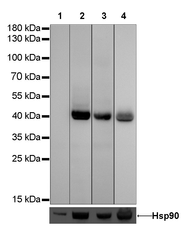
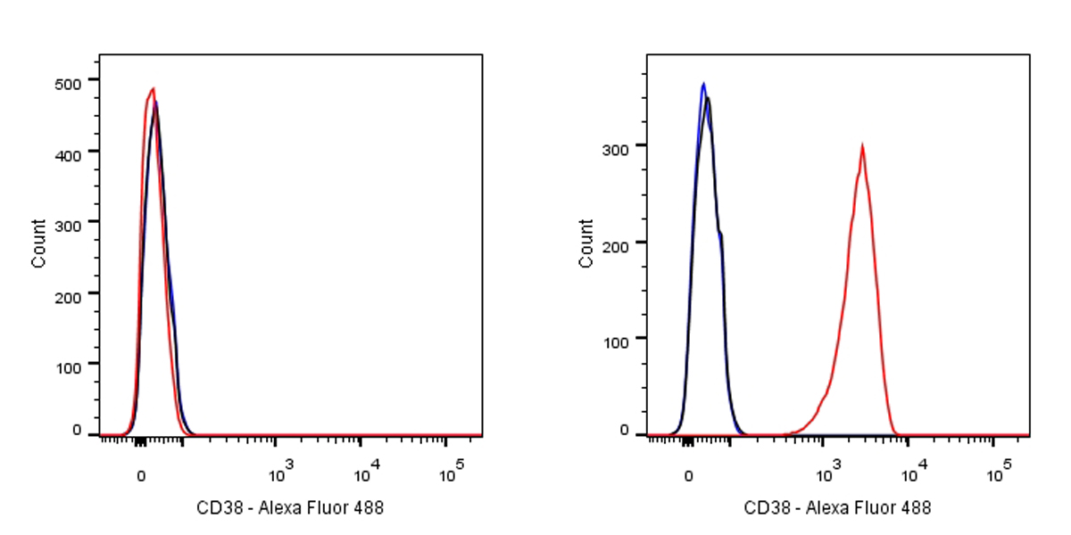
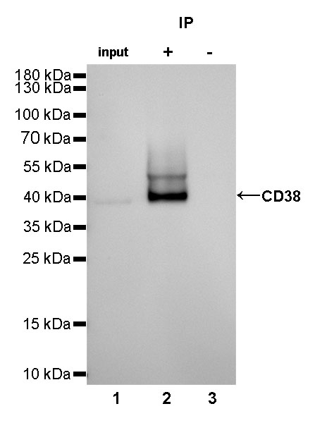
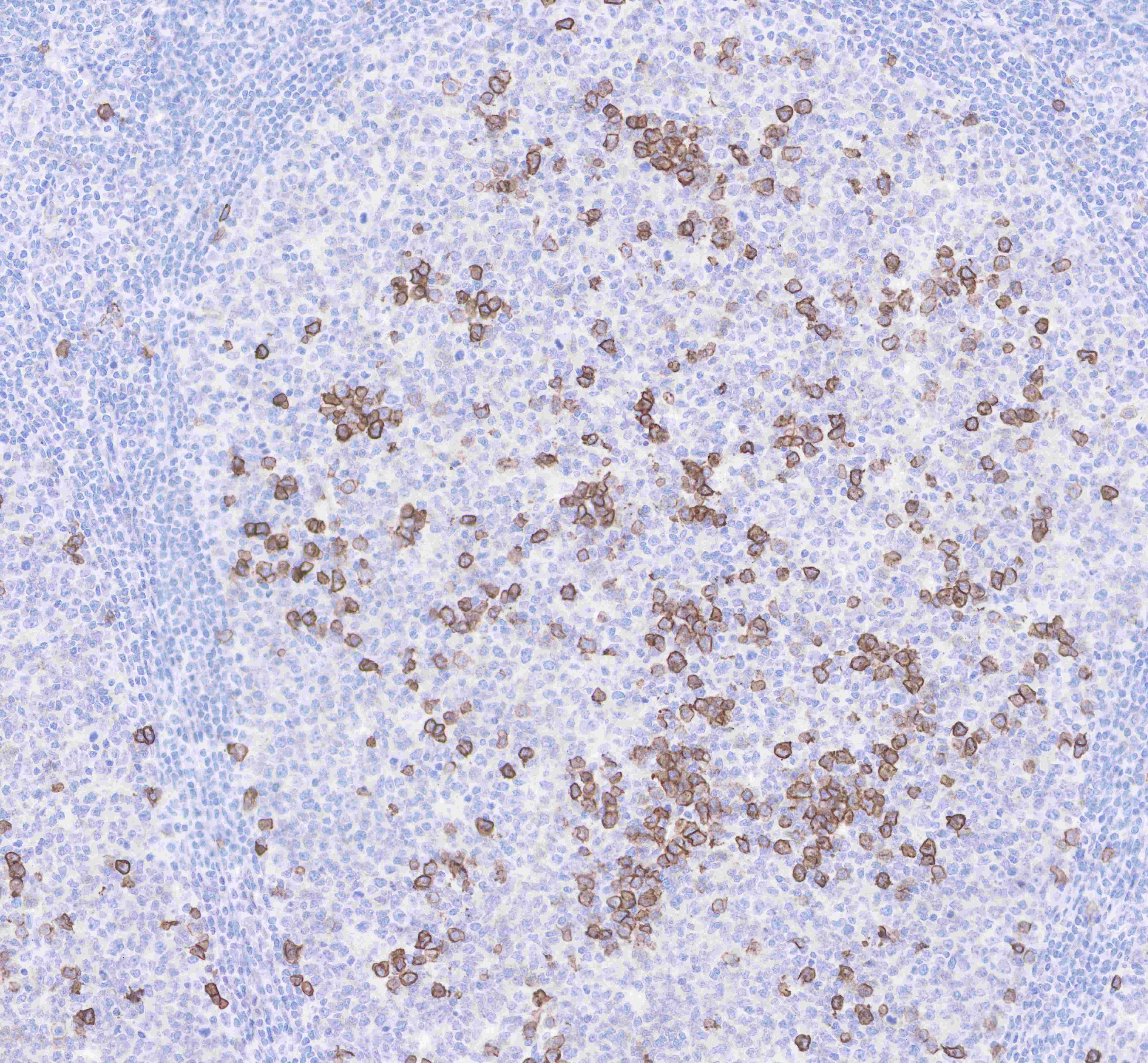
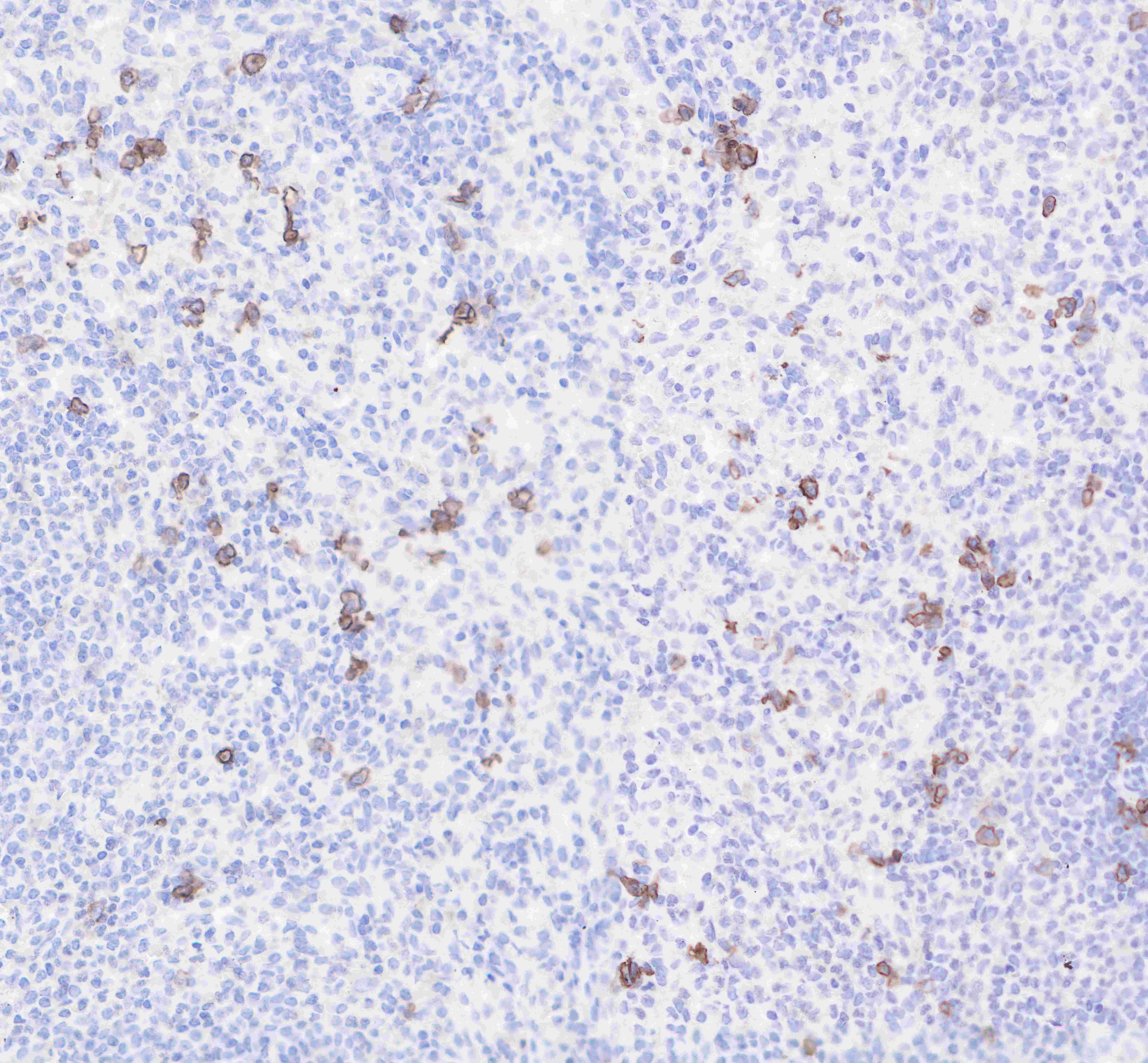
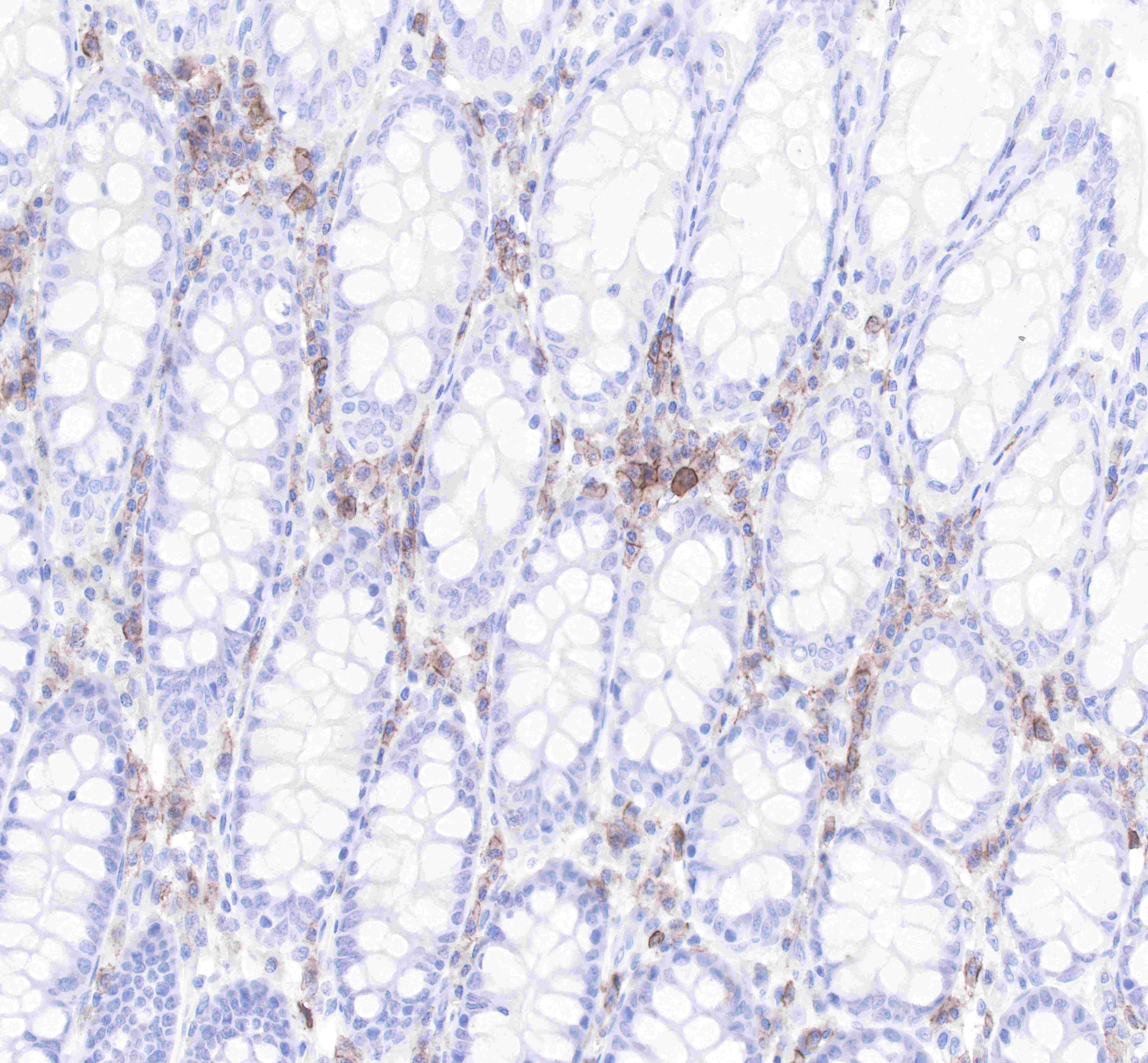
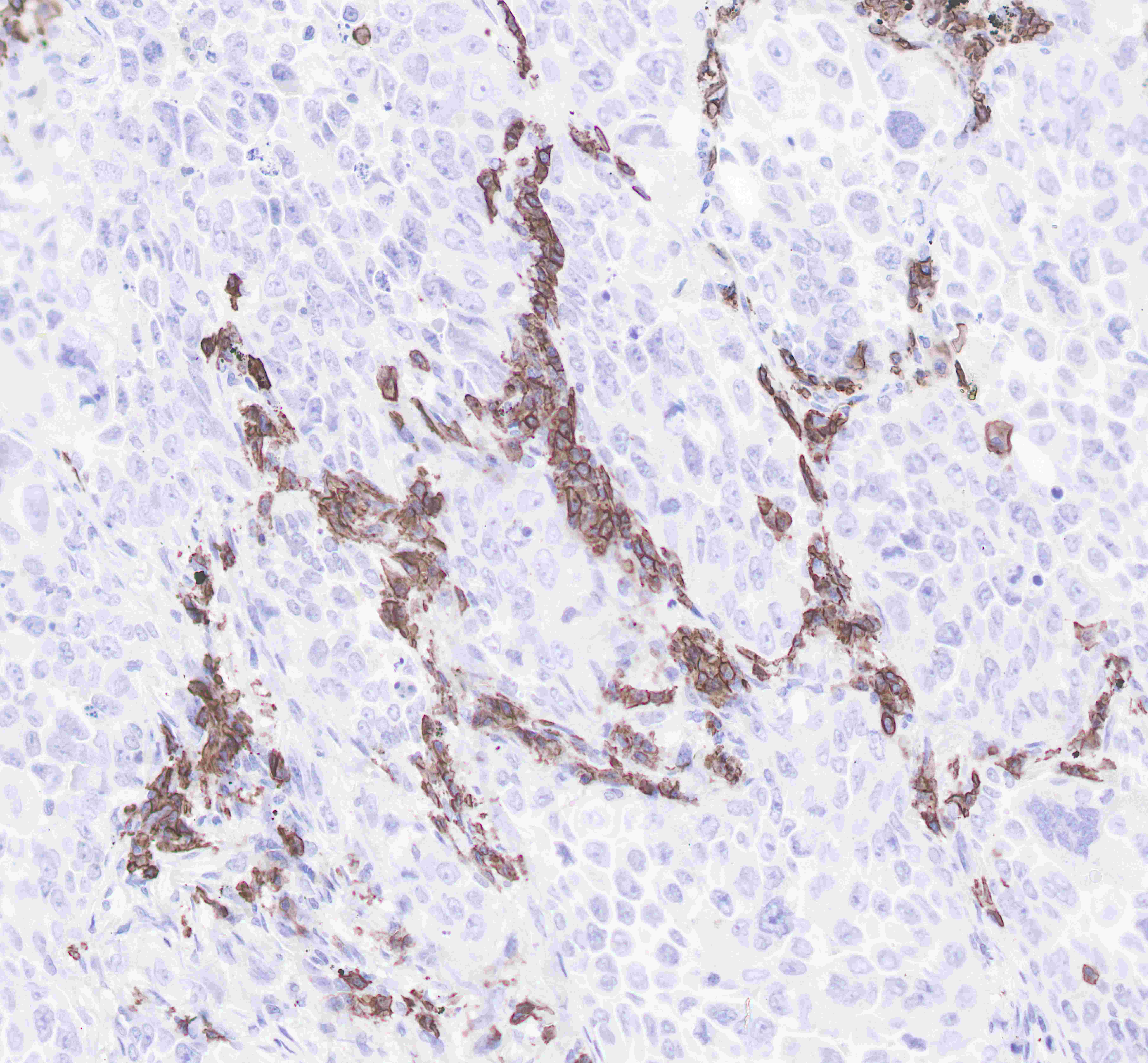
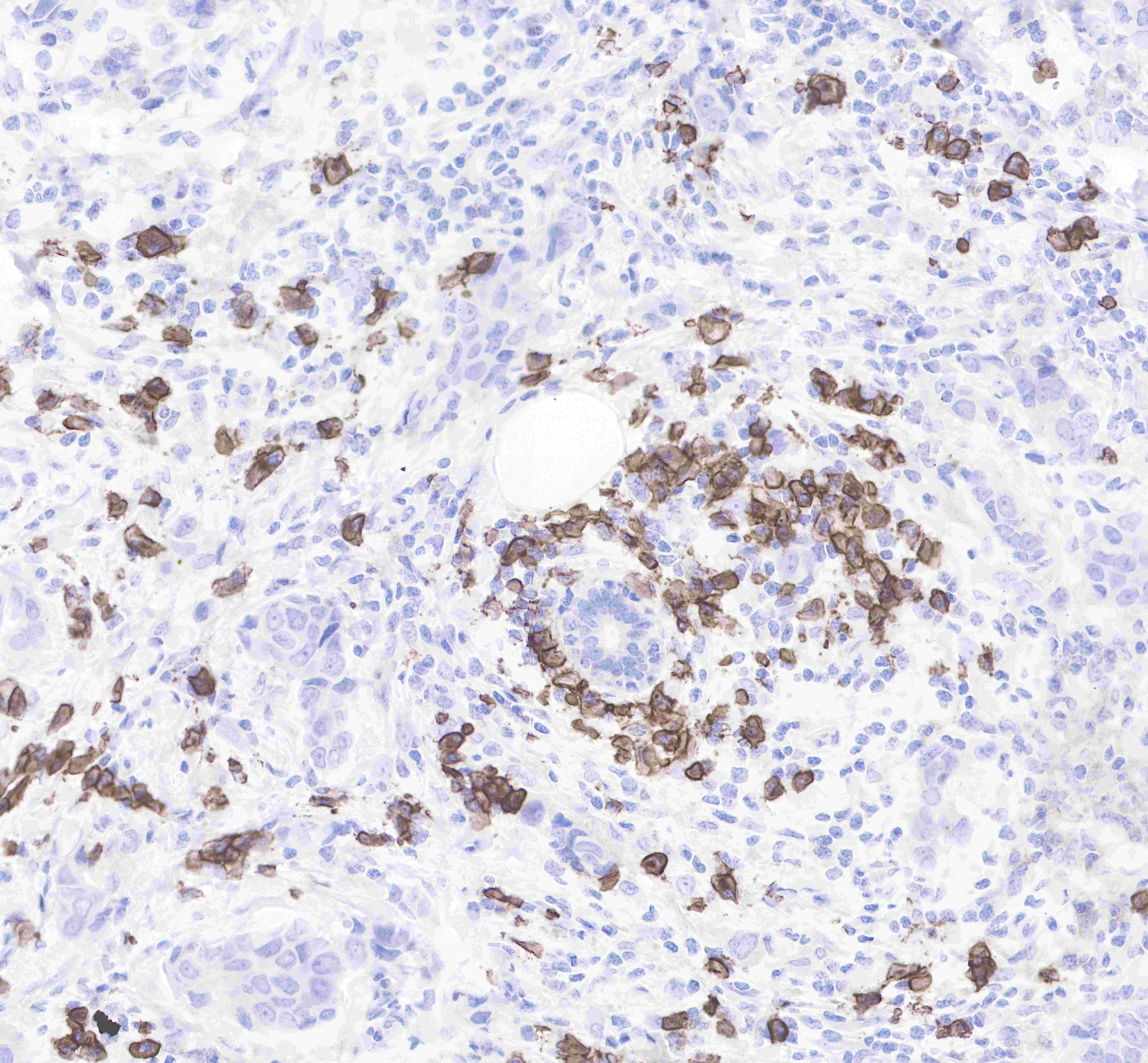
| Application |
WB, IHC-P, ICC, FCM, IP |
| Reactivity |
Hu |
| Purification |
Protein A |
| Concentration |
0.5mg/ml |
| Molecular Weight |
34-45kDa |
| Conjugation |
Unconjugated |
| Physical Appearance |
Liquid |
| Storage Buffer |
PBS, 40% Glycerol, 0.05%BSA, 0.03% Proclin 300 |
| Stability & Storage |
12 months from date of receipt / reconstitution, -20 °C as supplied |
Dilution
| application |
dilution |
species |
| IP |
1:25 |
null |
| IHC-P |
1:2000 |
null |
| ICC |
1:100 |
null |
| WB |
1:1000 |
null |
| FCM |
1:500 |
null |
Background
CD38 (cluster of differentiation 38), also known as cyclic ADP ribose hydrolase is a glycoprotein found on the surface of many immune cells (white blood cells), including CD4+, CD8+, B lymphocytes and natural killer cells. CD38 functions in cell adhesion, signal transduction and calcium signaling. The loss of CD38 function is associated with impaired immune responses, metabolic disturbances, and behavioral modifications including social amnesia possibly related to autism. The CD38 protein is a marker of cell activation. It has been connected to HIV infection, leukemias, myelomas, solid tumors, type II diabetes mellitus and bone metabolism, as well as some genetically determined conditions.