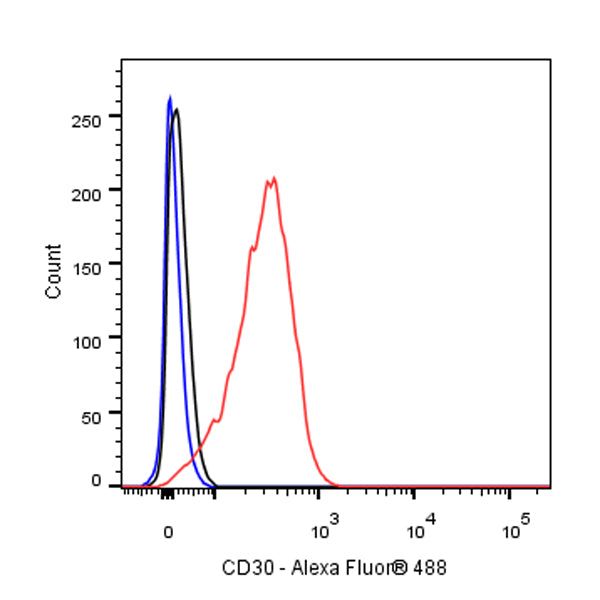
Flow cytometric analysis of 4% PFA fixed 90% methanol permeabilized Jurkat (Human T cell leukemia T lymphocyte) labelling CD30 antibody at 1/25 dilution (1 μg) / (Red) compared with a Rabbit monoclonal IgG (Black) isotype control and an unlabelled control (cells without incubation with primary antibody and secondary antibody) (Blue). Goat Anti - Rabbit IgG Alexa Fluor® 488 was used as the secondary antibody.
CD30 Recombinant Rabbit mAb (SDT-216-2)
CD30 Recombinant Rabbit mAb (SDT-216-2)
Price:
Regular price
$129.00 SGD
Regular price
Sale price
$129.00 SGD
Unit price
per
For shipping services or bulk orders, you may request a quotation.
Secure checkout with

View full details
Product Details
Product Details
Product Specification
| Host | Rabbit |
| Antigen | CD30 |
| Synonyms | Ki-1, KI-30 Ber-H2, Tumor necrosis factor receptor superfamily member 8, TNFRSF8, CD30L receptor, Ki-1 antigen, Lymphocyte activation antigen CD30 |
| Immunogen | Synthetic Peptide |
| Location | Cytoplasm, Cell membrane |
| Accession | P28908 |
| Clone Number | SDT-216-2 |
| Antibody Type | Recombinant mAb |
| Application | IHC-P, ICC, ICFCM |
| Reactivity | Hu, Ms |
| Purification | Protein A |
| Concentration | 0.25 mg/ml |
| Physical Appearance | Liquid |
| Storage Buffer | PBS, 40% Glycerol, 0.05% BSA, 0.03% Proclin 300 |
| Stability & Storage | 12 months from date of receipt / reconstitution, -20 °C as supplied |
Dilution
| application | dilution | species |
| IHC-P | 1:500 | |
| ICC | 1:250 | |
| ICFCM | 1:25 |
Background
CD30 is a transmembrane protein from the tumour necrosis factor receptor superfamily. It is expressed on a small subset of activated T and B lymphocytes, and various lymphoid neoplasms. CD30 is a particularly treatment target because its levels are high in tumours but low in healthy tissues. Several therapeutic strategies targeting CD30 have been developed, including monoclonal antibodies, conjugated antibodies (combination of brentuximab vedotin with chemotherapy or immunotherapy), bispecific antibodies and cell and gene therapies, such as anti-CD30 CAR-T cells in particular [PMID: 37170397].
Picture
Picture
FC
Immunohistochemistry

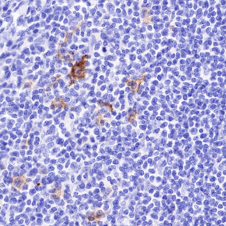
IHC shows positive staining in paraffin-embedded human tonsil. Anti-CD30 antibody was used at 1/1000 dilution, followed by a HRP Polymer for Mouse & Rabbit IgG (ready to use). Counterstained with hematoxylin. Heat mediated antigen retrieval with Tris/EDTA buffer pH9.0 was performed before commencing with IHC staining protocol.
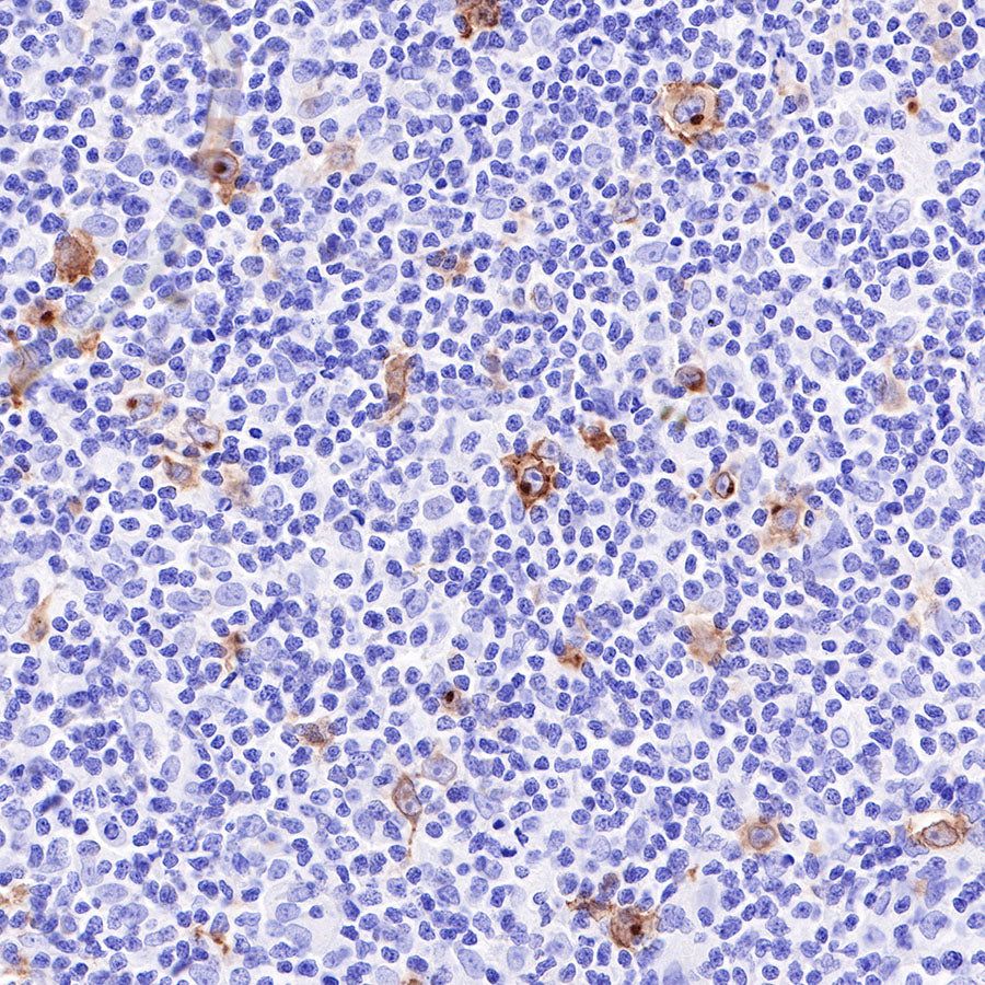
IHC shows positive staining in paraffin-embedded human Hodgkin's lymphoma. Anti-CD30 antibody was used at 1/1000 dilution, followed by a HRP Polymer for Mouse & Rabbit IgG (ready to use). Counterstained with hematoxylin. Heat mediated antigen retrieval with Tris/EDTA buffer pH9.0 was performed before commencing with IHC staining protocol.
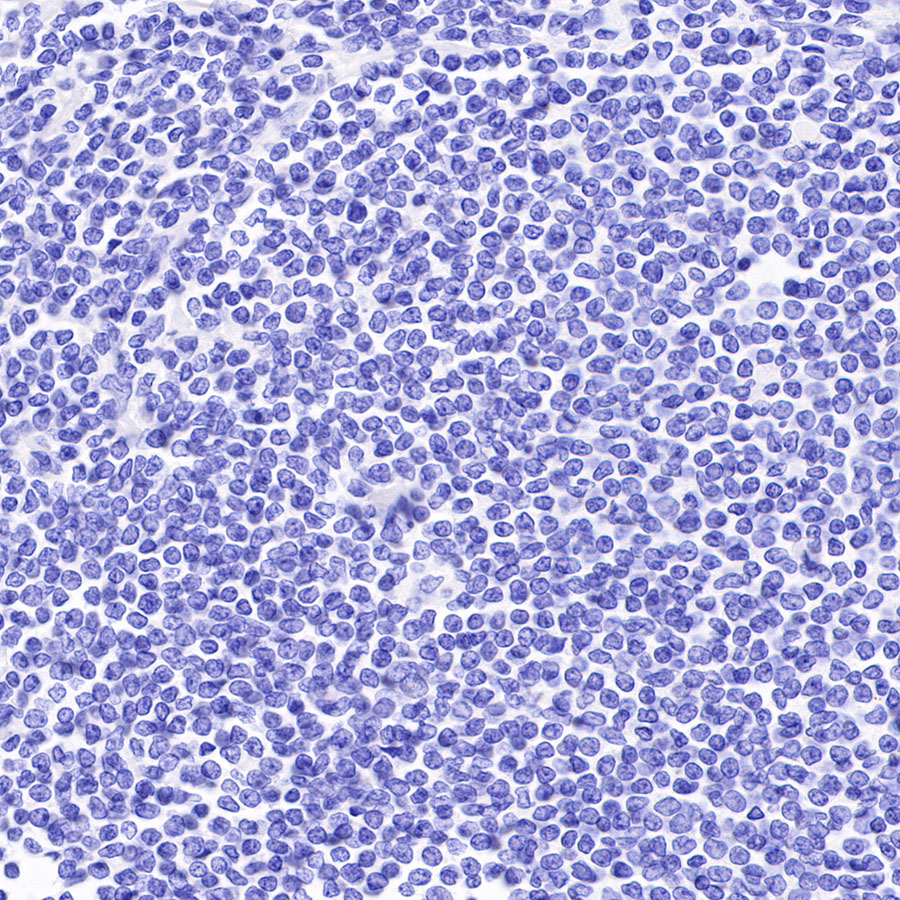
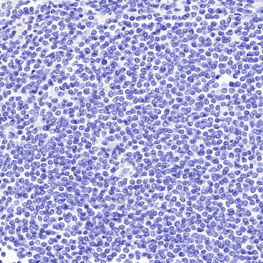
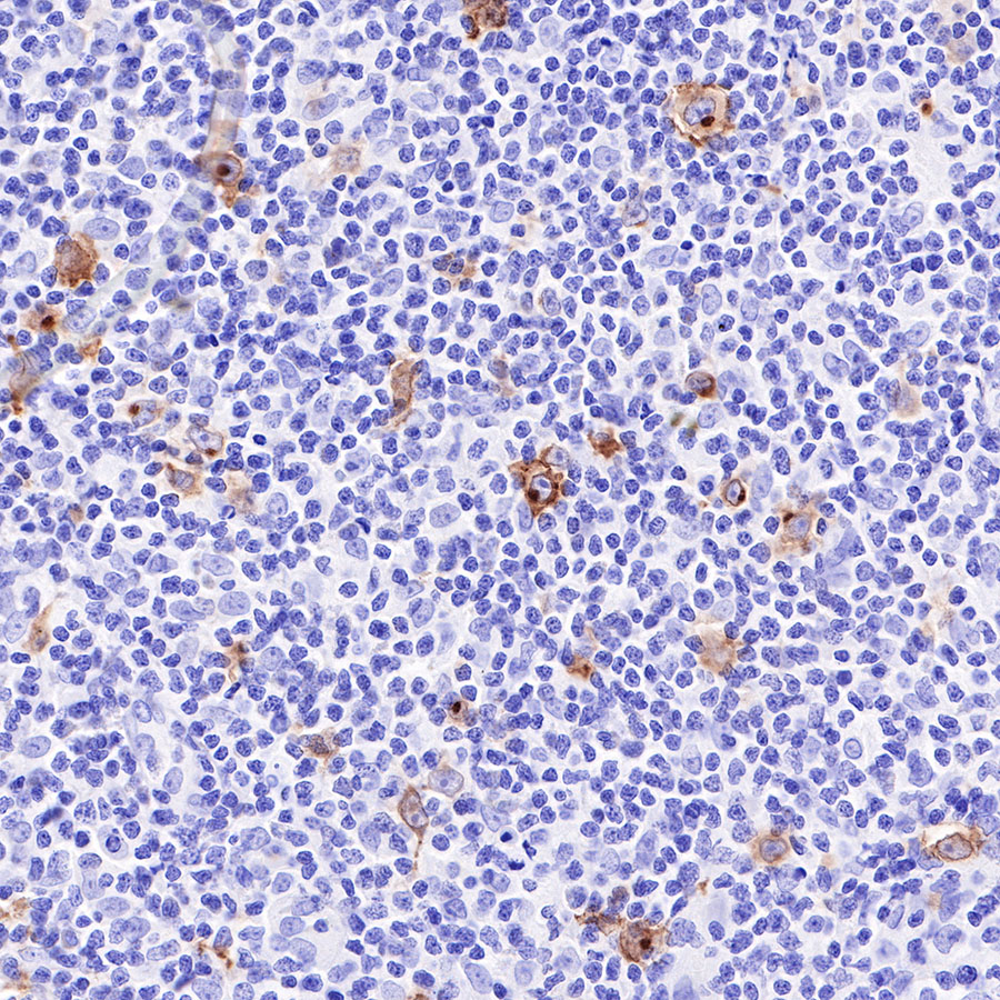
Negative control: IHC shows negative staining in paraffin-embedded human mantle cell lymphoma. Anti-CD30 antibody was used at 1/1000 dilution, followed by a HRP Polymer for Mouse & Rabbit IgG (ready to use). Counterstained with hematoxylin. Heat mediated antigen retrieval with Tris/EDTA buffer pH9.0 was performed before commencing with IHC staining protocol.

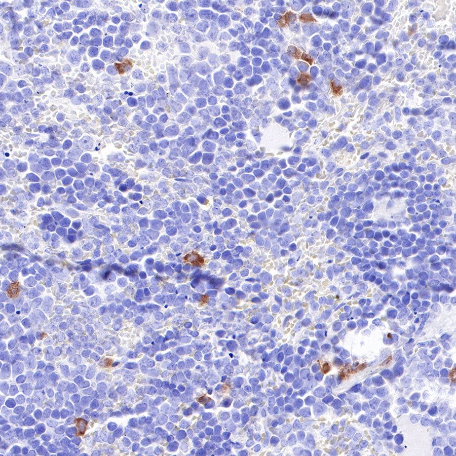
IHC shows positive staining in paraffin-embedded mouse spleen. Anti-CD30 antibody was used at 1/1000 dilution, followed by a HRP Polymer for Mouse & Rabbit IgG (ready to use). Counterstained with hematoxylin. Heat mediated antigen retrieval with Tris/EDTA buffer pH9.0 was performed before commencing with IHC staining protocol.
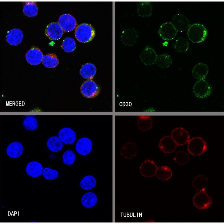
Immunocytochemistry
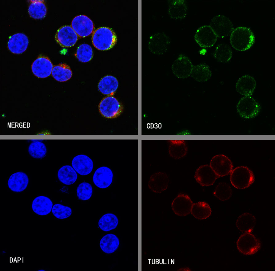
ICC shows positive staining in Jurkat cells. Anti-CD30 antibody was used at 1/250 dilution (Green) and incubated overnight at 4°C. Goat polyclonal Antibody to Rabbit IgG - H&L (Alexa Fluor® 488) was used as secondary antibody at 1/1000 dilution. The cells were fixed with 4%PFA and permeabilized with 0.1% PBS-Triton X-100. Nuclei were counterstained with DAPI (Blue). Counterstain with tubulin (red).