Androgen Receptor Recombinant Rabbit mAb (SDT-R157)
Androgen Receptor Recombinant Rabbit mAb (SDT-R157)
Product Details
Product Details
Product Specification
| Host | Rabbit |
| Antigen | Androgen Receptor |
| Synonyms | AR, Dihydrotestosterone receptor, Nuclear receptor subfamily 3 group C member 4 |
| Immunogen | N/A |
| Location | Nucleus |
| Accession | P10275 |
| Clone Number | SDT-R157 |
| Antibody Type | Recombinant mAb |
| Application | WB, IHC-P, ICC, ICFCM |
| Reactivity | Hu, Ms, Rt |
| Purification | Protein A |
| Concentration | 0.5 mg/ml |
| Physical Appearance | Liquid |
| Storage Buffer | PBS, 40% Glycerol, 0.05% BSA, 0.03% Proclin 300 |
| Stability & Storage | 12 months from date of receipt / reconstitution, -20 °C as supplied |
Dilution
| application | dilution | species |
| WB | 1:1000 | null |
| IHC-P | 1:1000 | null |
| ICFCM | 1:500 | null |
| ICC | 1:500 | null |
Background
The androgen receptor (AR), ligand-induced transcription factor, is expressed in primary prostate cancer and in metastases. AR regulates multiple cellular events, proliferation, apoptosis, migration, invasion, and differentiation. Its expression in prostate cancer cells is regulated by steroid and peptide hormones [PMID: 24384911]. The elucidation of the structures of the AR DNA binding domain (DBD) and ligand binding domain (LBD) provides a new framework for understanding the functions of this receptor and leads to the development of rational drug design for the treatment of prostate cancer [PMID: 24909511].
Picture
Picture
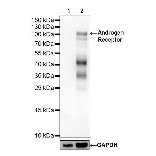
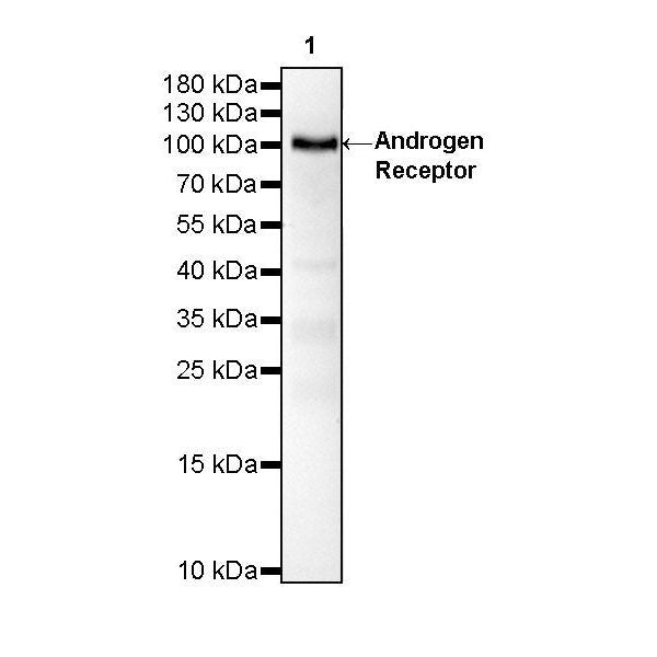
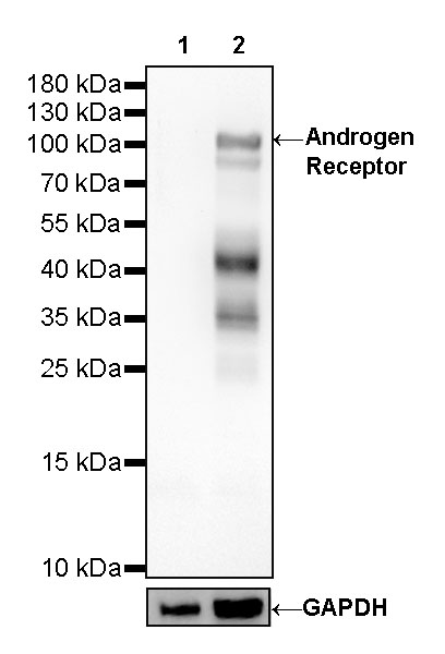
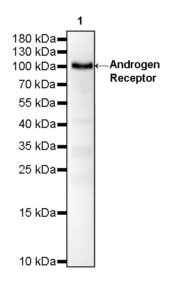
Western Blot
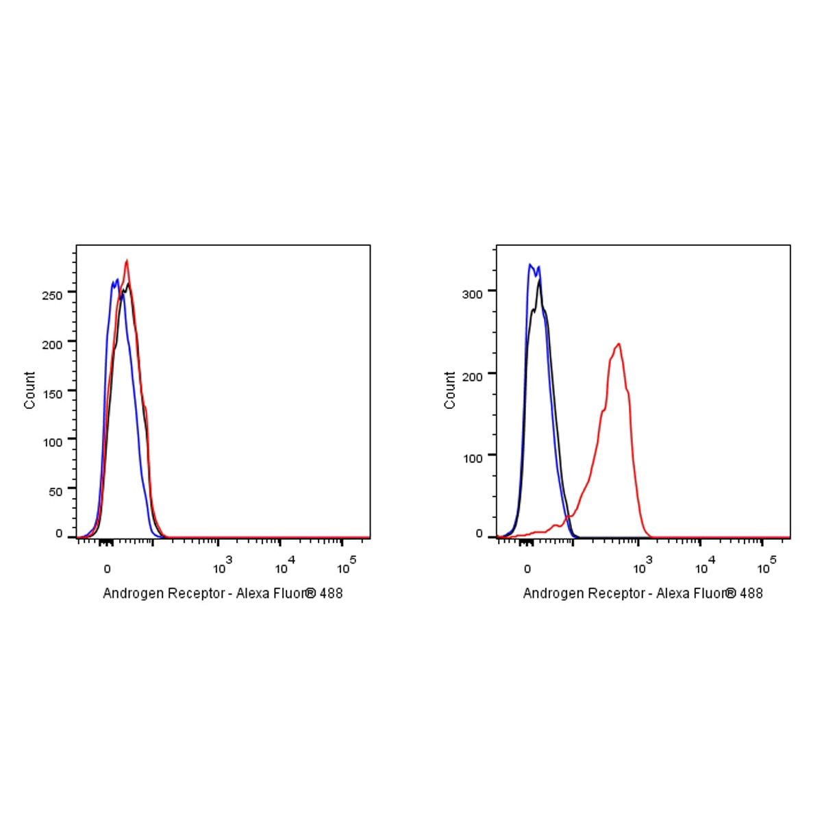
FC
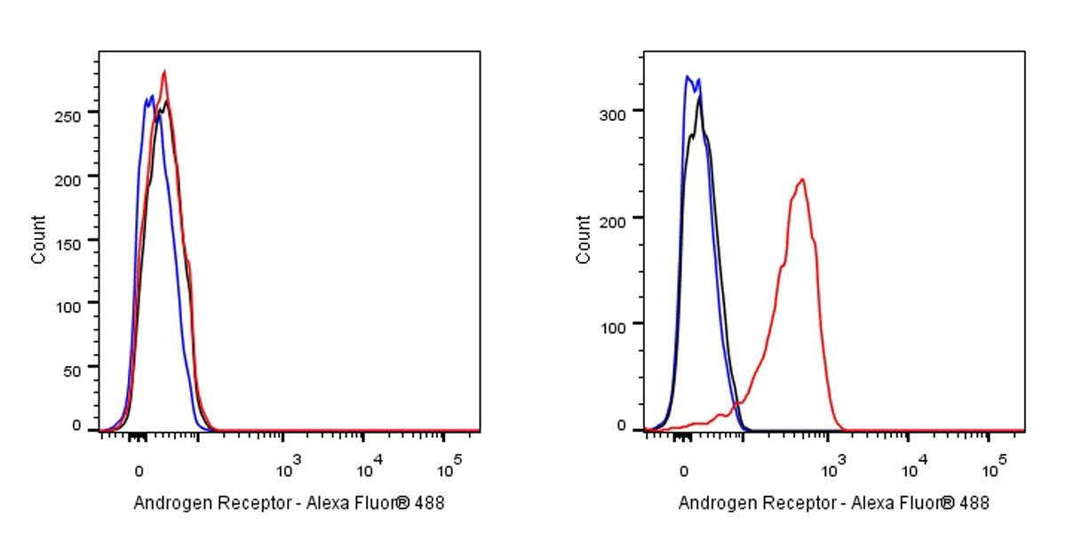
Flow cytometric analysis of 4% PFA fixed 90% methanol permeabilized Jurkat (Human T cell leukemia T lymphocyte, left) / LNCAP (Human prostate carcinoma epithelial cell, right) cells labelling Androgen Receptor antibody at 1/500 dilution (0.1 μg) / (red) compared with a Rabbit monoclonal IgG (Black) isotype control and an unlabelled control (cells without incubation with primary antibody and secondary antibody) (Blue). Goat Anti - Rabbit IgG Alexa Fluor® 488 was used as the secondary antibody.
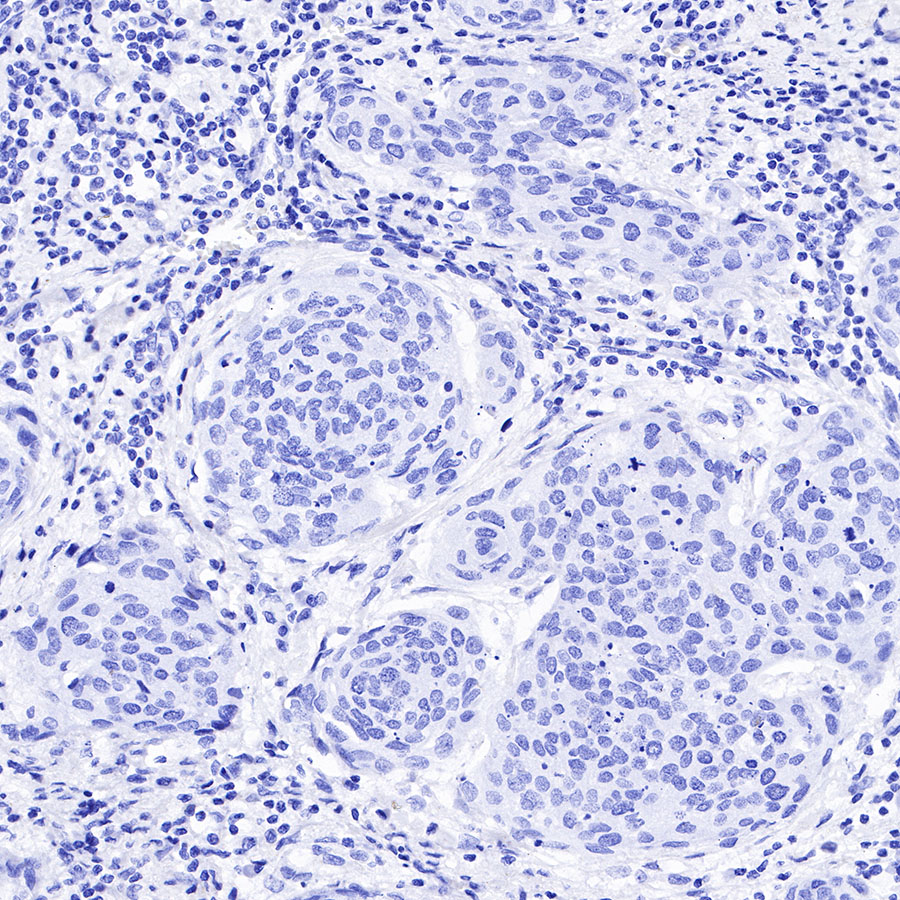
Negative control: Jurkat
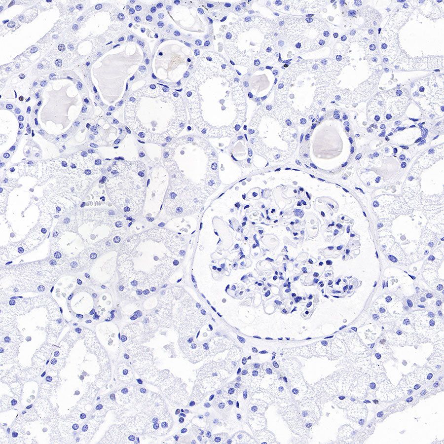
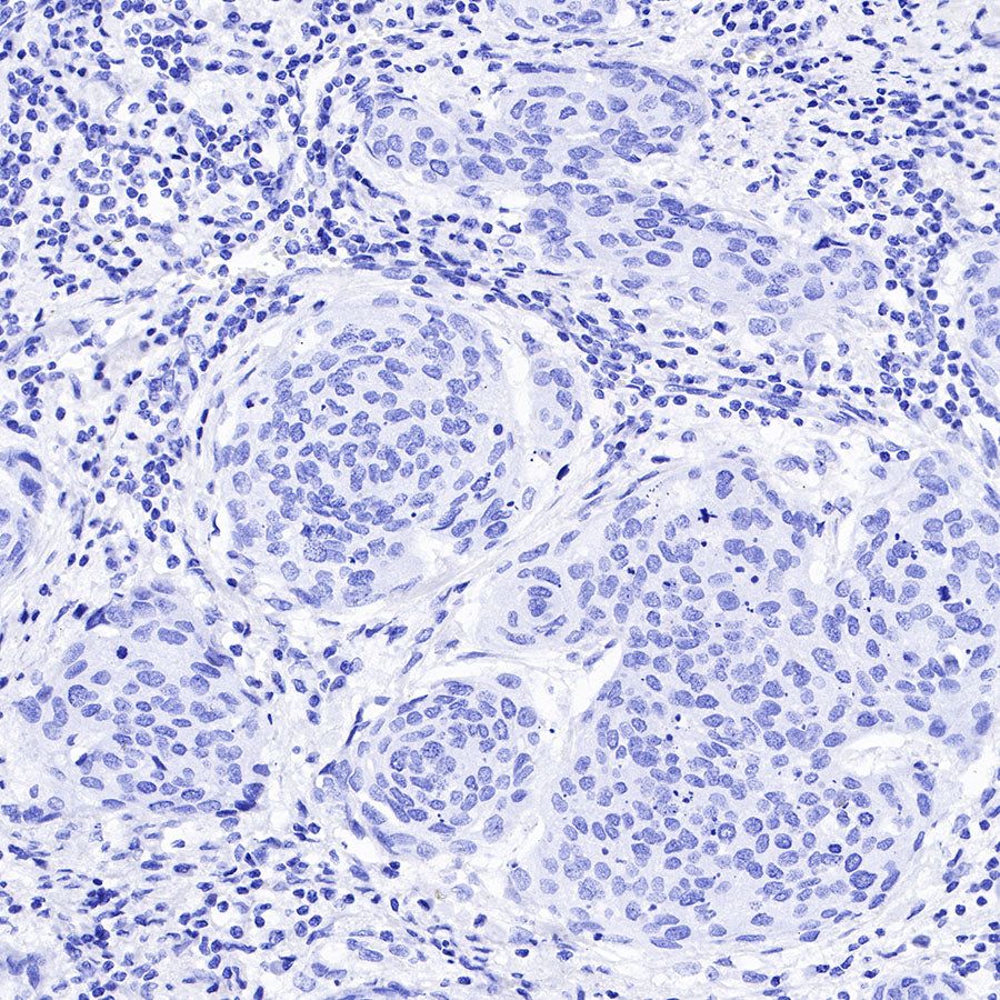
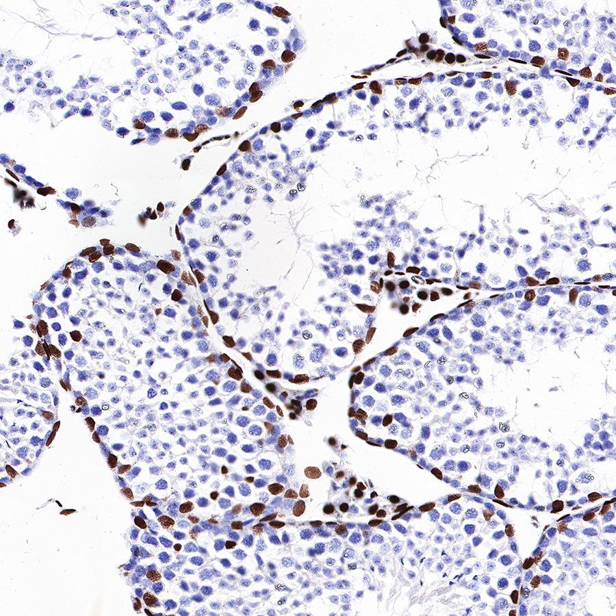
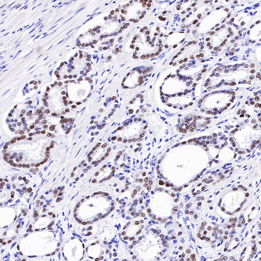
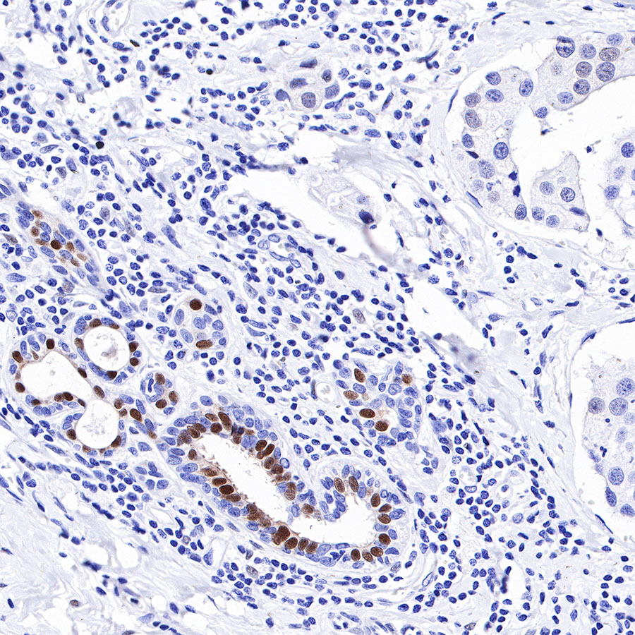
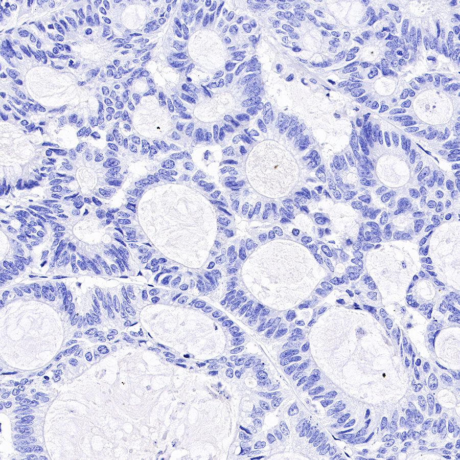
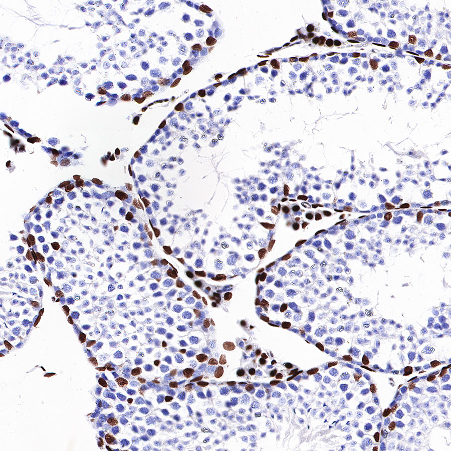
Immunohistochemistry

Immunocytochemistry
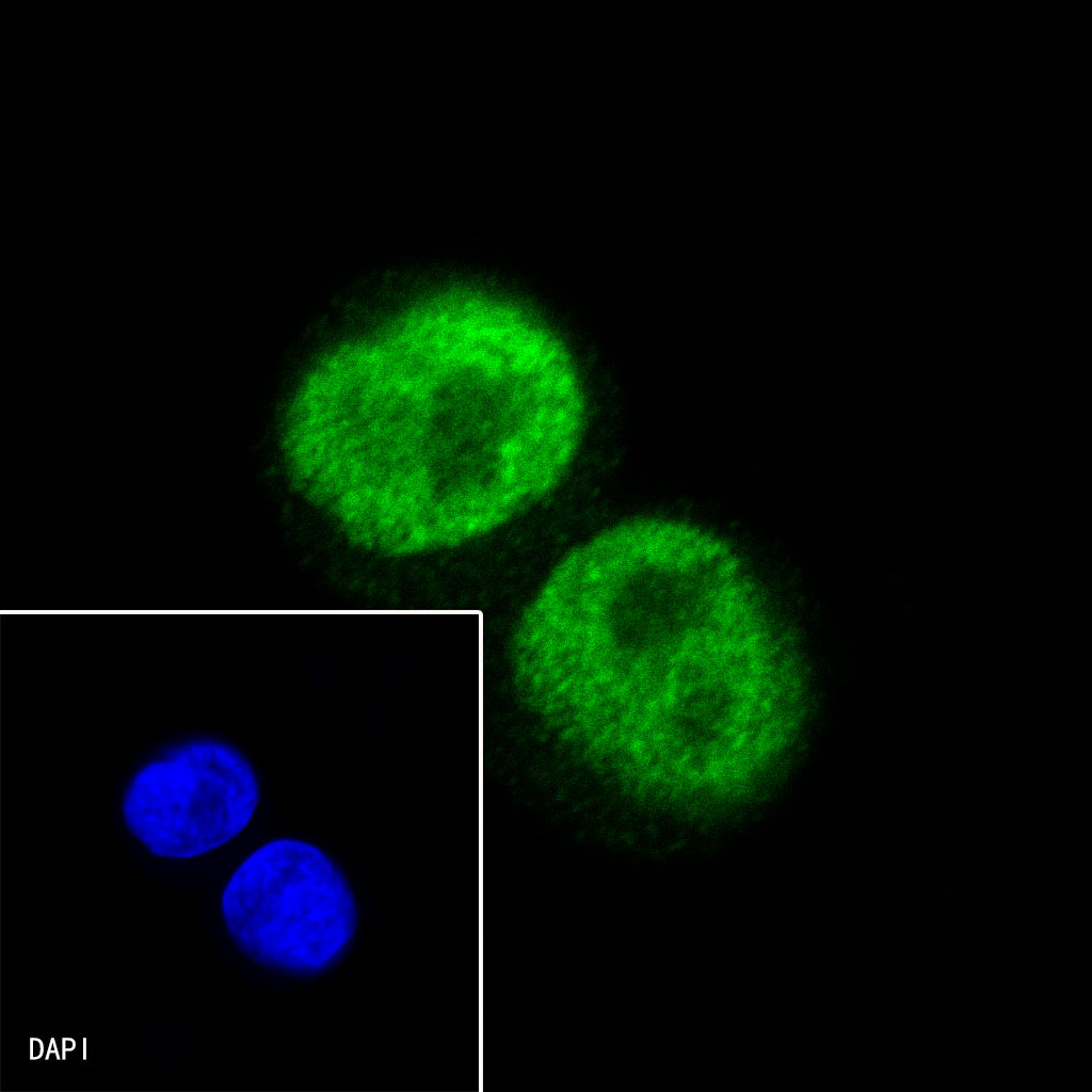
ICC shows positive staining in LnCap cells. Anti-Androgen Receptor antibody was used at 1/500 dilution (Green) and incubated overnight at 4°C. Goat polyclonal Antibody to Rabbit IgG - H&L (Alexa Fluor® 488) was used as secondary antibody at 1/1000 dilution. The cells were fixed with 100% ice-cold methanol and permeabilized with 0.1% PBS-Triton X-100. Nuclei were counterstained with DAPI (Blue).