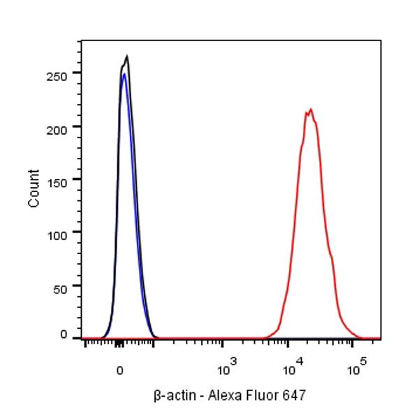
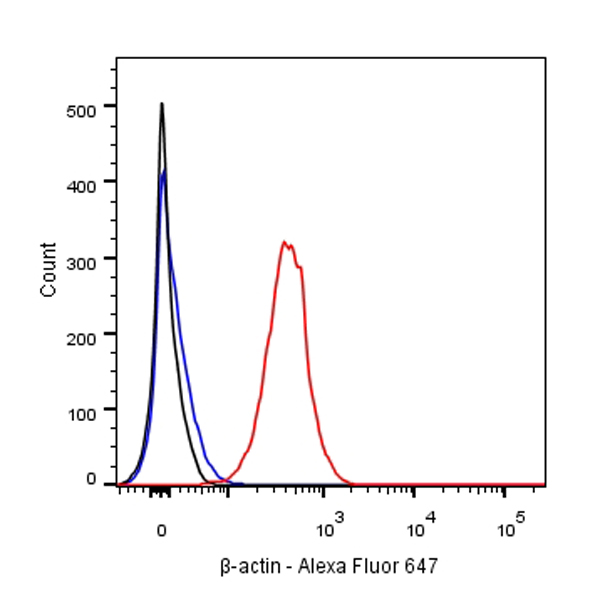
Flow cytometric analysis of NIH/3T3 cells labelling β-actin (Alexa Fluor® 647 Conjugate) antibody at 1/400 (1 μg) dilution/ (red) compared with a Rabbit monoclonal IgG (Black) isotype control and an unlabelled control (cells without incubation with primary antibody and secondary antibody) (Blue).
β-actin Recombinant Rabbit mAb (Alexa Fluor® 647 Conjugate) (SDT-R015)
β-actin Recombinant Rabbit mAb (Alexa Fluor® 647 Conjugate) (SDT-R015)
Price:
Regular price
$105.00 SGD
Regular price
Sale price
$105.00 SGD
Unit price
per
For shipping services or bulk orders, you may request a quotation.
Secure checkout with

View full details
Product Details
Product Details
Product Specification
| Host | Rabbit |
| Antigen | β-Actin |
| Synonyms | ACTB |
| Immunogen | N/A |
| Location | Cytoplasm, Cytoskeleton |
| Accession | P60709 |
| Clone Number | SDT-R015 |
| Antibody Type | Recombinant mAb |
| Application | WB, Functional Assay |
| Reactivity | Hu, Ms, Rt |
| Purification | Protein A |
| Concentration | 4 mg/ml |
| Conjugation | Alexa Fluor® 647 |
| Physical Appearance | Liquid |
| Storage Buffer | PBS, 0.1% BSA, 0.01% Proclin 300 |
| Stability & Storage | 12 months from date of receipt / reconstitution, 2 to 8 °C as supplied. |
Dilution
| application | dilution | species |
| ICC | 1:100 | null |
| ICFCM | 1:400-1:4000 | null |
Background
Beta-actin (human gene and protein abbreviation ACTB/ACTB) is one of six different actin isoforms which have been identified in humans. This is one of the two nonmuscle cytoskeletal actins. Actins are highly conserved proteins that are involved in cell motility, structure and integrity. Beta actin is often used in Western blotting as a loading control, to normalize total protein amounts and check for eventual protein degradation in the samples. Its transcript is also commonly used as a housekeeping gene standard in qPCR. Its molecular weight is approximately 42 kDa.
Picture
Picture
FC
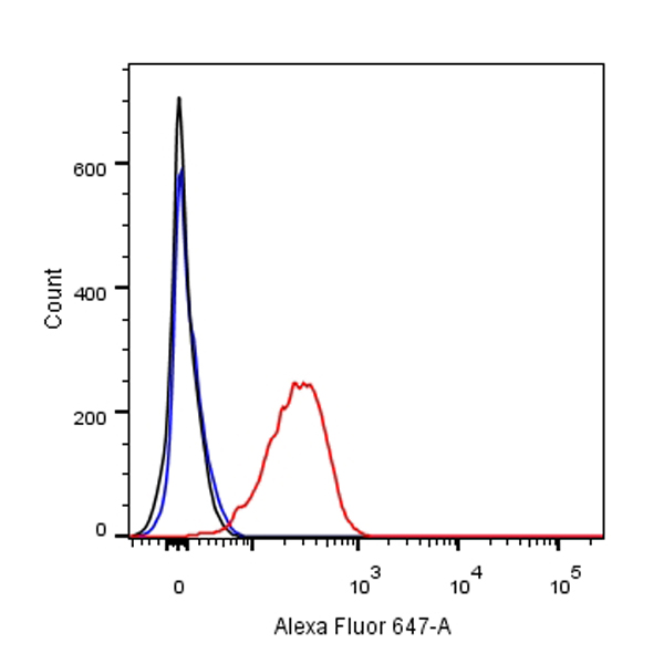
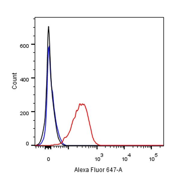
Flow cytometric analysis of C6 cells labelling β-actin (Alexa Fluor® 647 Conjugate) antibody at 1/4000 (0.1 μg) dilution/ (red) compared with a Rabbit monoclonal IgG (Black) isotype control and an unlabelled control (cells without incubation with primary antibody and secondary antibody) (Blue).
Immunohistochemistry
ICC shows positive staining in HeLa cells. Anti-β-actin (Alexa Fluor® 647 Conjugate) antibody was used at 1/100 dilution (magenta) and incubated overnight at 4°C. The cells were fixed with 100% ice-cold methanol and permeabilized with 0.1% PBS-Triton X-100. Nuclei were counterstained with DAPI (Blue).
Immunocytochemistry

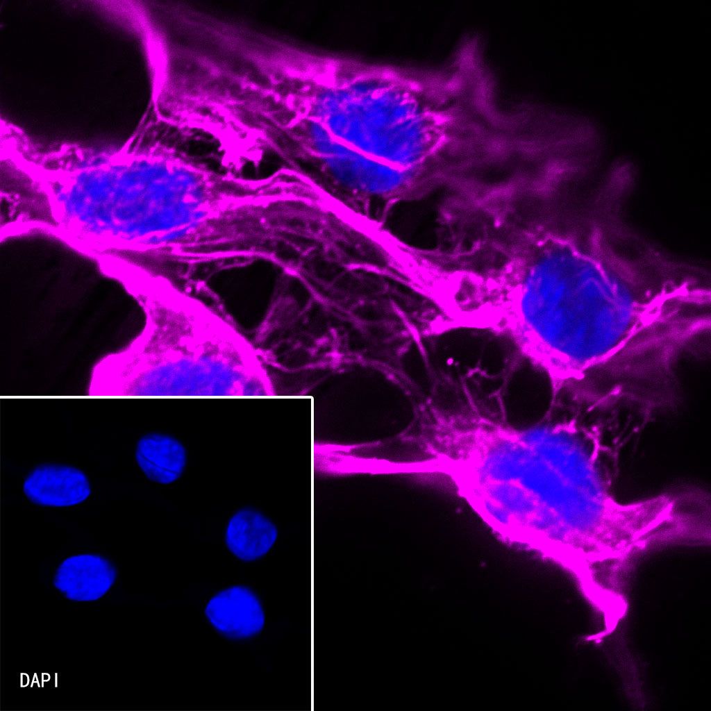
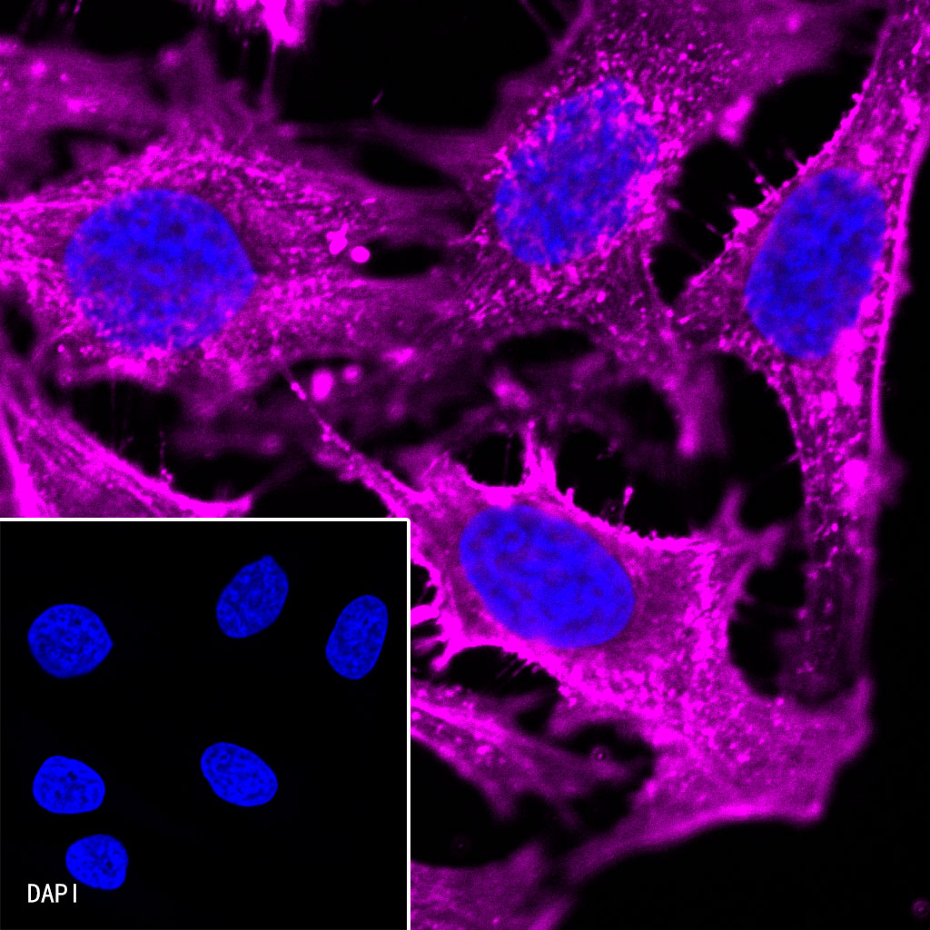
ICC shows positive staining in NIH/3T3 cells. Anti-β-actin (Alexa Fluor® 647 Conjugate) antibody was used at 1/100 dilution (magenta) and incubated overnight at 4°C. The cells were fixed with 100% ice-cold methanol and permeabilized with 0.1% PBS-Triton X-100. Nuclei were counterstained with DAPI (Blue).

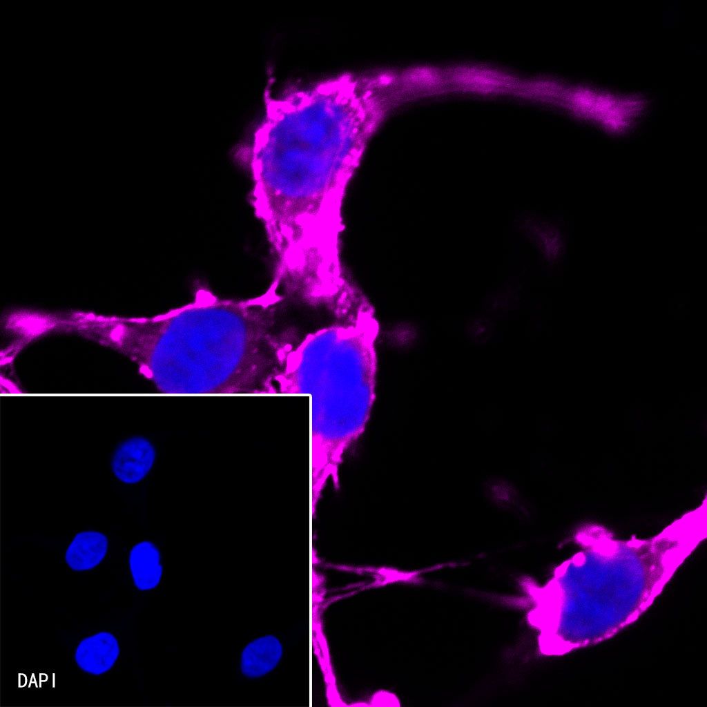
ICC shows positive staining in C6 cells. Anti-β-actin (Alexa Fluor® 647 Conjugate) antibody was used at 1/100 dilution (magenta) and incubated overnight at 4°C. The cells were fixed with 100% ice-cold methanol and permeabilized with 0.1% PBS-Triton X-100. Nuclei were counterstained with DAPI (Blue).