Product Specification
| Host |
Rabbit |
| Antigen |
β-Actin |
| Synonyms |
ACTB |
| Immunogen |
Recombinant Protein |
| Location |
Cytoplasm, Cytoskeleton |
| Accession |
P60709 |
| Clone Number |
SDT-R015 |
| Antibody Type |
Rabbit mAb |
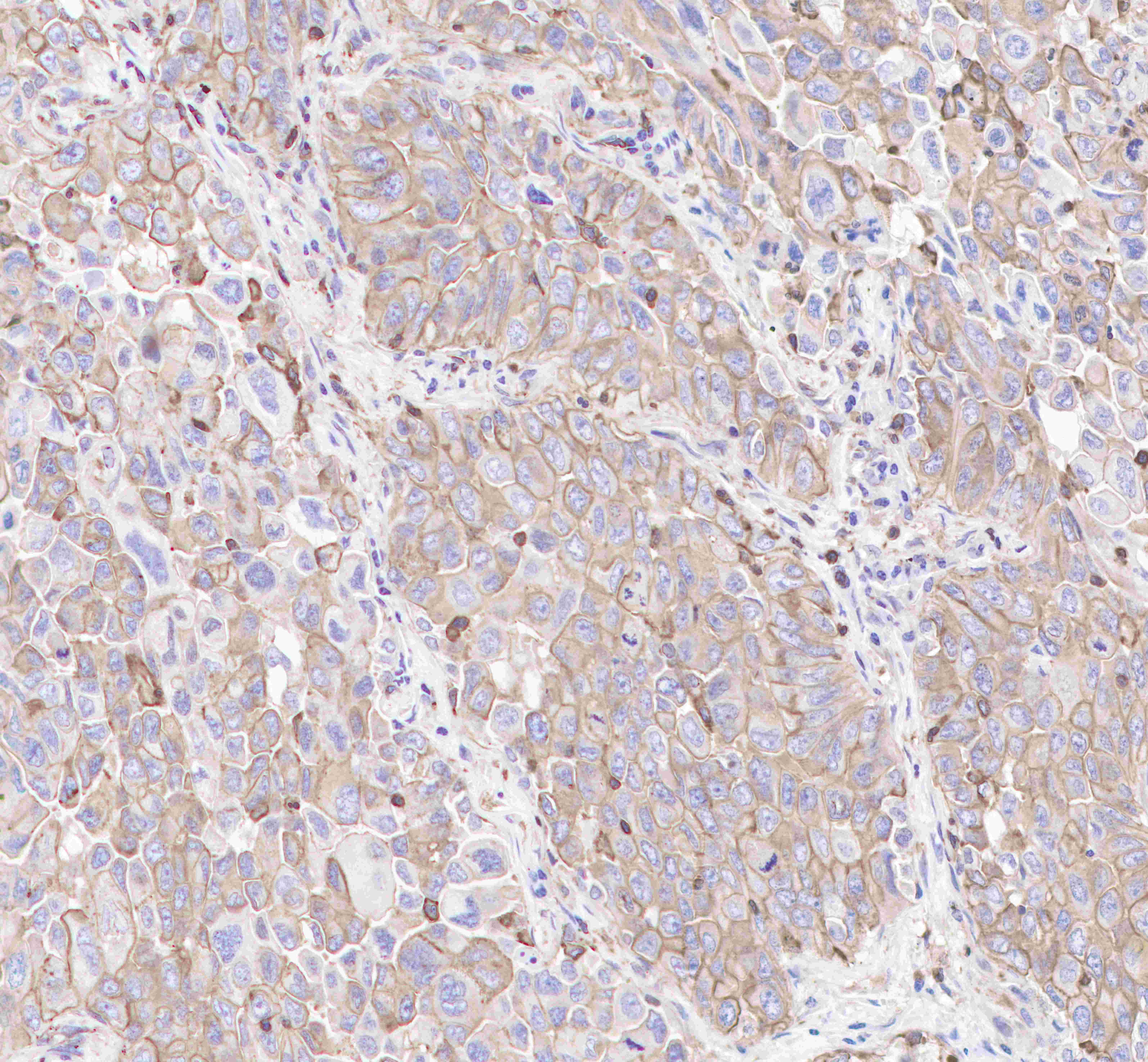
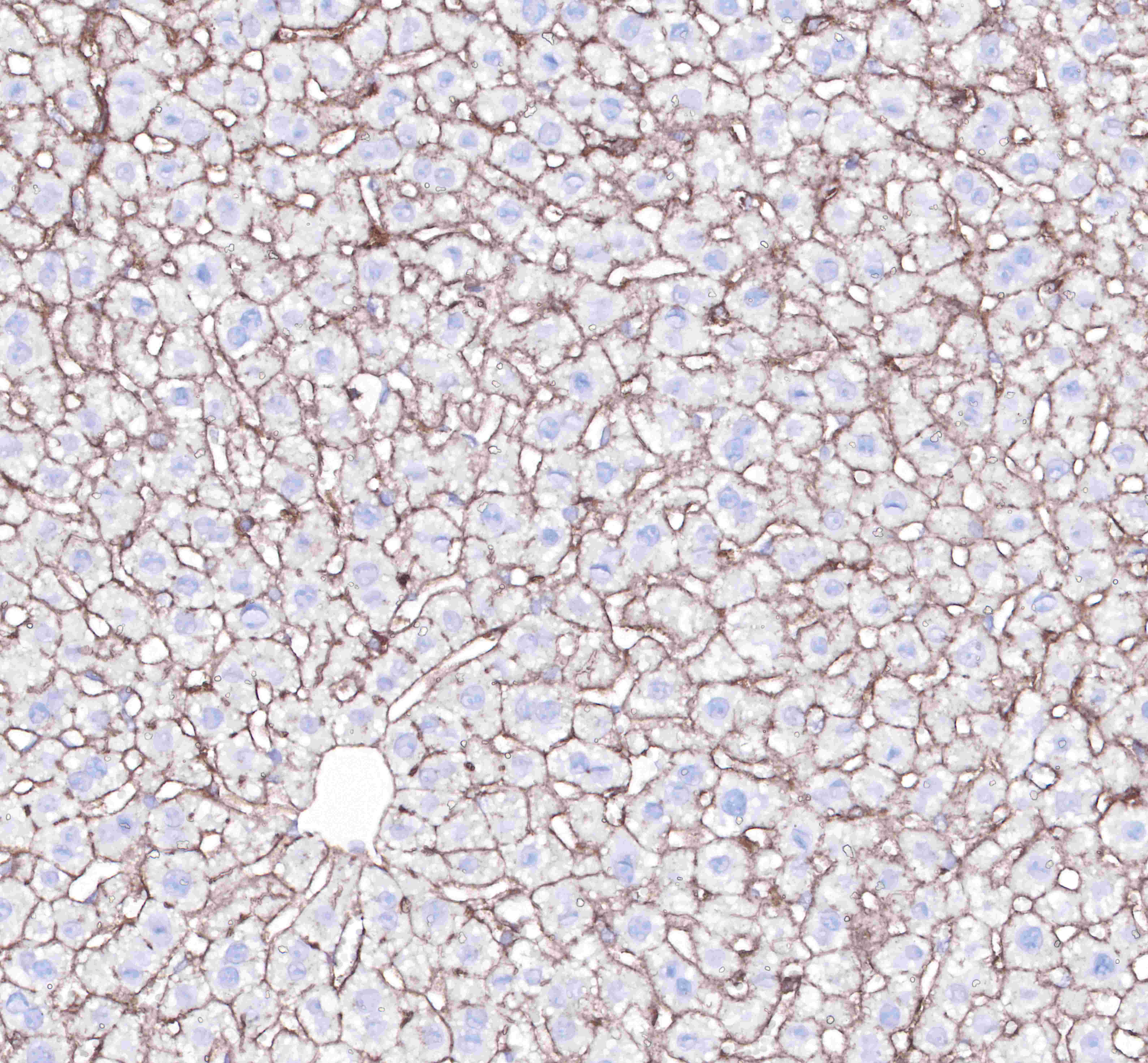
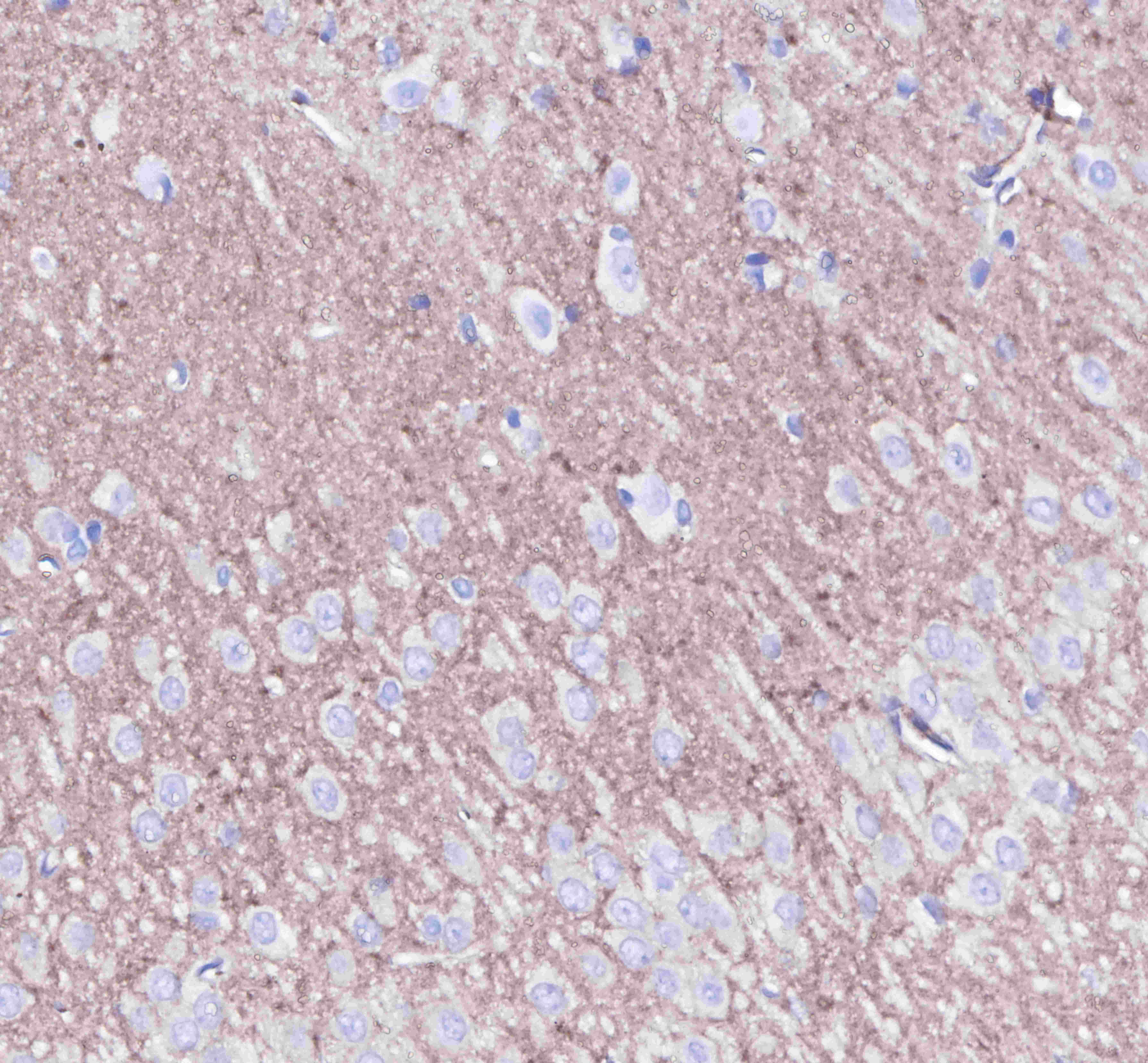
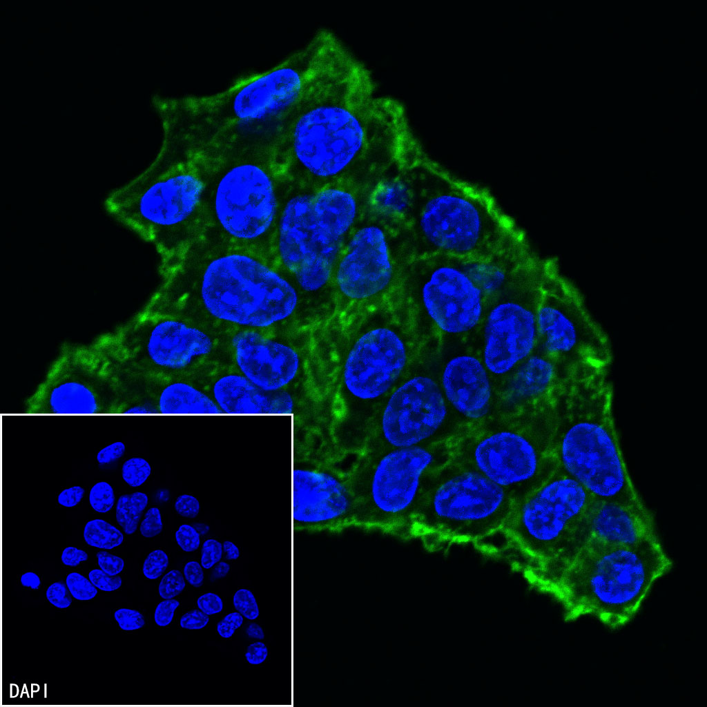
| Application |
WB, IHC-P, ICC, ICFCM |
| Reactivity |
Hu, Ms, Rt |
| Purification |
Protein A |
| Concentration |
0.5mg/ml |
| Molecular Weight |
42kDa |
| Conjugation |
Unconjugated |
| Physical Appearance |
Liquid |
| Storage Buffer |
PBS, 40% Glycerol, 0.05%BSA, 0.03% Proclin 300 |
| Stability & Storage |
12 months from date of receipt / reconstitution, -20 °C as supplied. |
Dilution
| application |
dilution |
species |
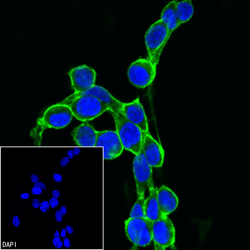
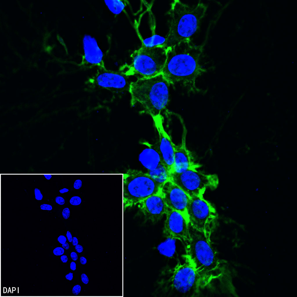
| ICC |
1:500 |
|
| WB |
1:1000-1:5000 |
|
| ICFCM |
1:500 |
|
| IHC-P |
1:2000 |
|
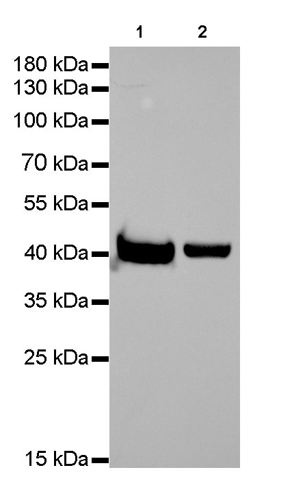
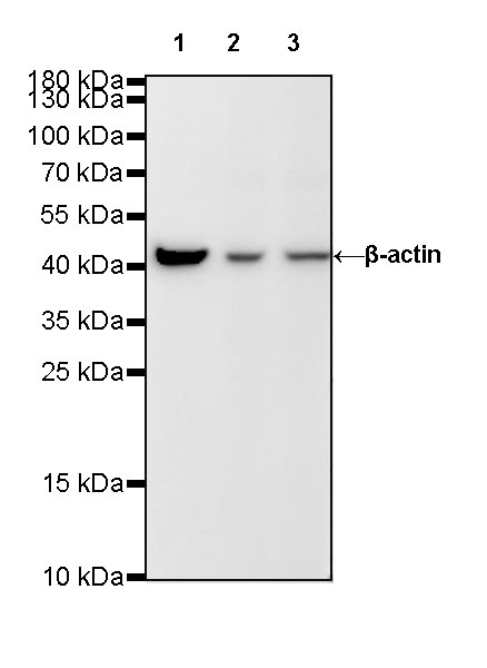
Background
Beta-actin (human gene and protein abbreviation ACTB/ACTB) is one of six different actin isoforms which have been identified in humans. This is one of the two nonmuscle cytoskeletal actins. Actins are highly conserved proteins that are involved in cell motility, structure and integrity. Beta actin is often used in Western blotting as a loading control, to normalize total protein amounts and check for eventual protein degradation in the samples. Its transcript is also commonly used as a housekeeping gene standard in qPCR. Its molecular weight is approximately 42 kDa.