EdU-488 Cell Proliferation Assay Kit (6-well plate)
EdU-488 Cell Proliferation Assay Kit (6-well plate)
Price:
Regular price
$440 USD
Regular price
Sale price
$440 USD
Unit price
per
For shipping services or bulk orders, you may request a quotation.
Secure checkout with

View full details
Product Details
Product Details
Product Specification
| Usage | Before the trial guidelines: 1, the corresponding animal experiment, EdU suggested initial dosage of 50 mg/kg (50 mu g/g), dilute concentration is 0.5 1 mg/mL. Are injected into mice weight 20 g, mg, 1 to 1 mg/mL, need injection of 1 mL. 2, for the cells, the recommended 10 microns, EdU work for in vitro culture cell, specific EdU use concentration, incubation time along with the samples and research purpose is different, can be adjusted appropriately. 3. This kit can be used for EdU experiments of about 200T in 6-well plates, and EdU experiments of about 2000T in 96-well plates or paraffin sections. The amount of use should be calculated according to the sample conditions during the experiment. I. Sample processing 1. Animal experiments (1) EdU injection in animals For mice, EdU can be prepared with PBS at a dose of 10 to 200mg/kg and injected intraperitoneally, locally into specific tissues or organs, or added to drinking water. The specific dosage is related to the type, weight and mode of use of animals, and can be referred to the relevant literature. Therefore, it is recommended to conduct a certain exploration of the use concentration of EdU for the first use, or directly use the concentration of 50mg/kg for testing. Injection methods: according to customer experiments, such as intraperitoneal injection, subcutaneous injection, intramuscular injection, tail vein injection, etc., among which intraperitoneal injection is the most common. After 6 hours or according to the specific experiments to determine the appropriate time, quickly kill mice, take out the organization, in accordance with the general steps to make frozen section and paraffin section. EdU tag when also can consult relevant literature to adjust. Small intestinal epithelial cells proliferate rapidly, and positive information can be detected 6 hours after EdU injection in adult mice, which can be used as a positive control for pre-experiments. (2) slice processing Before slicing processing: best cleaning tissues and organs, to remove residues in the tissues of the blood, EdU, reduce the background. It is advisable to slice thickness: 3-10 microns, biopsy had thick could affect the background and dyeing efficiency. Slice post-processing: Paraffin dewaxing: dimethyl benzene dewaxing 2 times, 15 minutes per times, gradient ethanol (100%, 95%, 85%, 75%), 5 minutes per times, deionized water elution 1 times. Processing of frozen sections: After 30 min at room temperature, the sections were fixed for 10 min and washed three times with PBS for 5 min each. 2, cell experiment (1) EdU labeling of cells a.with cell complete medium and EdU mother liquor by 2000:1 the ratio of dilution, preparation of a moderate amount of 100 microns EdU medium; b.each hole adding suitable amount of 10 microns EdU medium 2 hours incubation, abandon culture medium (optimal incubation time for 1/10 of the cell cycle) commonly, EdU incubation medium and EdU reaction liquid volume may refer to the table below; c.Cells were washed 3 times with PBS for 5 minutes each time
(2) cells fixed fully a.Cell fixative (PBS containing 4% paraformaldehyde) was added to each well and incubated at room temperature for 15 minutes. The fixative was discarded and washed with PBS for 3 times, 5 minutes each time. b.Each well was decolorized with an osmotic agent (0.5% TritonX-100 in PBS) and incubated for 15 minutes. PBS was washed three times for 5 min each. Second, EdU response
The EdU reaction solution was prepared according to the ratio of PB: CU: AC: 488 chromogen = 855:40:100:5 (the reaction solution was prepared on the spot). add 50-500&mu per sample dropfold; L EdU dyeing liquid evenly cover samples (reaction), reaction liquid at room temperature away from light incubation 15 to 60 minutes, abandon the dyeing liquid reaction, PBS wash three times, each time for 5 minutes. < br / > 3, DAPI staining & have spent < br / > DAPI (100 x), with PBS according to the scale of 1:100 dilution into ready-to-use DAPI staining fluid, each sample in 50-500 & mu; L DAPI staining solution, staining in the dark for 15 min, and washing with PBS three times for 5 min each. < br / > 4, image photos & have spent < br / > dyeing advice immediately after the completion of observation, using fluorescence microscopy, confocal microscope or film scanner to capture images, after dyed dark glass slides 4 ℃. 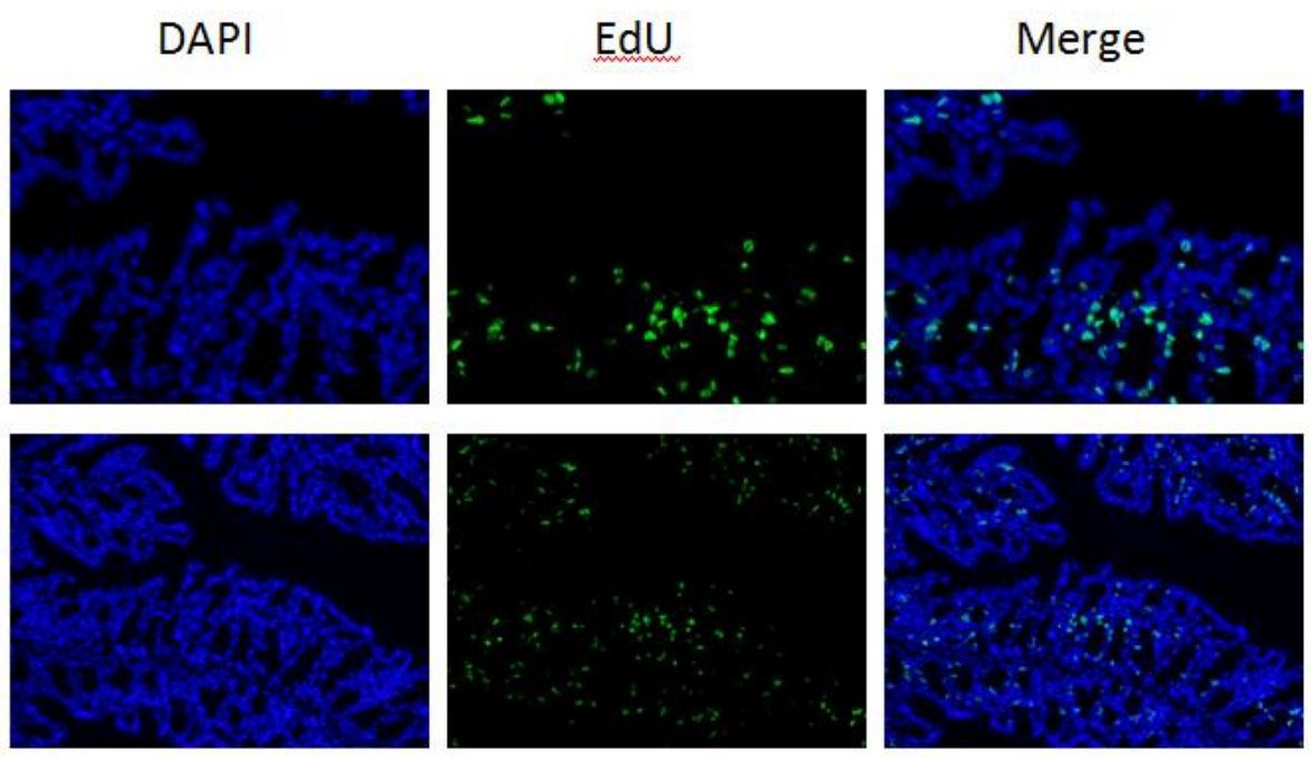 Above are stained images of 50mg/kg labeled mice for 6 hours |
|||||||||||||||||||||||||||||||||
| Description | EdU(5-ethynyl-2' -deoxyuridine), also known as 5-ethynyl-2' -deoxyuridine (5-ethynyl-2' -deoxyuridine), is a thymidine analogue with an alkynyl hydroxyl group that is rare in natural compounds and can replace thymidine (thymidine, 5-ethynyl-2' -deoxyuridine) in cell proliferation. thymidine (thymidine) is inserted into the replicating DNA molecule, the ethynyl group on EdU can react covalently with the fluorescently labeled small molecule Azide probe (Azide Alexa Fluor 488, etc.) through the catalysis of copper monovalent ion to form a stable triazole ring. "It is called the Clickreaction, which allows for an efficient and rapid measurement of cell proliferation, especially the percentage of cells in S phase." Compared with the traditional immunofluorescence staining (BrdU) detection method, EdU is only 1/500 of the size of BrdU antibody, and it is easier to diffuse in cells. It does not require strict sample denaturation (acid hydrolysis, pyolysis, and enzymatic hydrolysis), which effectively avoids sample damage and helps to observe the real situation of cell proliferation at the overall level of tissues and organs. Has higher sensitivity and faster detection speed.
According to PB, CU: AC: 488 chromogen = 855: spoken, Catalan than column configuration EdU reaction liquid (liquid of reaction is used). Each sample add 50-500 mu L EdU staining reaction liquid liquid evenly cover samples (reaction), 15 to 60 minutes at room temperature away from light incubation, abandon the dyeing liquid reaction, PBS wash 3 times, each time for 5 minutes. 3. DAPI staining DAPI(100x) was diluted with PBS at a ratio of 1:100 to make ready-to-use DAPI staining solution. 50-500μL of DAPI staining solution was added to each sample and stained in the dark for 15 minutes, followed by three washes with PBS for 5 minutes each. 4. Take pictures Fluorescence microscope, confocal microscope or whole-film scanner were used to collect images. After staining, the slides were stored in a dark place at 4℃. Above are stained images of 50mg/kg labeled mice for 6 hours. |
|||||||||||||||||||||||||||||||||
| Storage Temp. | -20 ° C, valid for 12 months. |