Anti-tumor efficacy of S0B0594 in B16-OVA tumor-bearing mice
Invivo anti-mouse PD-1 Recombinant mAb (D265A)
Invivo anti-mouse PD-1 Recombinant mAb (D265A)
Price:
Regular price
$120 USD
Regular price
Sale price
$120 USD
Unit price
per
For shipping services or bulk orders, you may request a quotation.
Secure checkout with

View full details
Product Details
Product Details
Product Specification
| Host | Mouse |
| Antigen | PD-1 |
| Synonyms | Programmed cell death protein 1, mPD-1, CD279 |
| Location | Cell membrane |
| Accession | Q02242 |
| Clone Number | S-5001 |
| Antibody Type | Recombinant mAb |
| Isotype | Mouse IgG1 D265A, κ |
| Isotype Control | Invivo mouse IgG1 isotype control (D265A) |
| Mutations | D265A |
| Application | In vivo blocking of PD-1/PD-L signaling |
| Reactivity | Ms |
| Purification | Protein G |
| Concentration | Lot specific* (generally 5 to 20 mg/ml)* |
| Endotoxin | <1EU/mg |
| Conjugation | Unconjugated |
| Physical Appearance | Liquid |
| Storage Buffer | PBS pH7.4, containing no preservative |
| Stability & Storage |
2 to 8 °C for 2 weeks under sterile conditions; -20 °C for 3 months under sterile conditions; -80 °C for 24 months under sterile conditions.
Please avoid repeated freeze-thaw cycles.
|
Background
This antibody recognizes the same epitope as clone RMP1-14.
Programmed cell death protein 1, also known as PD-1 and CD279 (cluster of differentiation 279), is a protein on the surface of T and B cells that has a role in regulating the immune system's response to the cells of the human body by down-regulating the immune system and promoting self-tolerance by suppressing T cell inflammatory activity. This prevents autoimmune diseases, but it can also prevent the immune system from killing cancer cells. PD-1 is an immune checkpoint and guards against autoimmunity through two mechanisms. First, it promotes apoptosis (programmed cell death) of antigen-specific T-cells in lymph nodes. Second, it reduces apoptosis in regulatory T cells (anti-inflammatory, suppressive T cells).
Picture
Picture
Validation Data
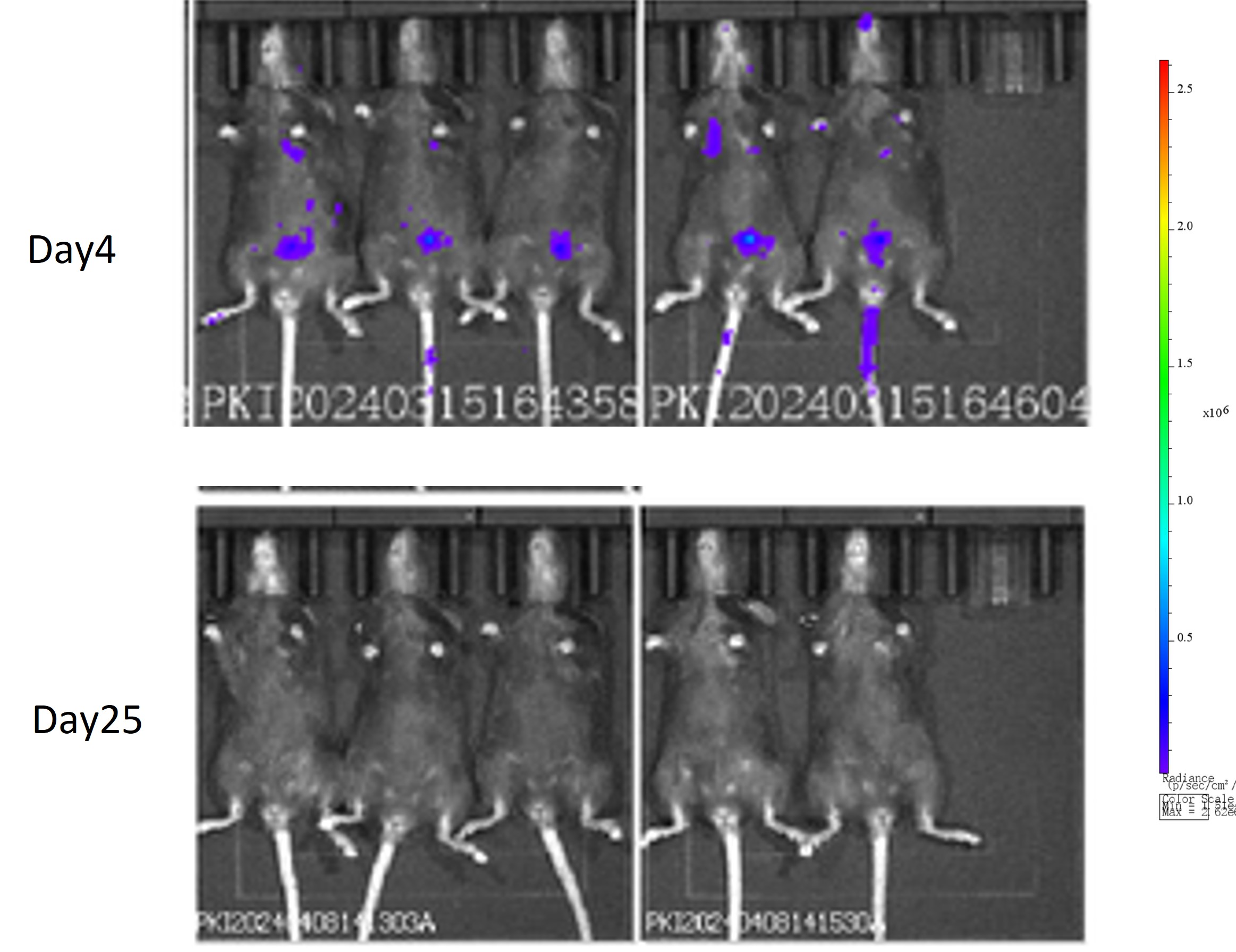
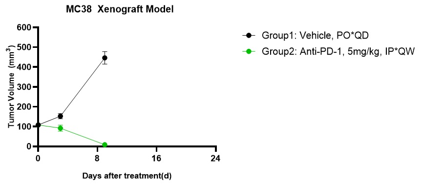
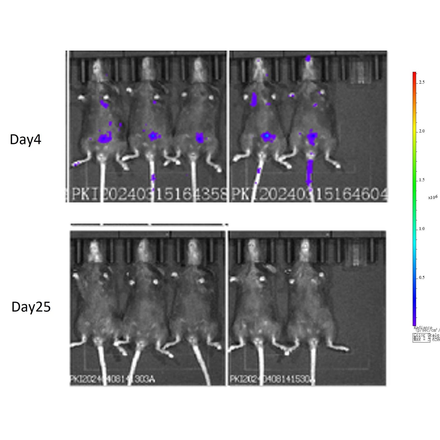
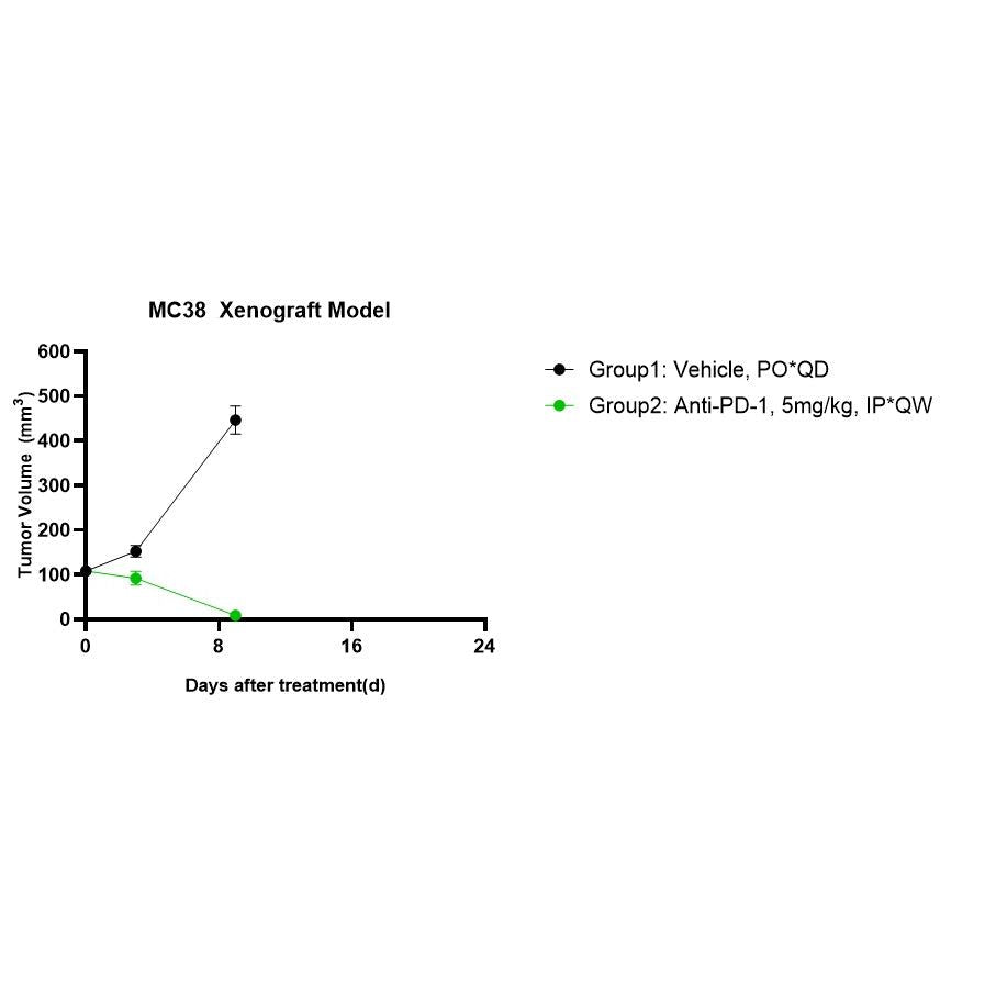
Anti-tumor efficacy of S0B0594 in MC38 Xenograft Model