HSP60 Recombinant Rabbit mAb (SDT-076-6)
HSP60 Recombinant Rabbit mAb (SDT-076-6)
Product Details
Product Details
Product Specification
| Host | Rabbit |
| Antigen | HSP60 |
| Synonyms | 60 kDa chaperonin, Chaperonin 60 (CPN60), Heat shock protein 60 (HSP-60; Hsp60) HuCHA60, Mitochondrial matrix protein P1, P60 lymphocyte protein, HSPD1 |
| Immunogen | Synthetic Peptide |
| Location | Intracellular |
| Accession | P10809 |
| Clone Number | SDT-076-6 |
| Antibody Type | Rabbit mAb |
| Application | WB, IHC-P, ICC, ICFCM |
| Reactivity | Hu, Ms, Rt |
| Predicted Reactivity | Hm |
| Purification | Protein A |
| Concentration | 0.5mg/ml |
| Conjugation | Unconjugated |
| Physical Appearance | Liquid |
| Storage Buffer | PBS, 40% Glycerol, 0.05%BSA, 0.03% Proclin 300 |
| Stability & Storage | 12 months from date of receipt / reconstitution, -20 °C as supplied. |
Dilution
| application | dilution | species |
| WB | 1:1000-1:20000 | |
| IHC-P | 1:2000 | |
| ICFCM | 1:500 | |
| ICC | 1:1000 |
Background
HSP60 is primarily a mitochondrial protein and is important for the folding of key proteins upon entry into the mitochondria. Studies have shown that a large amount of HSP60 is also present in the cytoplasm of many cells, and the inducers of HSP60 are stress, inflammation and immune response.
Picture
Picture
Western Blot
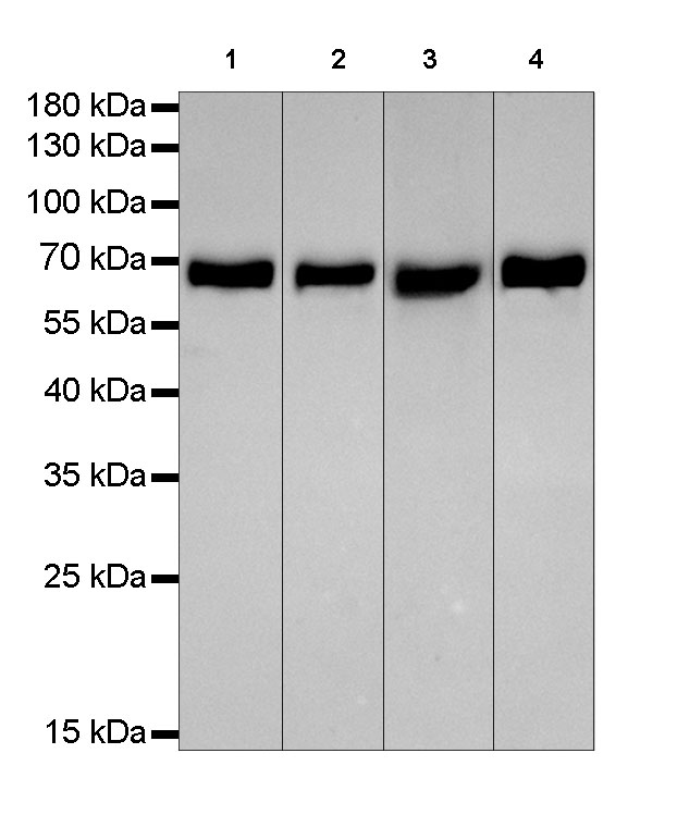
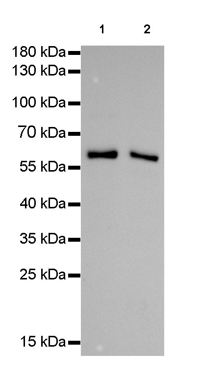
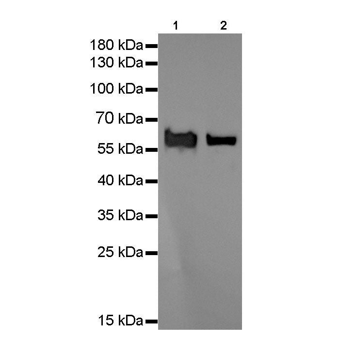
WB result of Hsp60 Rabbit mAb
Primary antibody : Hsp60 Rabbit mAb at 1/1000 dilution
Lane 1: NIH3T3 whole cell lysate 20 µg
Lane 2: mouse heart lysate 20 µg
Secondary antibody: Goat Anti-Rabbit IgG, (H+L), HRP conjugated at 1/10000 dilution
Predicted MW: 60 kDa
Observed MW: 60 kDa
Exposure time: 10s
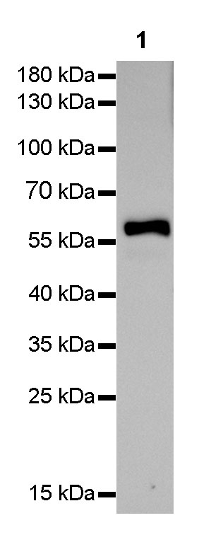
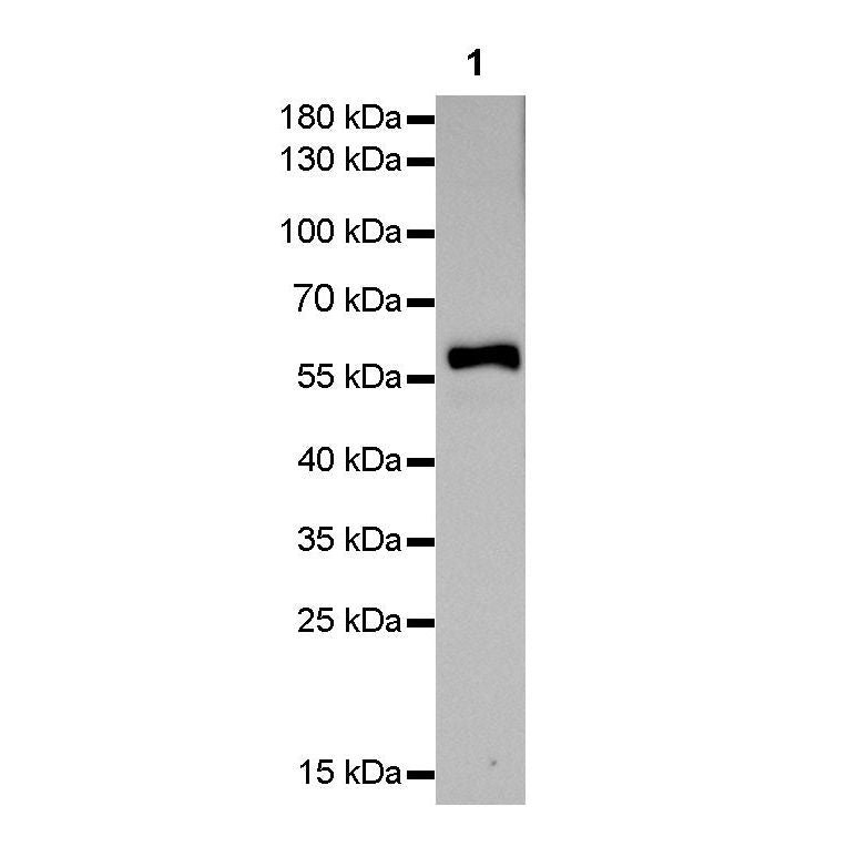
WB result of Hsp60 Rabbit mAb
Primary antibody : Hsp60 Rabbit mAb at 1/1000 dilution
Lane 1: rat heart lysate 20 µg
Secondary antibody: Goat Anti-Rabbit IgG, (H+L), HRP conjugated at 1/10000 dilution
Predicted MW: 60 kDa
Observed MW: 60 kDa
Exposure time: 10s
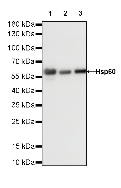
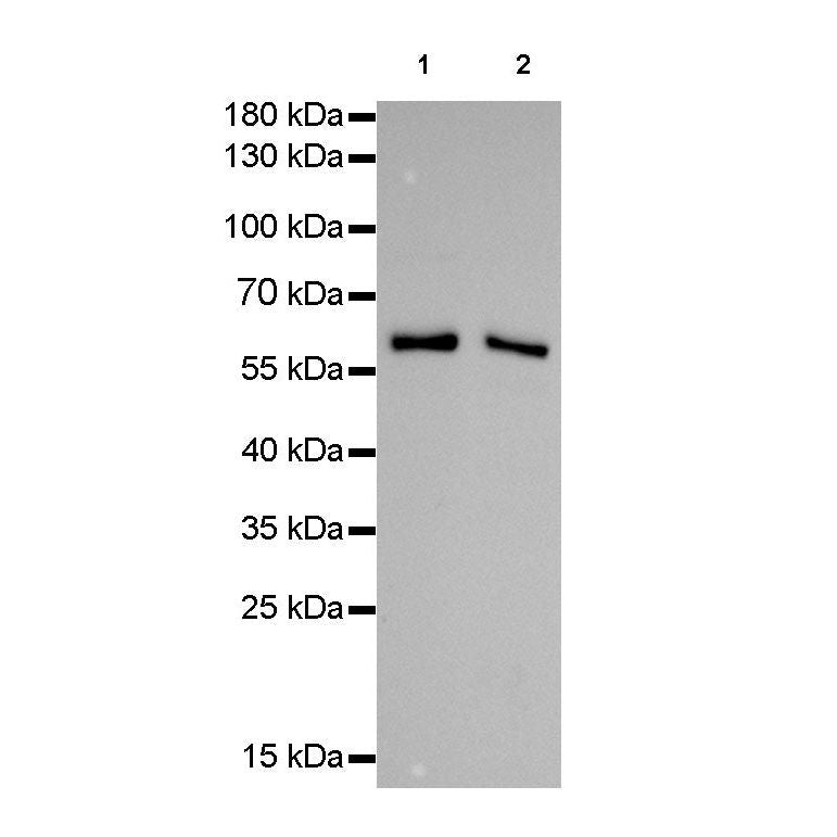
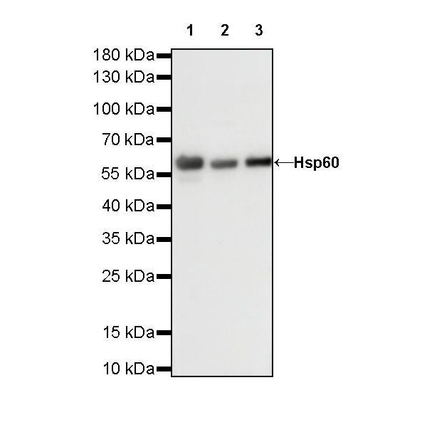
WB result of Hsp60 Rabbit mAb
Primary antibody: Hsp60 Rabbit mAb at 1/10000 dilution
Lane 1: HeLa whole cell lysate 20 µg
Lane 2: NIH/3T3 whole cell lysate 20 µg
Lane 3: rat heart lysate 20 µg
Secondary antibody: Goat Anti-Rabbit IgG, (H+L), HRP conjugated at 1/10000 dilution
Predicted MW: 60 kDa
Observed MW: 60 kDa
Exposure time: 60 s
FC

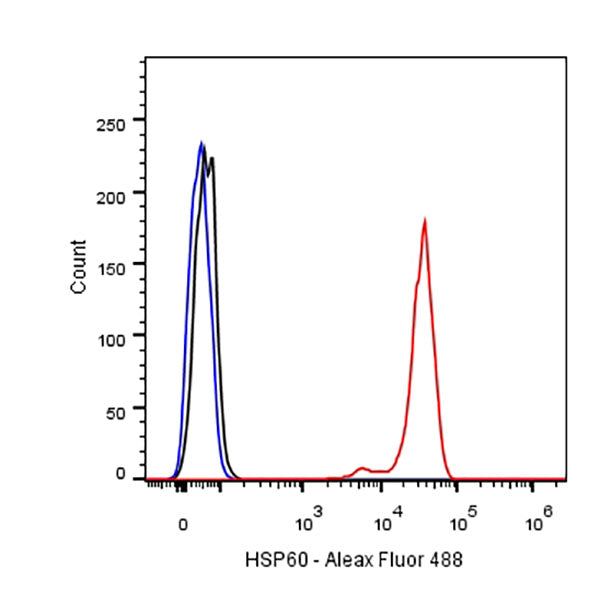
Flow cytometric analysis of HeLa cells labelling Hsp 60 antibody at 1/500(0.1ug) dilution/ (red) compared with a Rabbit monoclonal IgG (Black) isotype control and an unlabelled control (cells without incubation with primary antibody and secondary antibody) (Blue). Goat Anti-Rabbit IgG Alexa Fluor® 488 was used as the secondary antibody.
Immunohistochemistry
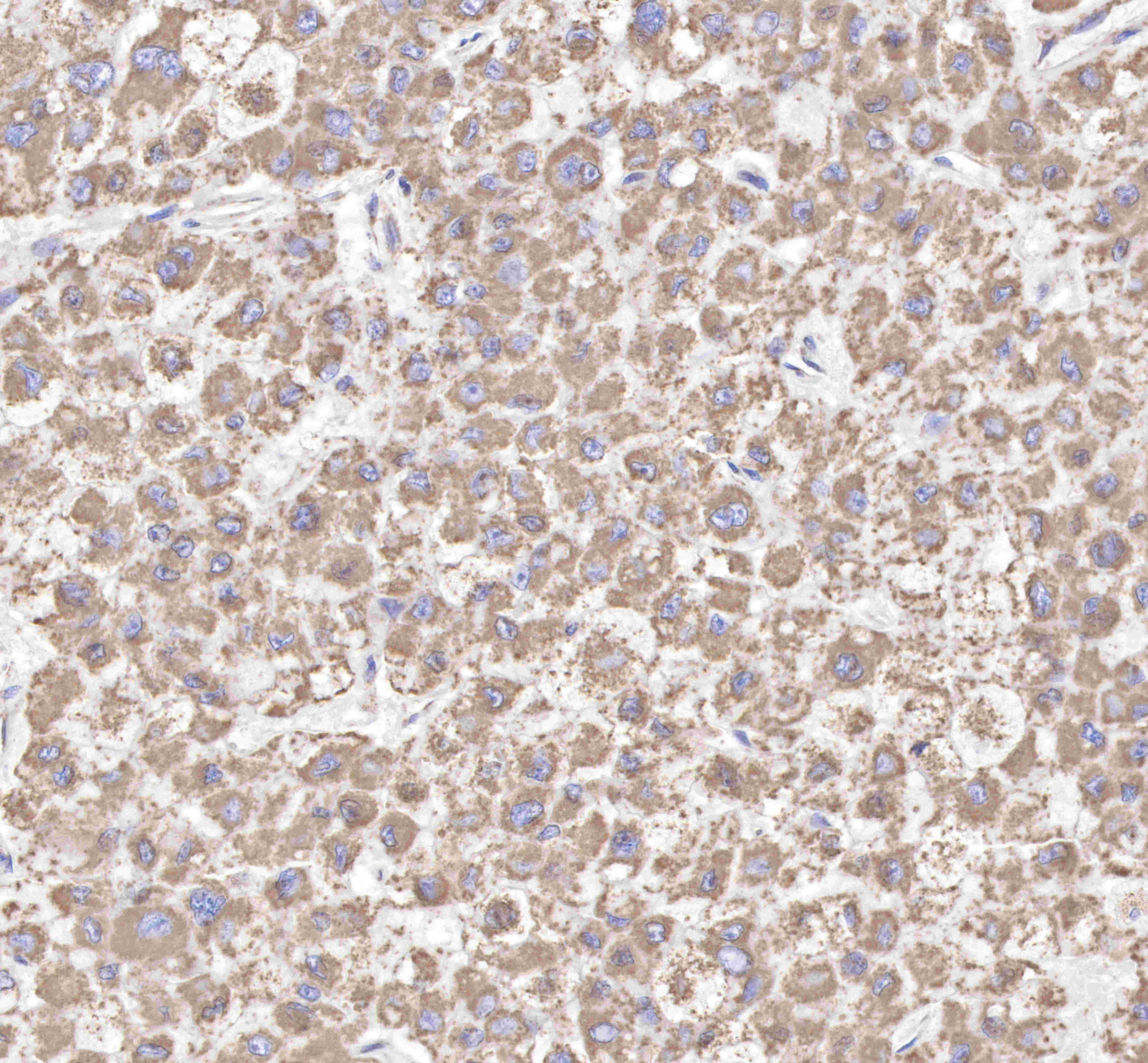
IHC shows positive staining in paraffin-embedded human liver cancer.
Anti-Hsp60 antibody was used at 1/2000 dilution, followed by a Goat Anti-Rabbit IgG H&L (HRP) ready to use.
Counterstained with hematoxylin.
Heat mediated antigen retrieval with Tris/EDTA buffer pH9.0 was performed before commencing with IHC staining protocol.
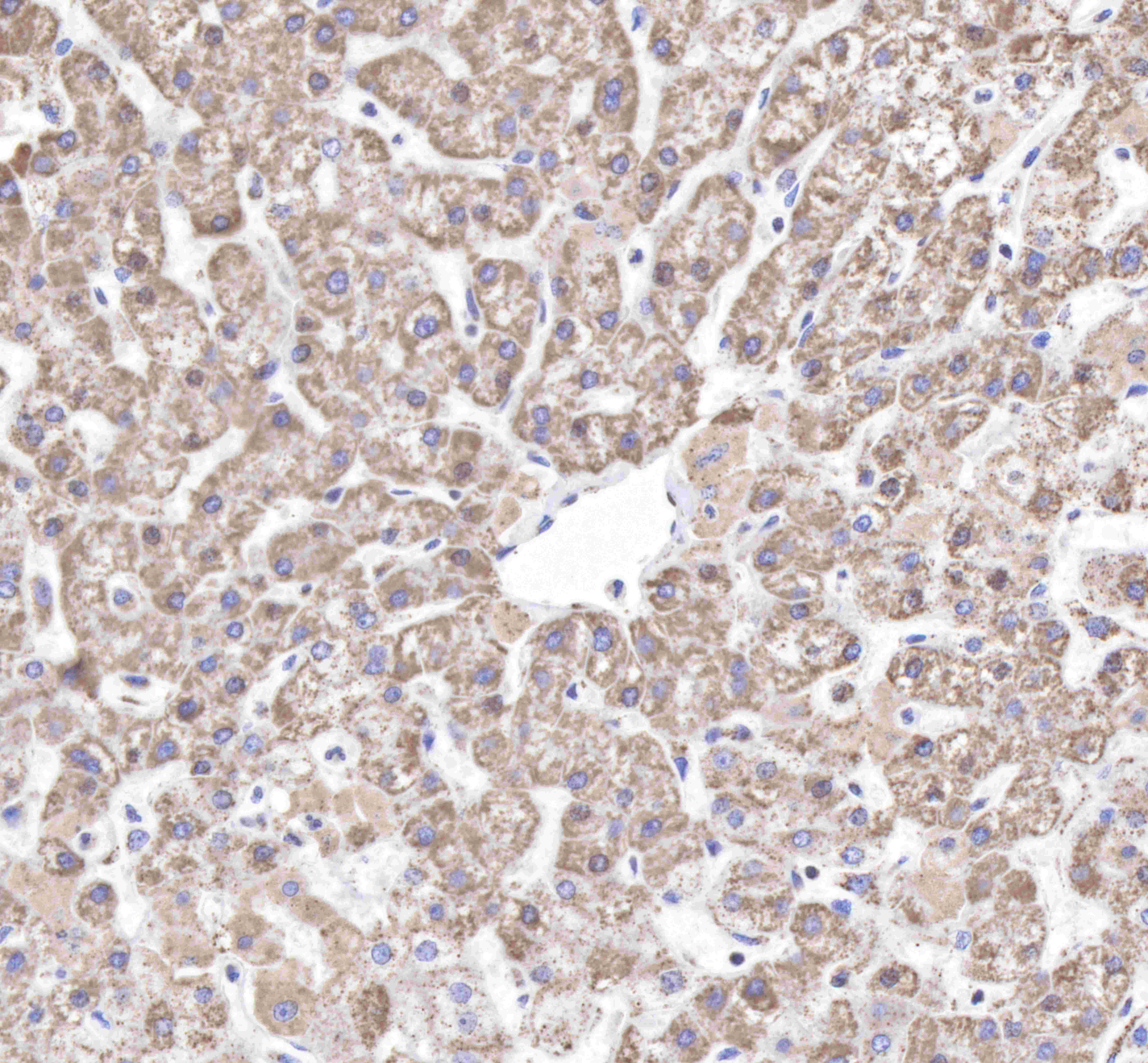
IHC shows positive staining in paraffin-embedded human liver.
Anti-Hsp60 antibody was used at 1/2000 dilution, followed by a Goat Anti-Rabbit IgG H&L (HRP) ready to use.
Counterstained with hematoxylin.
Heat mediated antigen retrieval with Tris/EDTA buffer pH9.0 was performed before commencing with IHC staining protocol.
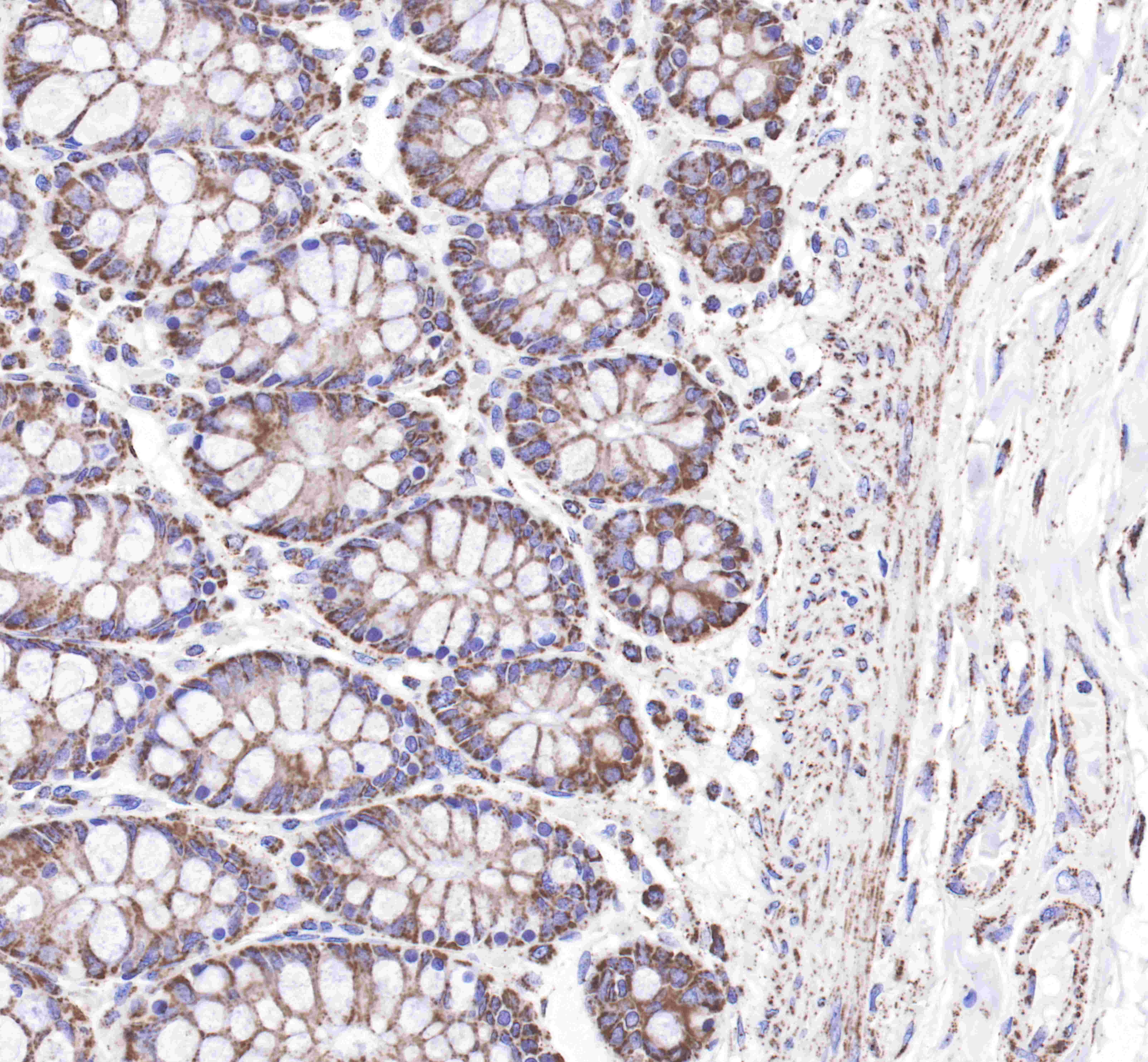
IHC shows positive staining in paraffin-embedded human colon.
Anti-Hsp60 antibody was used at 1/2000 dilution, followed by a Goat Anti-Rabbit IgG H&L (HRP) ready to use.
Counterstained with hematoxylin.
Heat mediated antigen retrieval with Tris/EDTA buffer pH9.0 was performed before commencing with IHC staining protocol.
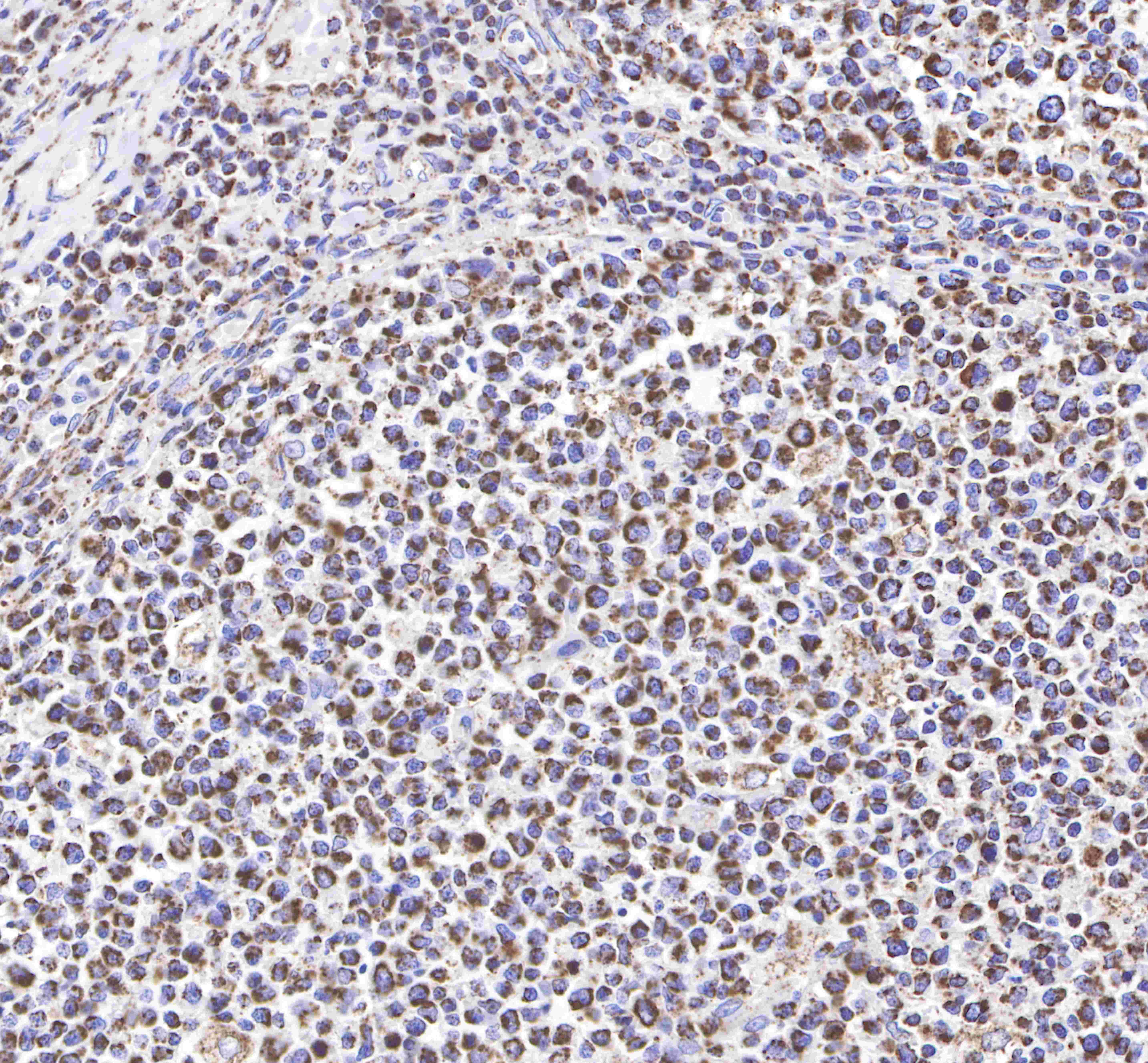
IHC shows positive staining in paraffin-embedded human tonsil.
Anti-Hsp60 antibody was used at 1/2000 dilution, followed by a Goat Anti-Rabbit IgG H&L (HRP) ready to use.
Counterstained with hematoxylin.
Heat mediated antigen retrieval with Tris/EDTA buffer pH9.0 was performed before commencing with IHC staining protocol.
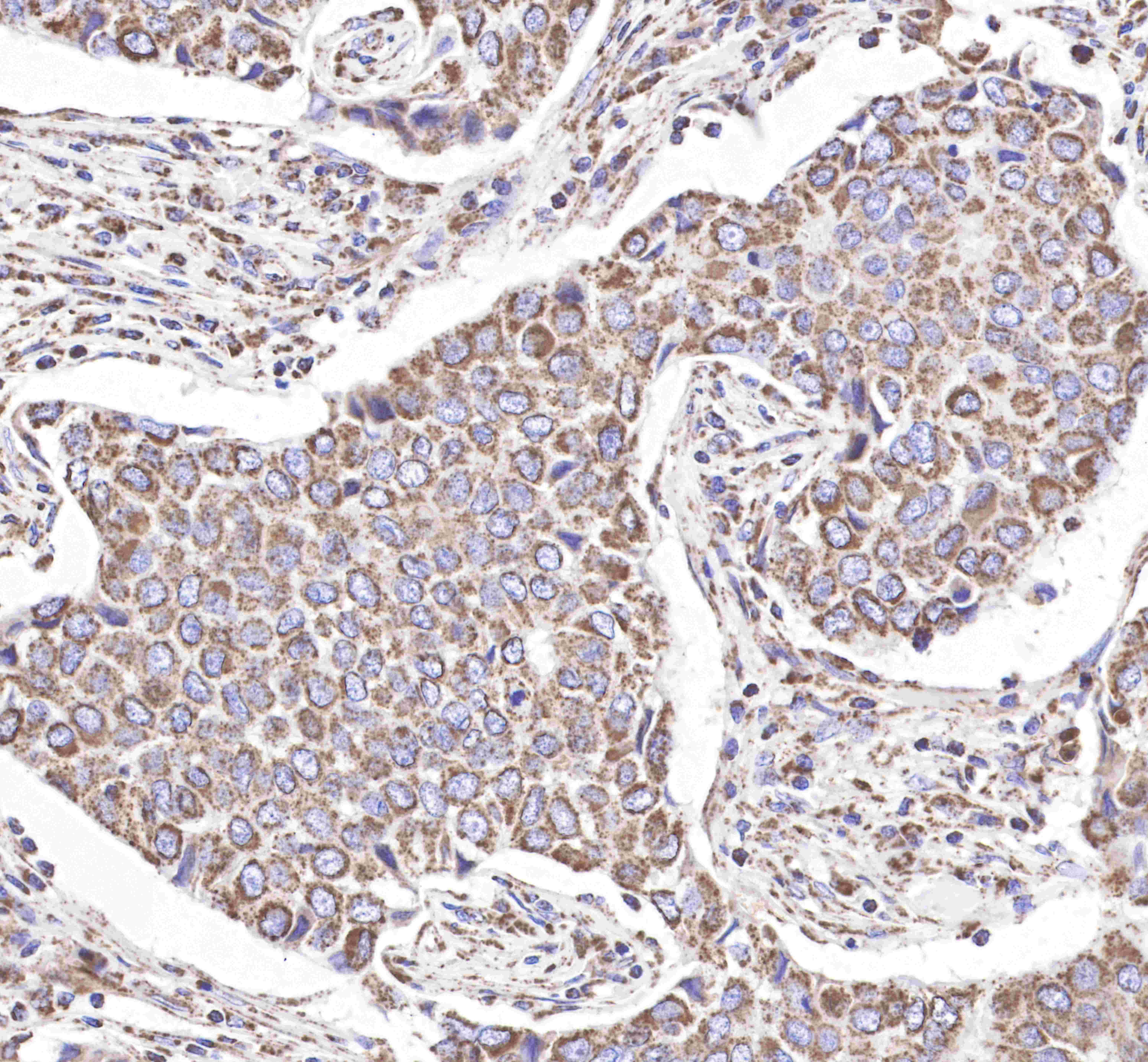
IHC shows positive staining in paraffin-embedded human breast cancer.
Anti-Hsp60 antibody was used at 1/2000 dilution, followed by a Goat Anti-Rabbit IgG H&L (HRP) ready to use.
Counterstained with hematoxylin.
Heat mediated antigen retrieval with Tris/EDTA buffer pH9.0 was performed before commencing with IHC staining protocol.
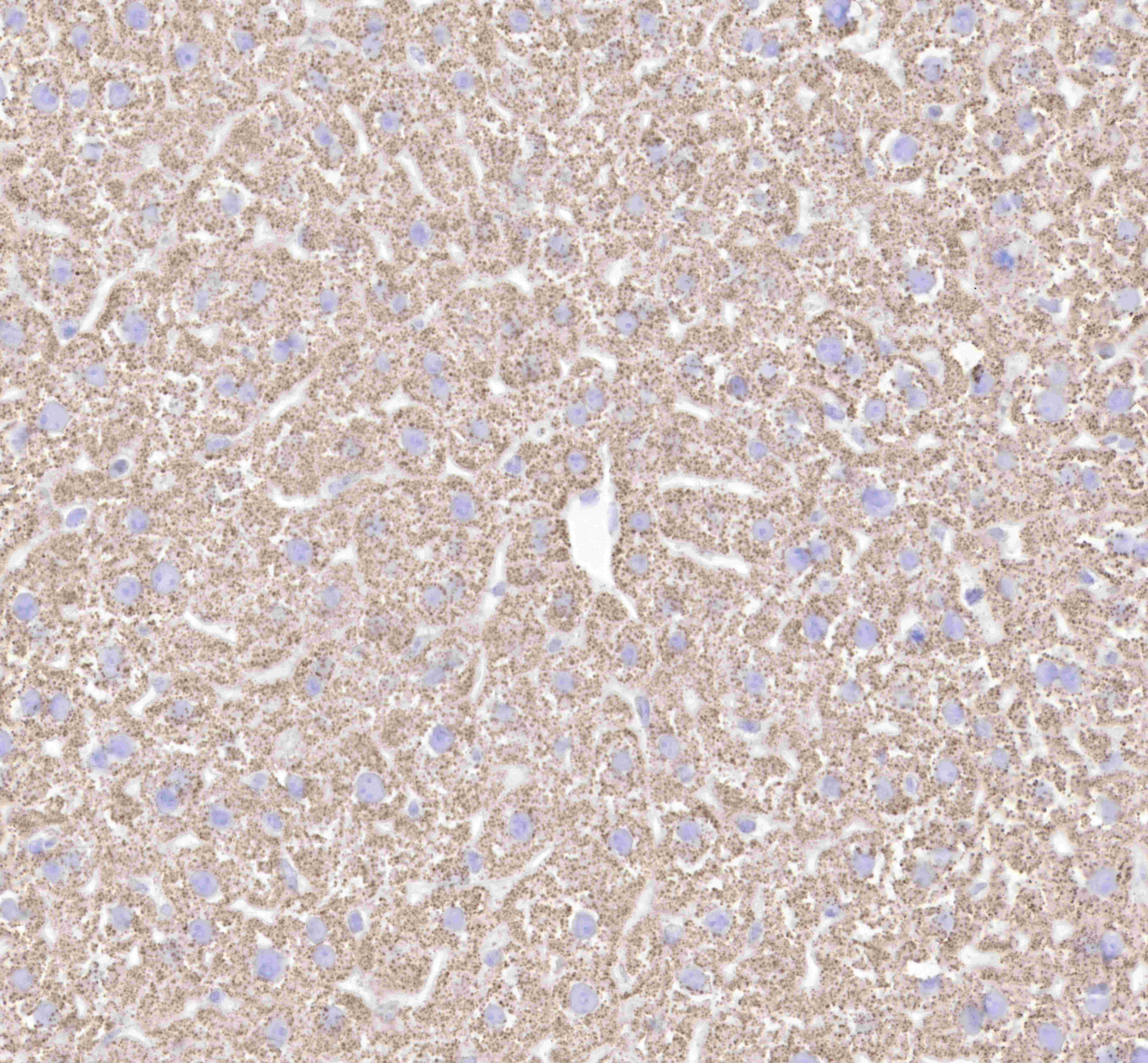
IHC shows positive staining in paraffin-embedded mouse liver.
Anti-Hsp60 antibody was used at 1/2000 dilution, followed by a Goat Anti-Rabbit IgG H&L (HRP) ready to use.
Counterstained with hematoxylin.
Heat mediated antigen retrieval with Tris/EDTA buffer pH9.0 was performed before commencing with IHC staining protocol.
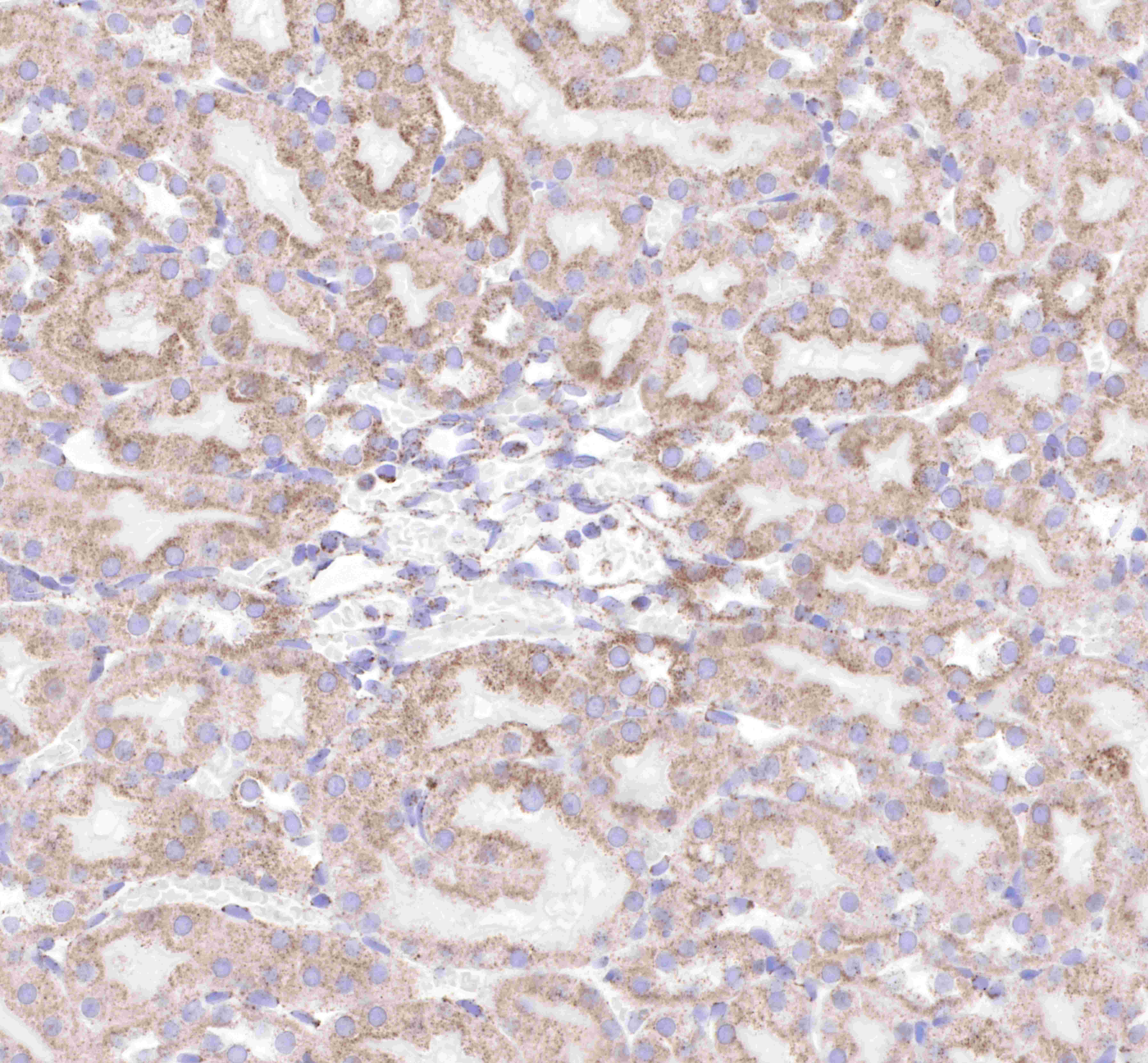
IHC shows positive staining in paraffin-embedded rat kindey.
Anti-Hsp60 antibody was used at 1/2000 dilution, followed by a Goat Anti-Rabbit IgG H&L (HRP) ready to use.
Counterstained with hematoxylin.
Heat mediated antigen retrieval with Tris/EDTA buffer pH9.0 was performed before commencing with IHC staining protocol.
Immunocytochemistry
ICC shows cytoplasm staining in HeLa cells.
Anti-Hsp60 antibody was used at 1/1000 dilution and incubated overnight at 4°C.
Goat polyclonal Antibody to Rabbit IgG - H&L (Alexa Fluor® 488) was used as secondary antibody at 1/1000 dilution.
The cells were fixed with 100% methanol and permeabilized with 0.1% PBS-Triton X-100.
Nuclei were counterstained with DAPI.
ICC shows cytoplasm staining in NIH/3T3 cells.
Anti-Hsp60 antibody was used at 1/1000 dilution and incubated overnight at 4°C.
Goat polyclonal Antibody to Rabbit IgG - H&L (Alexa Fluor® 488) was used as secondary antibody at 1/1000 dilution.
The cells were fixed with 100% methanol and permeabilized with 0.1% PBS-Triton X-100.
Nuclei were counterstained with DAPI.