Product Specification
| Host |
Rabbit |
| Antigen |
FOXP1 |
| Synonyms |
Forkhead box protein P1, MFH |
| Immunogen |
N/A |
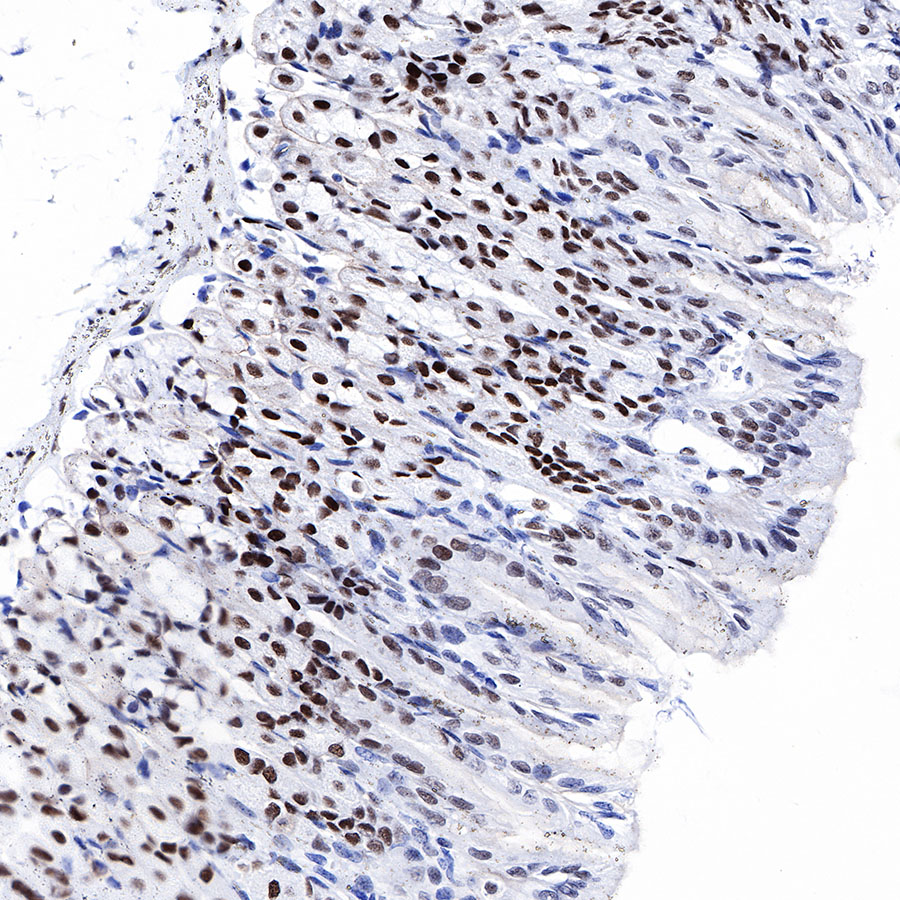
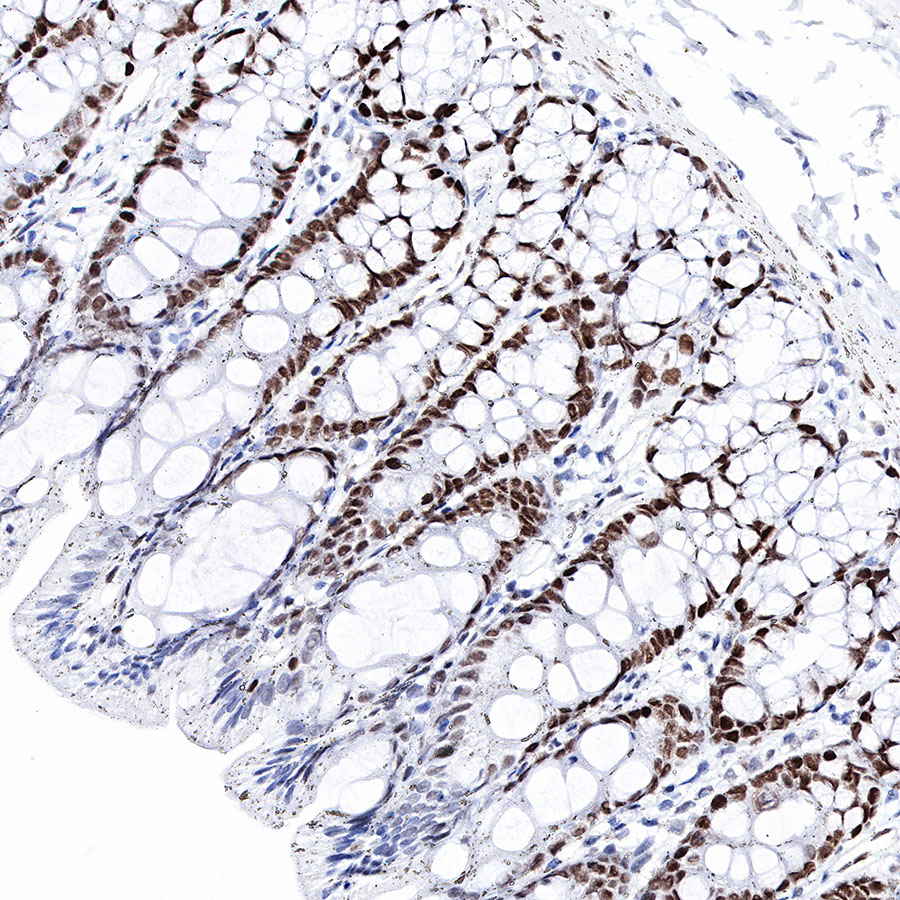
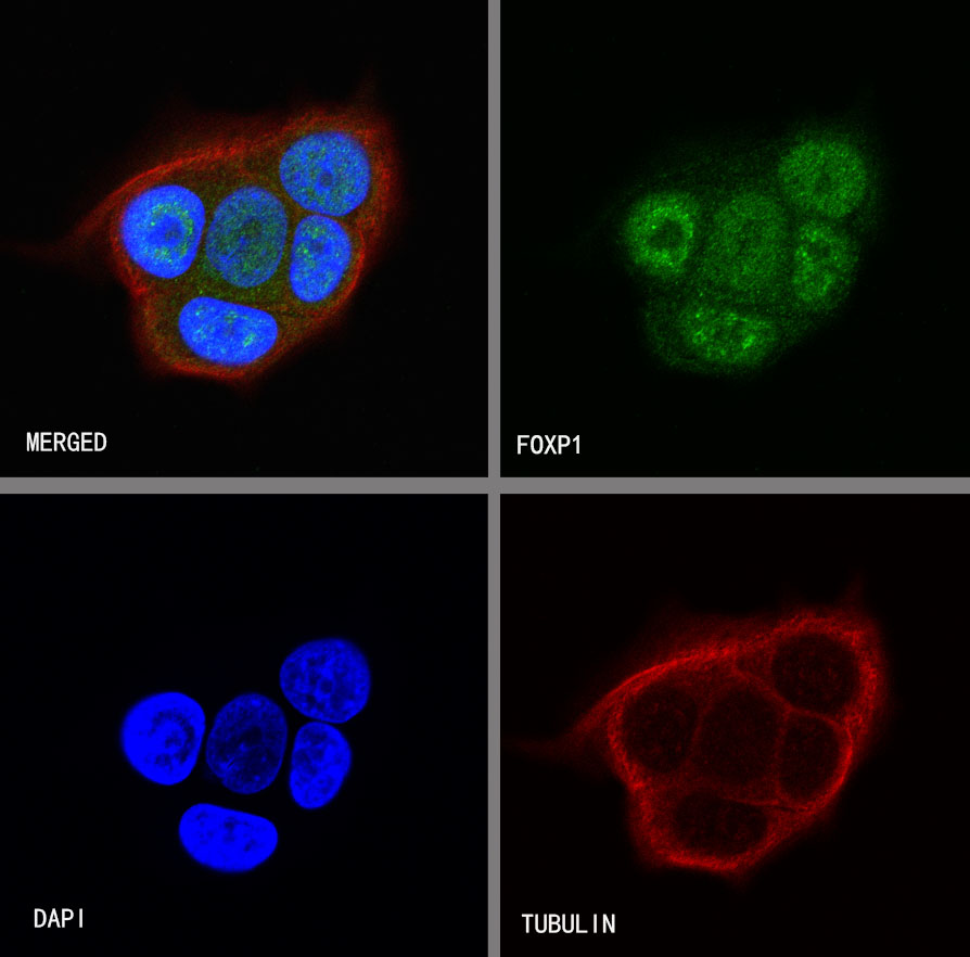
| Location |
Nucleus |
| Accession |
Q9H334 |
| Clone Number |
SDT-R053 |
| Antibody Type |
Rabbit mAb |
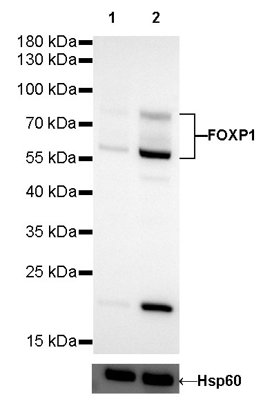
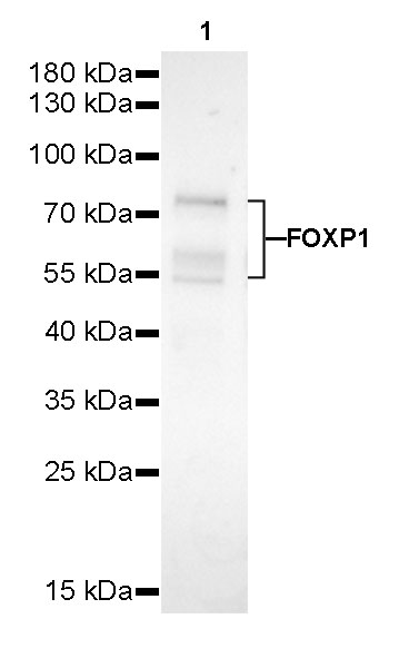
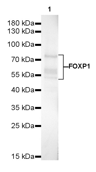
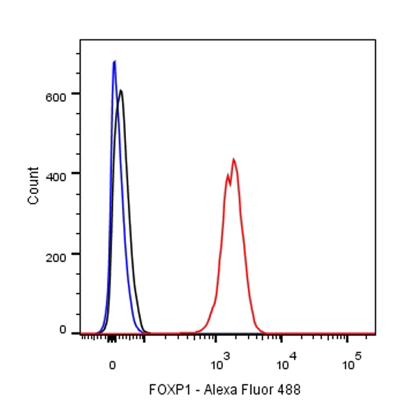
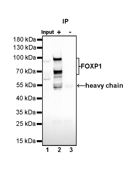
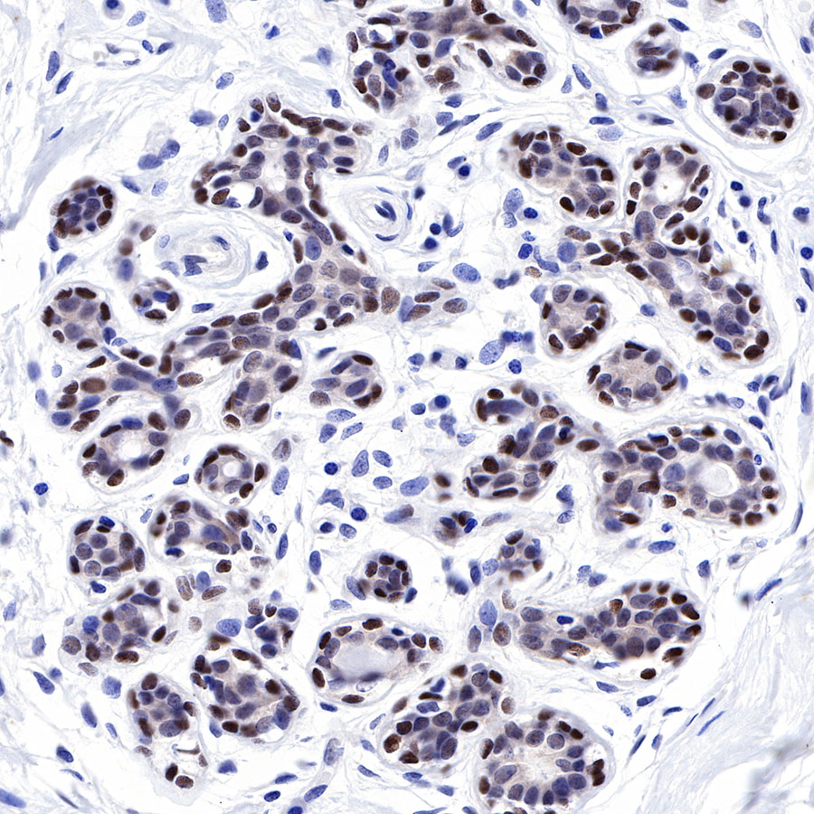
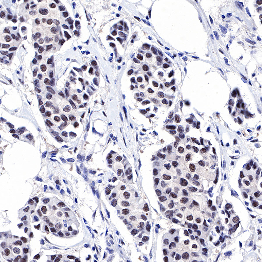
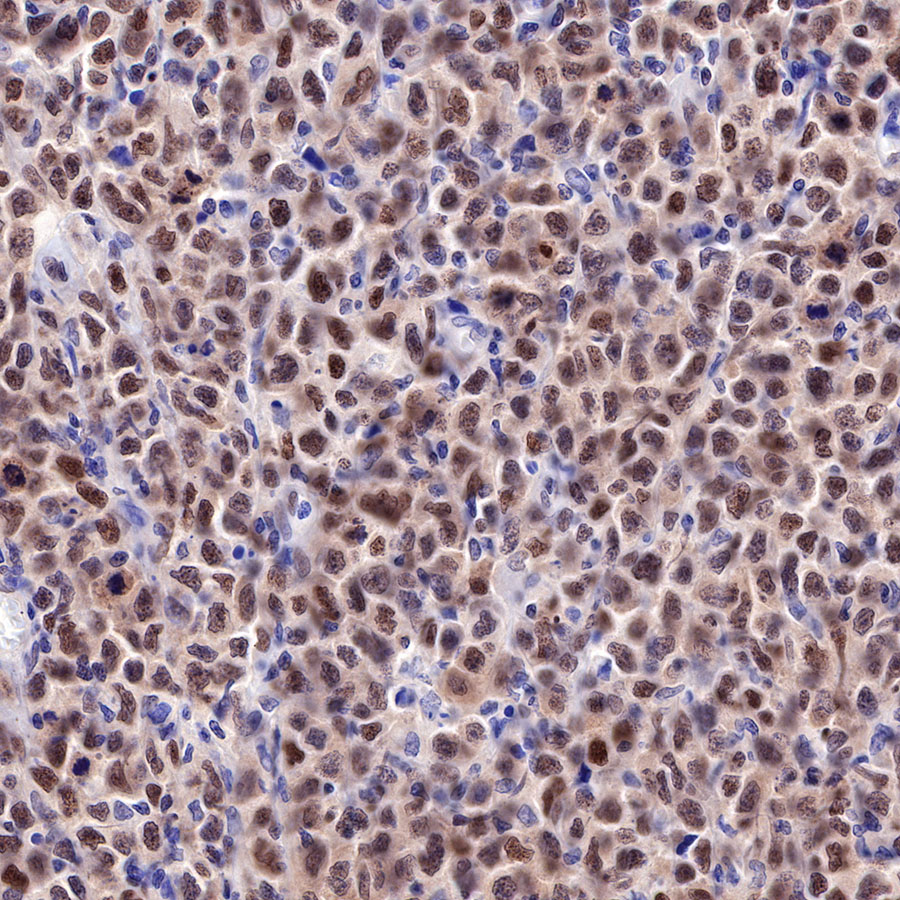
| Application |
WB, IHC-P, ICC, IP, ICFCM |
| Reactivity |
Hu, Ms, Rt |
| Purification |
Protein A |
| Concentration |
0.5 mg/ml |
| Physical Appearance |
Liquid |
| Storage Buffer |
PBS, 40% Glycerol, 0.05%BSA, 0.03% Proclin 300 |
| Stability & Storage |
12 months from date of receipt / reconstitution, -20 °C as supplied |
Dilution
| application |
dilution |
species |
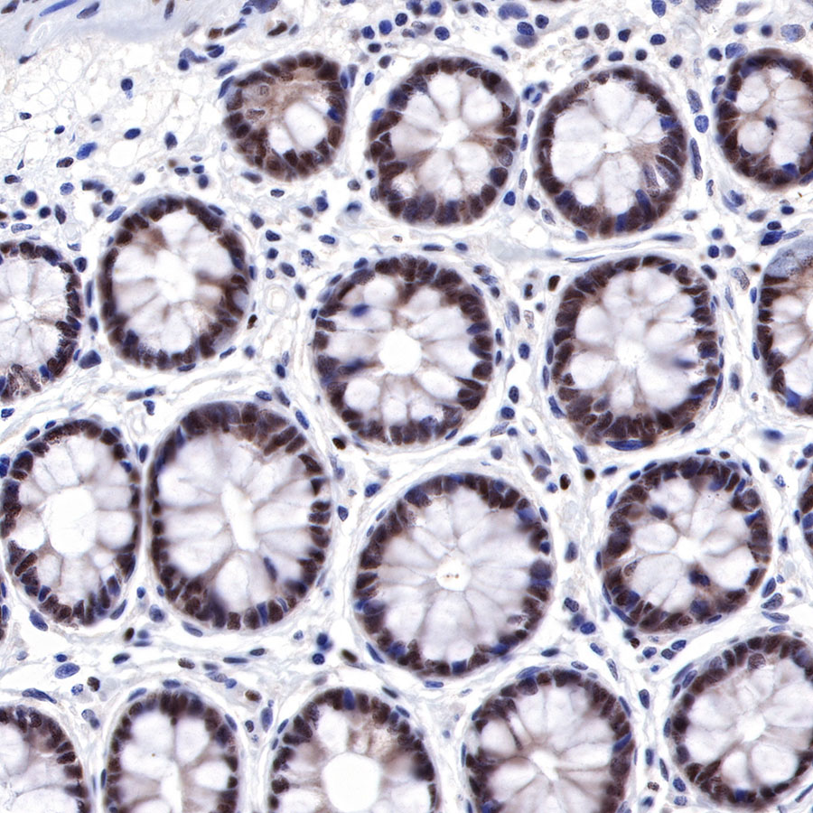
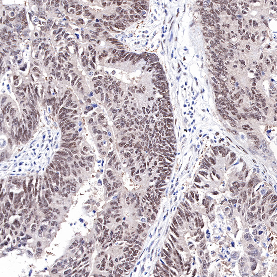
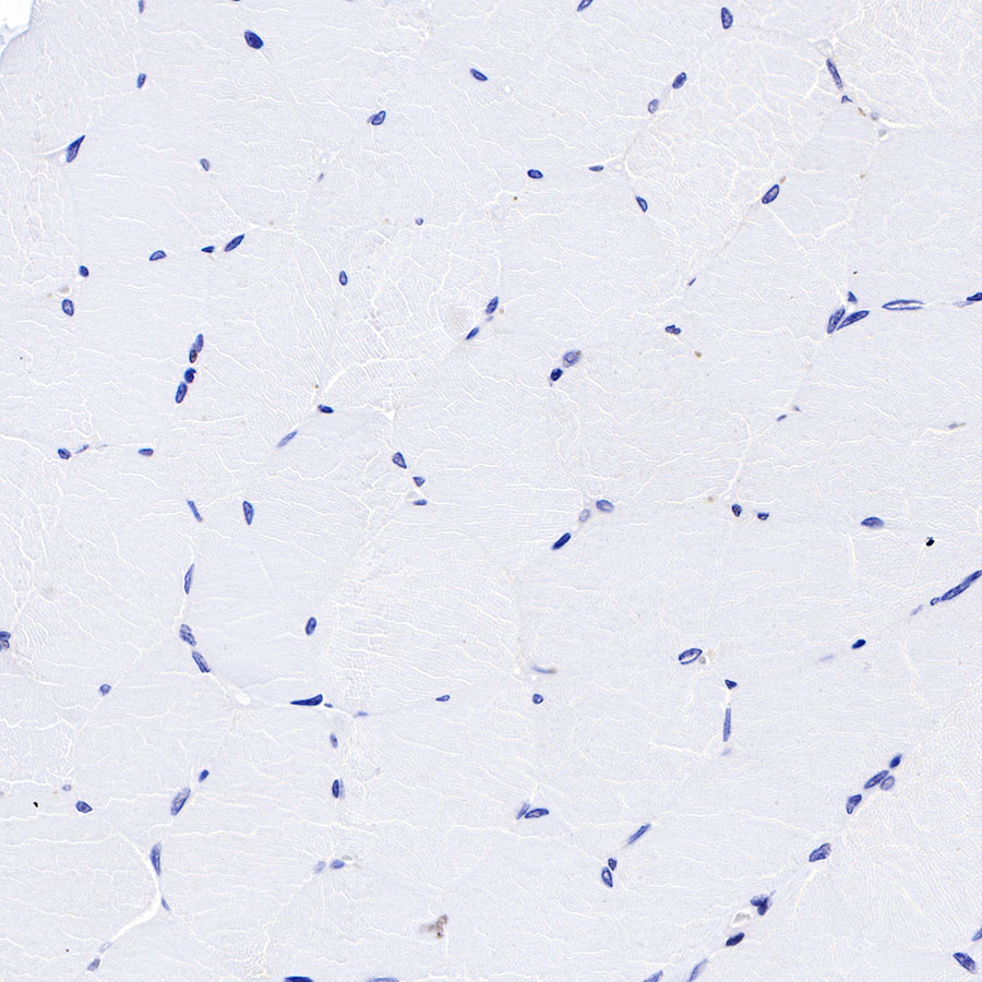
| IHC-P |
1:500-1:1000 |
|
| ICFCM |
1:500 |
|
| WB |
1:1000 |
|
| ICC |
1:500 |
|
| IP |
1:25 |
|
Background
The FOXP subfamily is defined by four members (FOXP1–FOXP4). FOXPs function as transcription factors and possess distinct transcriptional regulatory and DNA-binding domains. The N terminus of FOXPs contains a transcriptional repressor region with a zinc-finger/leucine zipper motif. The DNA-binding domain of FOXPs is uniquely positioned among forkhead proteins near the C terminus. FOXP1 is widely expressed in human tissues and regulates the development of many tissues, including heart, thymus, and lung. FOXP1-deficient embryos have severe defects in cardiac morphogenesis, including outflow tract septation and cushion defects, resulting in embryonic death.