WB result of CD44 Mouse mAb
Primary antibody: CD44 Mouse mAb at 1/2000 dilution
Lane 1: LNCaP whole cell lysate 20 µg
Lane 2: HeLa whole cell lysate 20 µg
Lane 3: PANC-1 whole cell lysate 20 µg
Lane 4: A549 whole cell lysate 20 µg
Negative control: LNCaP whole cell lysate
Secondary antibody: Goat Anti-Mouse IgG, (H+L), HRP conjugated at 1/10000 dilution
Predicted MW: 81 kDa
Observed MW: 70~90 kDa
CD44 Mouse mAb (SDT-630-174)
CD44 Mouse mAb (SDT-630-174)
Price:
Regular price
$100 USD
Regular price
Sale price
$100 USD
Unit price
per
For shipping services or bulk orders, you may request a quotation.
Secure checkout with

View full details
Product Details
Product Details
Product Specification
| Host | Mouse |
| Antigen | CD44 |
| Synonyms | CDw44, Epican, Extracellular matrix receptor III (ECMR-III), GP90 lymphocyte homing/adhesion receptor, HUTCH-I, Heparan sulfate proteoglycan, Hermes antigen, Hyaluronate receptor, Phagocytic glycoprotein 1 (PGP-1), Phagocytic glycoprotein I (PGP-I), LHR, MDU2, MDU3, MIC4 |
| Immunogen | Recombinant Protein |
| Location | Cell membrane |
| Accession | P16070 |
| Clone Number | SDT-630-174 |
| Antibody Type | Mouse mAb |
| Isotype | IgG1,k |
| Application | WB, IHC-P, ICC, FCM, IF |
| Reactivity | Hu |
| Purification | Protein G |
| Concentration | 2 mg/ml |
| Conjugation | Unconjugated |
| Physical Appearance | Liquid |
| Storage Buffer | PBS, 40% Glycerol, 0.05%BSA, 0.03% Proclin 300 |
| Stability & Storage | 12 months from date of receipt / reconstitution, -20 °C as supplied |
Dilution
| application | dilution | species |
| WB | 1:2000 | null |
| IHC | 1:1000-1:2000 | null |
| FCM | 1:2000 | null |
| ICC | 1:500 | null |
| IF | 1:500 | null |
Background
The CD44 antigen is a cell-surface glycoprotein involved in cell–cell interactions, cell adhesion and migration. CD44 is expressed in a large number of mammalian cell types. It participates in a wide variety of cellular functions including lymphocyte activation, recirculation and homing, hematopoiesis, and tumor metastasis. CD44, along with CD25, is used to track early T cell development in the thymus. CD44 expression is an indicative marker for effector-memory T-cells. Memory cell proliferation (activation) can also be assayed in vitro with CFSE chemical tagging. In addition, variations in CD44 are reported as cell surface markers for some breast and prostate cancer stem cells. In breast cancer research CD44+/CD24- expression is commonly used as a marker for breast CSCs and is used to sort breast cancer cells into a population enriched in cells with stem-like characteristics and has been seen as an indicator of increased survival time in epithelial ovarian cancer patients.
Picture
Picture
Western Blot
FC
Flow cytometric analysis of LNCaP (Human prostate carcinoma epithelial cell, left) / HeLa (Human cervix adenocarcinoma epithelial cell, right) cells labelling CD44 antibody at 1/2000 dilution (0.1 μg) / (red) compared with a Mouse monoclonal IgG (Black) isotype control and an unlabelled control (cells without incubation with primary antibody and secondary antibody) (Blue). Goat Anti - Mouse IgG Alexa Fluor® 488 was used as the secondary antibody.
Negative control: LNCaP
Immunohistochemistry
IHC shows positive staining in paraffin-embedded human bladder. Anti-CD44 antibody was used at 1/2000 dilution, followed by a HRP Polymer for Mouse & Rabbit IgG (ready to use). Counterstained with hematoxylin. Heat mediated antigen retrieval with Tris/EDTA buffer pH9.0 was performed before commencing with IHC staining protocol.
IHC shows positive staining in paraffin-embedded human liver. Anti-CD44 antibody was used at 1/1000 dilution, followed by a HRP Polymer for Mouse & Rabbit IgG (ready to use). Counterstained with hematoxylin. Heat mediated antigen retrieval with Tris/EDTA buffer pH9.0 was performed before commencing with IHC staining protocol.
IHC shows positive staining in paraffin-embedded human prostate. Anti-CD44 antibody was used at 1/1000 dilution, followed by a HRP Polymer for Mouse & Rabbit IgG (ready to use). Counterstained with hematoxylin. Heat mediated antigen retrieval with Tris/EDTA buffer pH9.0 was performed before commencing with IHC staining protocol.
IHC shows positive staining in paraffin-embedded human colon cancer. Anti-CD44 antibody was used at 1/1000 dilution, followed by a HRP Polymer for Mouse & Rabbit IgG (ready to use). Counterstained with hematoxylin. Heat mediated antigen retrieval with Tris/EDTA buffer pH9.0 was performed before commencing with IHC staining protocol.
IHC shows positive staining in paraffin-embedded human lung adenocarcinoma. Anti-CD44 antibody was used at 1/1000 dilution, followed by a HRP Polymer for Mouse & Rabbit IgG (ready to use). Counterstained with hematoxylin. Heat mediated antigen retrieval with Tris/EDTA buffer pH9.0 was performed before commencing with IHC staining protocol.
IHC shows positive staining in paraffin-embedded human thyroid cancer. Anti-CD44 antibody was used at 1/1000 dilution, followed by a HRP Polymer for Mouse & Rabbit IgG (ready to use). Counterstained with hematoxylin. Heat mediated antigen retrieval with Tris/EDTA buffer pH9.0 was performed before commencing with IHC staining protocol.
Immunocytochemistry
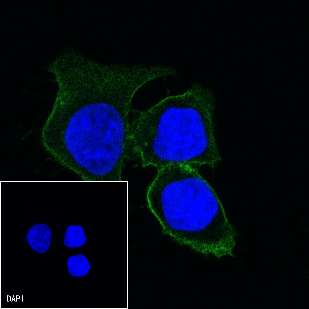
ICC shows positive staining in HeLa cells. Anti-CD44 antibody was used at 1/500 dilution (Green) and incubated overnight at 4°C. Goat polyclonal Antibody to Rabbit IgG - H&L (Alexa Fluor® 488) was used as secondary antibody at 1/1000 dilution. The cells were fixed with 100% ice-cold methanol and permeabilized with 0.1% PBS-Triton X-100. Nuclei were counterstained with DAPI (Blue).
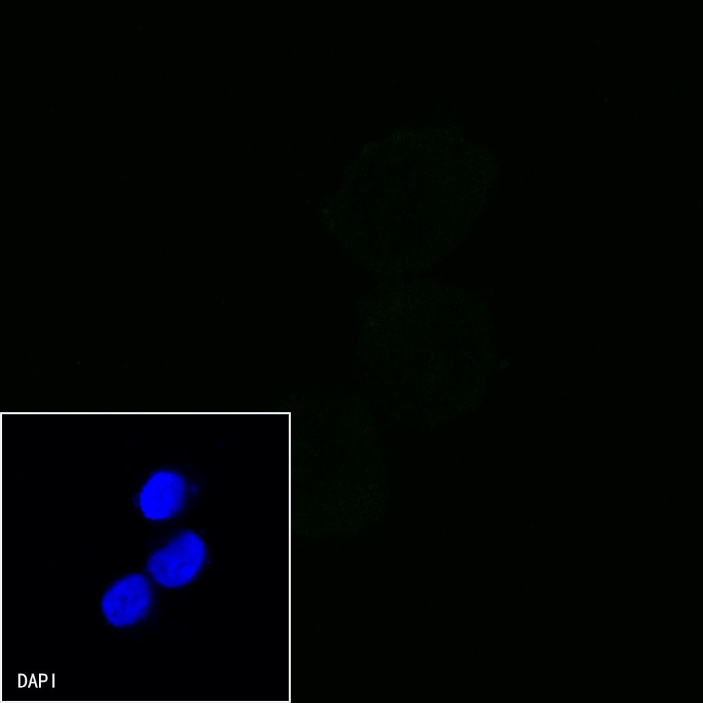
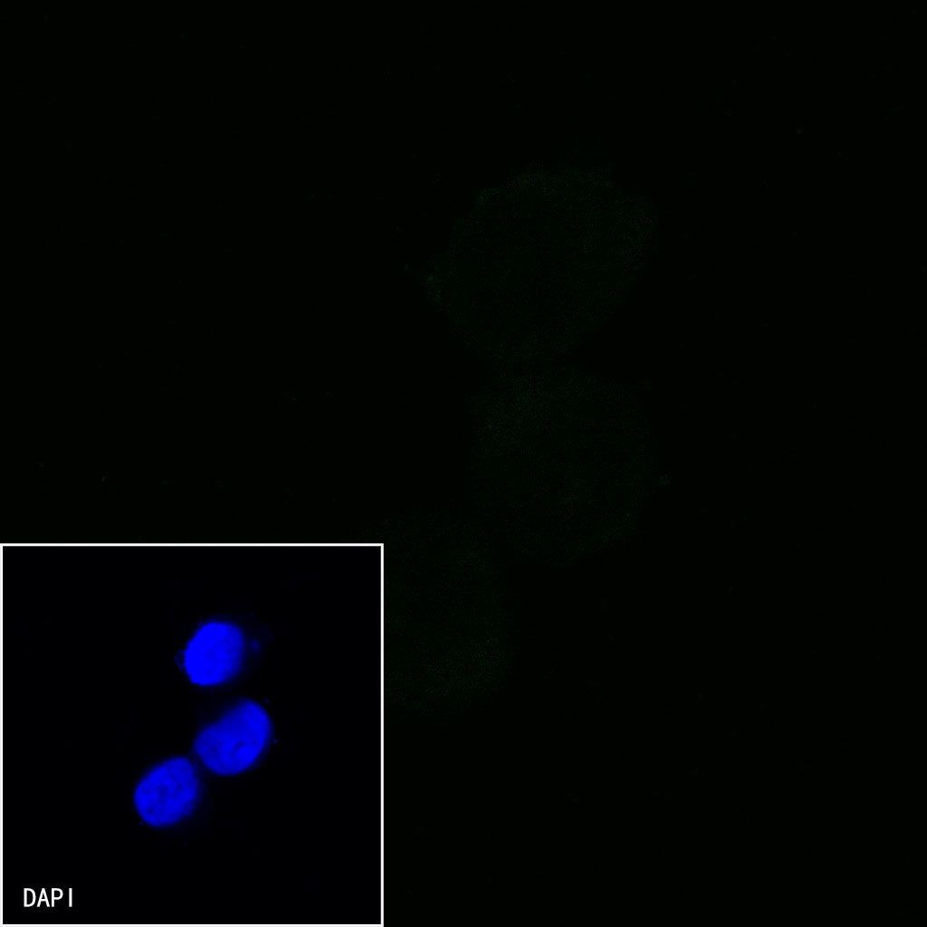
Negative control:ICC shows negative staining in Lncap cells. Anti-CD44 antibody was used at 1/500 dilution and incubated overnight at 4°C. Goat polyclonal Antibody to Rabbit IgG - H&L (Alexa Fluor® 488) was used as secondary antibody at 1/1000 dilution. The cells were fixed with 100% ice-cold methanol and permeabilized with 0.1% PBS-Triton X-100. Nuclei were counterstained with DAPI (Blue).
Immunofluorescence
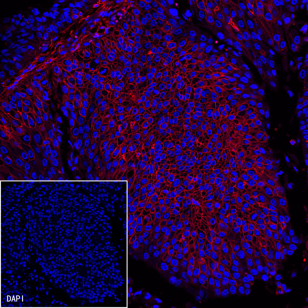
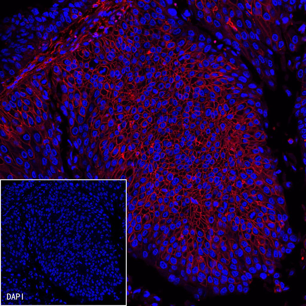
IF shows positive staining in paraffin-embedded human bladder cancer. Anti-CD44 antibody was used at 1/500 dilution (Red) and incubated overnight at 4°C.Goat anti-Mouse IgG(H+L) (Alexa Fluor® 594 Conjugate) (S0B4010) was used as secondary antibody at 1/1000 dilution.Counterstained with DAPI (Blue). Heat mediated antigen retrieval with EDTA buffer pH9.0 was performed before commencing with IF staining protocol.