Product Specification
| Host |
Rabbit |
| Antigen |
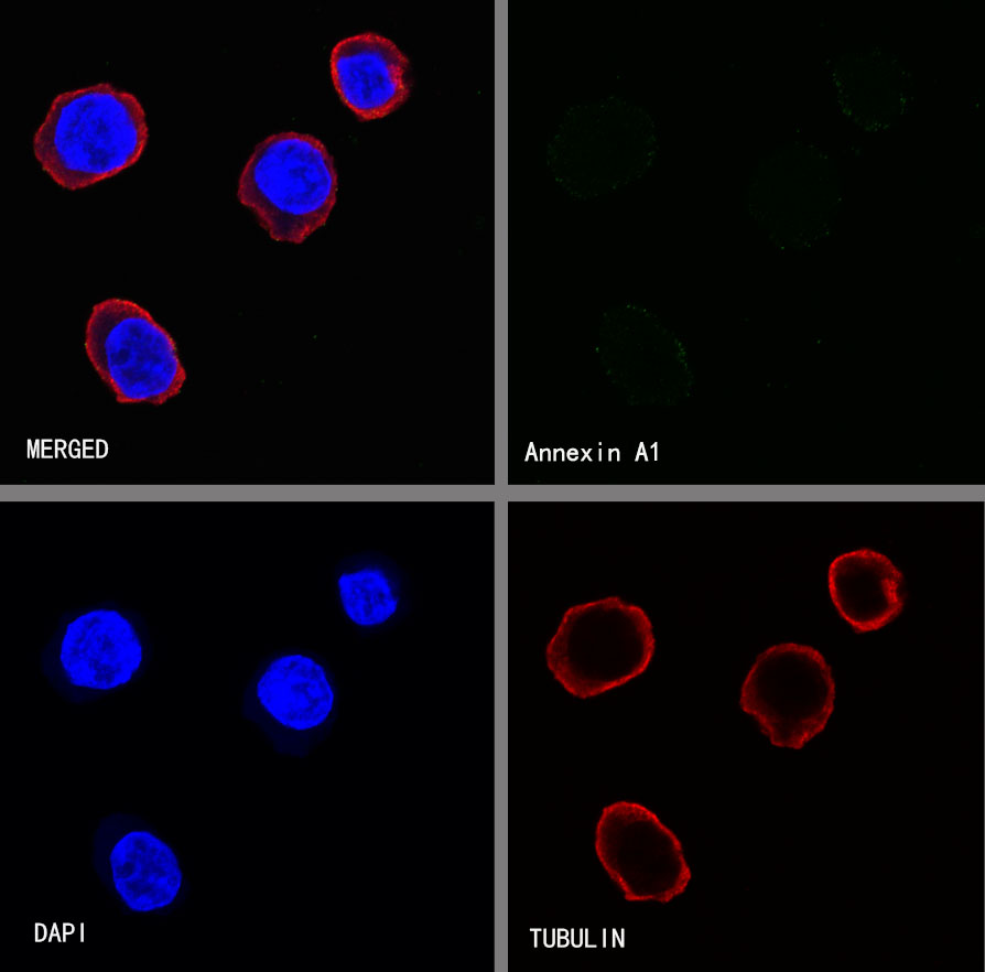
Annexin A1 |
| Synonyms |
Calpactin II,ANX1, LPC1, Annexin I, Annexin-1, Calpactin-2, Chromobindin-9 |
| Immunogen |
Recombinant Protein |
| Accession |
P4083 |
| Clone Number |
SDT-020-36 |
| Antibody Type |
Rabbit mAb |
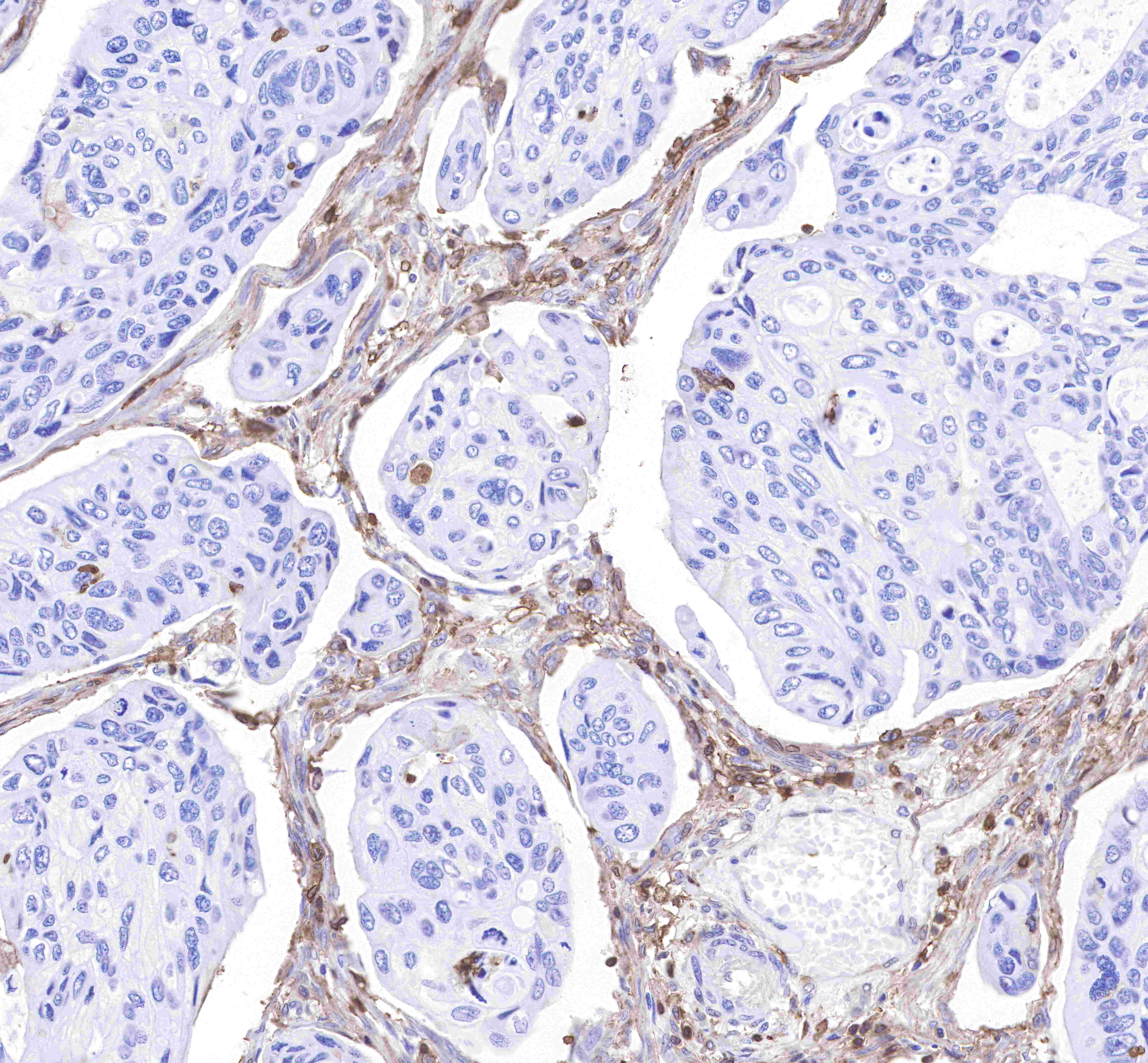
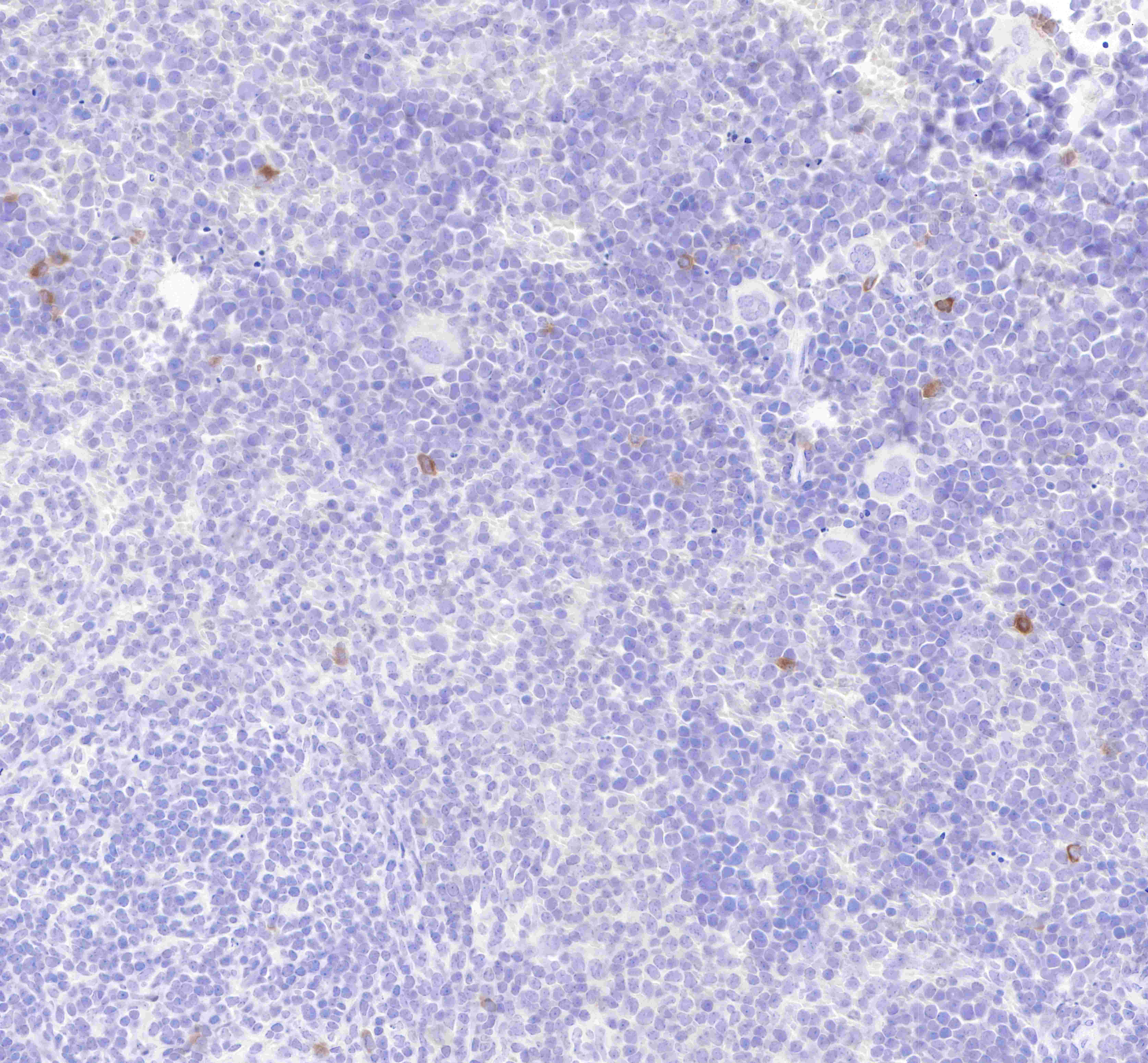
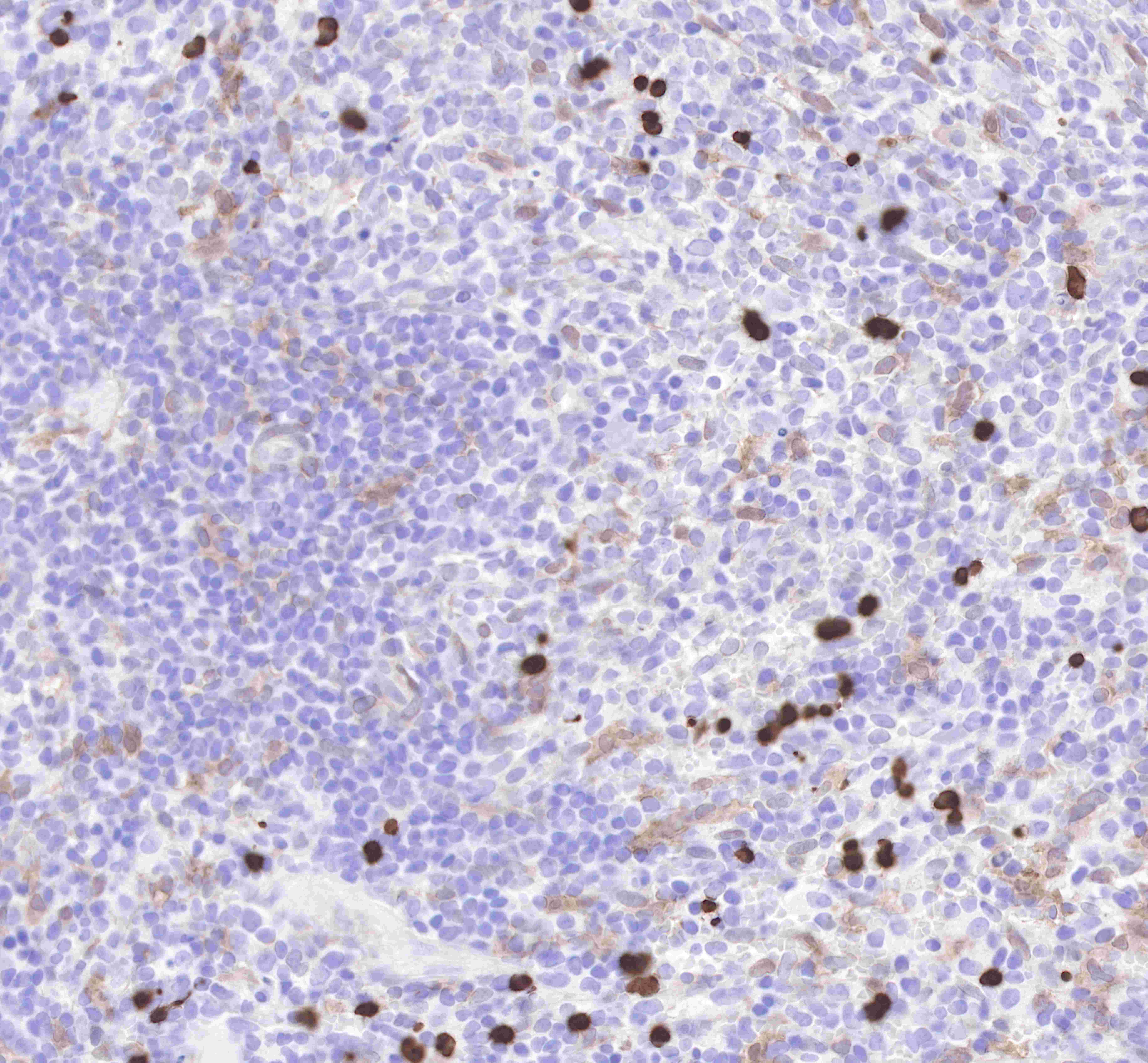
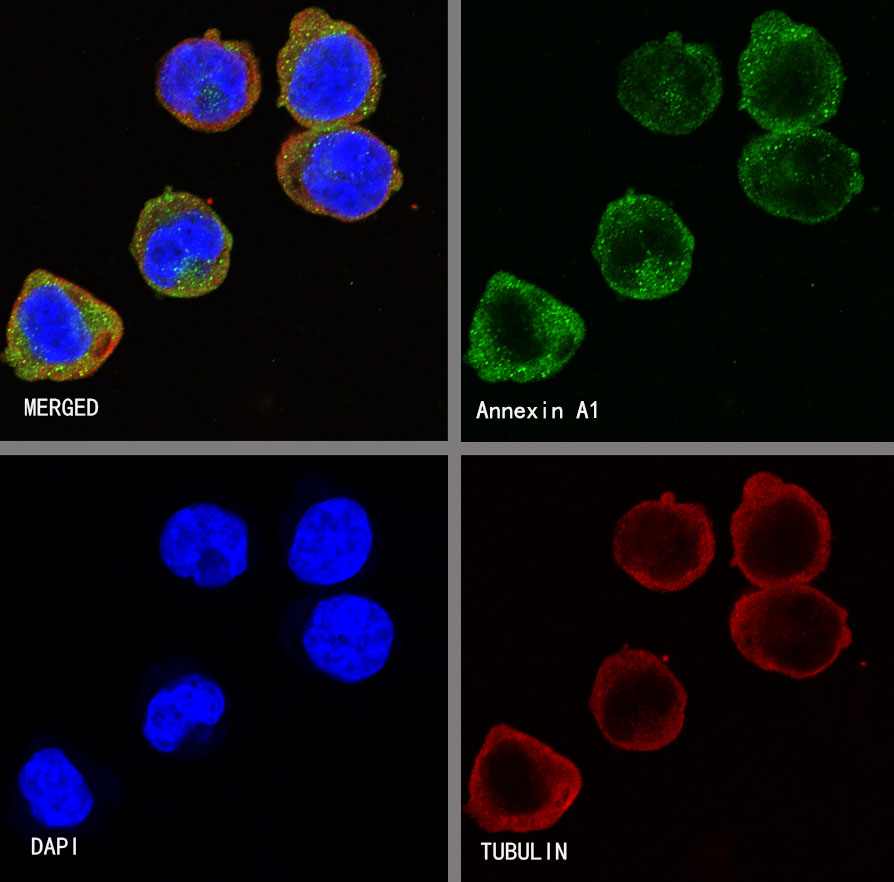
| Application |
WB, IHC-P, ICC, ICFCM |
| Reactivity |
Hu, Ms, Rt |
| Purification |
Protein A |
| Concentration |
0.5mg/ml |
| Conjugation |
Unconjugated |
| Physical Appearance |
Liquid |
| Storage Buffer |
PBS, 40% Glycerol, 0.05%BSA, 0.03% Proclin 300 |
| Stability & Storage |
12 months from date of receipt / reconstitution, -20 °C as supplied |
Dilution
| application |
dilution |
species |
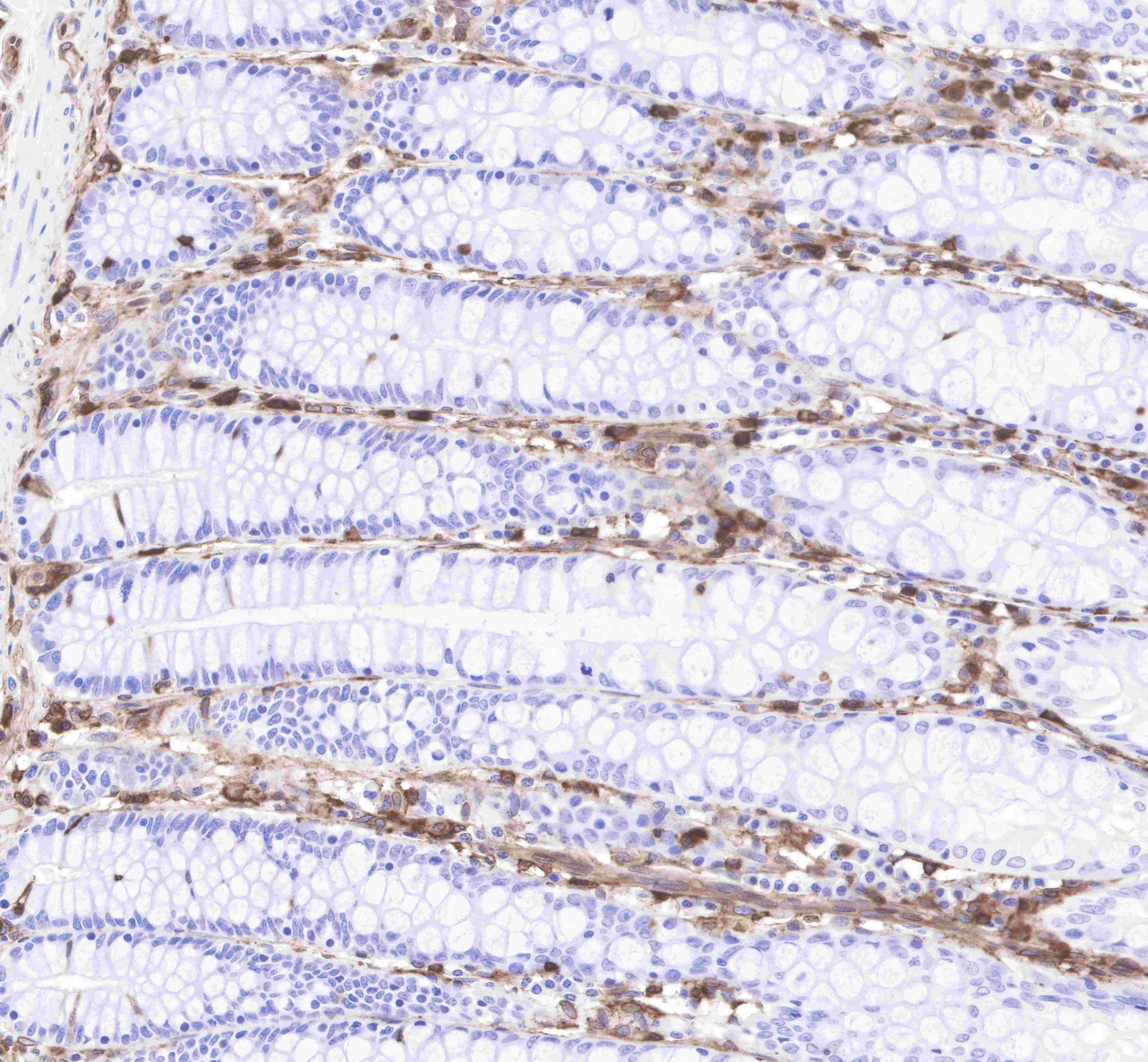
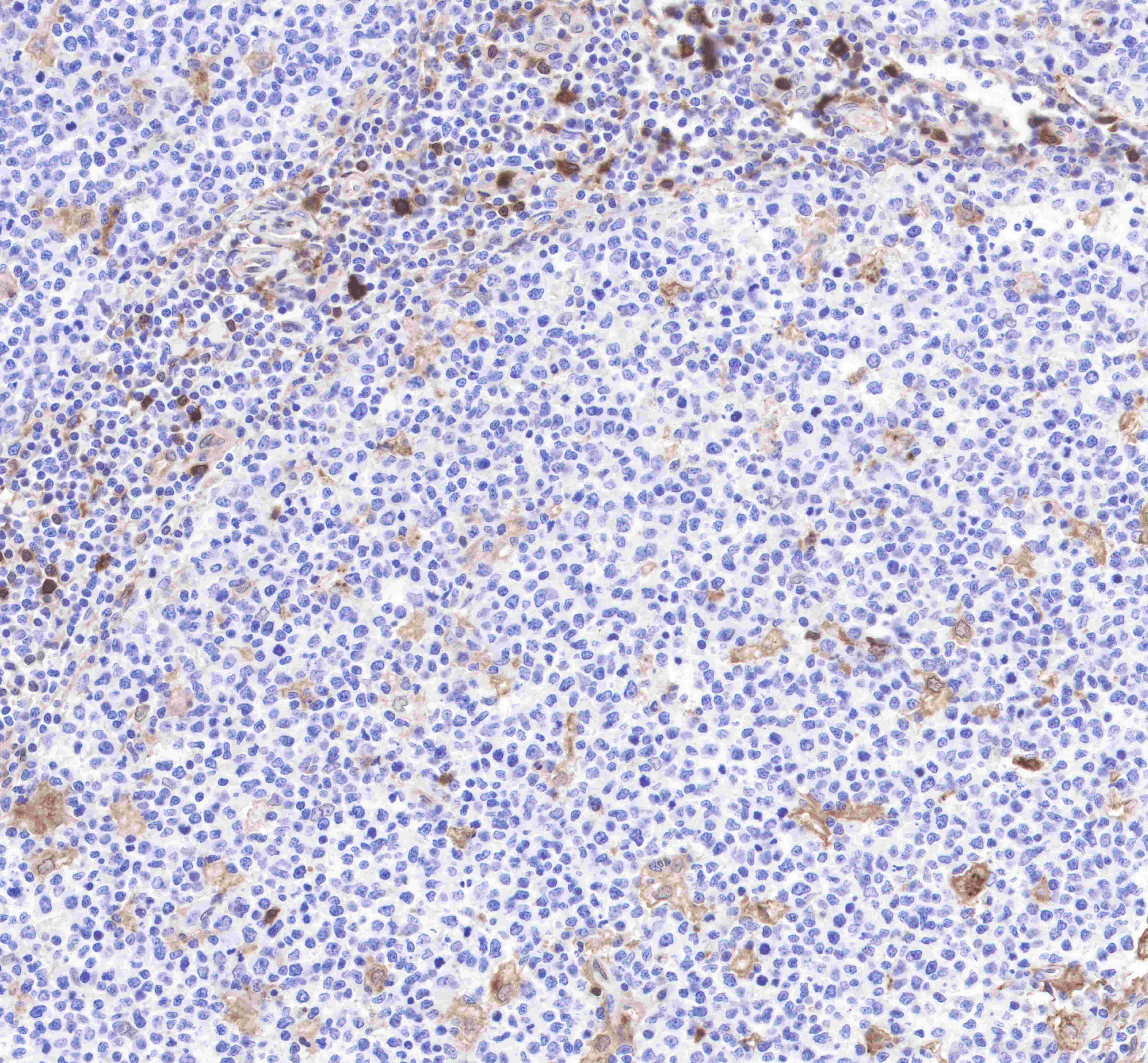
| IHC-P |
1:2000 |
null |
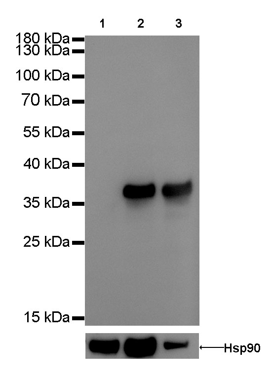
| WB |
1:1000 |
null |
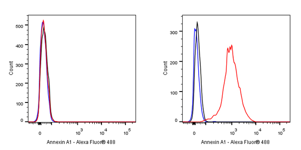
| ICFCM |
1:5000 |
null |
| ICC |
1:500 |
null |
Background
Annexin A1 belongs to the annexin family of Ca2+-dependent phospholipid-binding proteins that have a molecular weight of approximately 35,000 to 40,000 Dalton and are preferentially located on the cytosolic face of the plasma membrane. Annexin A1 protein has an apparent relative molecular mass of 40 kDa with phospholipase A2 inhibitory activity. In resting conditions, human and mouse immune cells such as neutrophils, monocytes, and macrophages contain high levels of annexin A1 in their cytoplasm. Following cell activation (for example, by neutrophil adhesion to endothelial-cell monolayers), annexin A1 is promptly mobilized to the cell surface and secreted. Annexin A1 promotes neutrophil detachment and apoptosis, and phagocytosis of apoptotic neutrophils by macrophages. On the other hand, it reduces the tendency of neutrophils to penetrate the endothelium of blood vessels. Higher expression of annexin A1 during pathological conditions could increase the strength of TCR signalling through the mitogen-activated protein kinase signalling pathway, thereby causing a state of hyperactivation of T cells.