Guidelines for the preparation of spleen single-cell suspensions (enzymatic digestion)

The mouse spleen is an important immune organ located in the left abdominal cavity of the mouse, near the left side of the stomach, in an oblong shape. The size will vary depending on the breed and health of the mouse, but in general, the spleen length of an adult mouse is approximately between 1.5 cm and 2.5 cm. The color and texture of the spleen may vary depending on the breed and health of the mouse, and it usually has a leaf-like shape and is dark red in color. In experimental research, the isolation, culture, and enumeration of mouse spleen cells is an important method for immunological research, and it is also a prerequisite for flow cytometry analysis.
1. Consumables and reagents
Table 1 Reference table of some reagent consumables for dissociation of mouse spleen
|
material |
reagent |
equipment |
|
15/50 mL centrifuge tubes |
1x PBS buffer |
Horizontal thermostatic shaker |
|
10 cm Petri dish |
RPMI 1640 medium |
Hemocytometer |
|
Ophthalmic surgical scissors, eye tweezers |
75% alcohol |
Electronic pipettes, 1mL tips |
|
100 μm cell sieve |
FBS |
High-speed centrifuge |
|
10 mL pipette |
HBSS equilibrium solution |
Vortex instrument |
|
5 mL flow cytometry tubes |
Absin Tissue Dissociation Kit |
4 °C freezer |
|
|
Cell staining buffer |
Flow cytometry |
Table 2 Product information
|
Catalog number |
Product name |
specification |
use |
|
abs50097 |
Mouse spleen tissue dissociation kit |
10T/25T |
Dissociate the tissue |
|
abs7303 |
5 mL flow cytometry tubes (sterile, enzyme-free, with caps) |
500pcs/carton |
Flow cytometry tubes |
|
abs9484 |
RPMI1640 medium |
500mL |
Preserve cell viability |
|
70um cell sieve |
100pcs/carton |
Supplies |
|
|
abs7102 |
15 mL centrifuge tubes (culet-bottom) |
100pcs/bag |
Supplies |
|
abs7100 |
50 mL centrifuge tubes (culet-bottom) |
40pcs/bag |
Supplies |
|
PBS buffer (1x) |
500mL |
reagent |
|
|
abs9475 |
Cell staining buffer |
500mL |
reagent |
|
abs9101 |
Red blood cell lysate (1x) |
100mL/500mL |
Split red liquor |
2. Experimental procedures
Preparation before the experiment: preheat the spleen mouse spleen tissue dissociation kit in a 37 degree water bath (after the spleen tissue dissociation reagent is received, it is recommended to divide and cryostore), if the red blood cell lysate is 10 ×, it needs to be diluted into 1 × with ultrapure water in advance. PBS pre-cooled in advance.
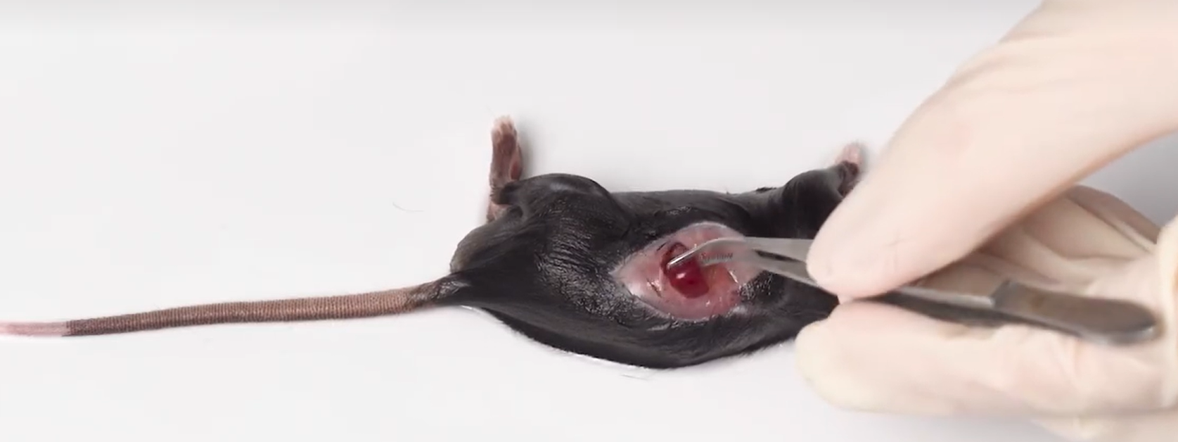
1. Dislocate the neck of the mouse to death, disinfect the surface of the mouse with 75% alcohol, as for the tray, the left abdomen is facing up, the tweezers assist the operation, lift the skin in the middle of the left abdomen, cut the skin, the dark red is visible to the naked eye, the long strip-like spleen, cut the peritoneum, lift the spleen with forceps, see the separation, remove the connective tissue and fat as much as possible, rinse with PBS, take about 100mg-200mg of fresh mouse spleen tissue (a 20g mouse, the spleen weight may be about 100mg, The specific situation shall prevail) add to a 50mL sterile centrifuge tube, and thoroughly chop the tissue to 1-2mm with ophthalmic scissors3, add 1mL of mouse spleen tissue dissociation reagent;
2. Place the centrifuge tube on a constant temperature shaker, incubate at 37°C/180rpm for about 30min, digest the spleen tissue of mice, and adjust the digestion time appropriately according to the state of the tissue (the spleen is relatively easy to digest).
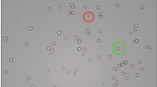
In the red circle: dead cells In the green circle: live cells
3. After the incubation, the tissue homogenate in the centrifuge tube is filtered with a 70um sterile filter, then the cell culture medium is added to flush the sterile filter, and the filtrate is collected with a 50mL centrifuge tube;
4. Centrifuge the collected filtrate at 1200rpm for 5min and discard the supernatant;
5. Red blood cell lysate can be used to remove red blood cells in the process of tissue dissociation, the specific operation is to add 3mL of 1× red blood cell lysate to the precipitate obtained in step 4, resuspend the precipitate, vortex to mix, incubate at room temperature in the dark for 3-5min, add 2mL of pre-cooled PBS, centrifuge at 1200rpm for 5min, and discard the supernatant. Repeat the wash once;
6. Discard the supernatant, resuspend the cell pellet in cell staining buffer, and adjust the cell concentration to 5-10×106cells/mL, dispense the cell suspension of 100 uL/tube into a 12×75 mm flow cytometry tube for follow-up experiments.
3. Problems and strategies
1. Low cell yield
In general, adult C57BL/6 mice can dissociate about 10×10 spleen8–1.4×10⁸cells, if the cell yield is found to be particularly low, it is necessary to pay attention to the complete removal of the fat around the tissue as much as possible during the sample acquisition stage, and to cut the tissue as much as possible to improve the cell yield. In the washing step involved, use a pipette to aspirate the upper layer of fat before decanting the supernatant (trim the tip of the tip to avoid clogging when aspirating)
2. The cell viability is low, and there are many dead cells/debris
First of all, the incubation time of the dissociation reagent should not be too long, long-term incubation will cause the reduction of cell viability and the loss of surface markers, too many dead cells, the release of DNA filaments entangled, resulting in viscous solution, secondly, do not shorten the cleaning steps, cleaning can help remove adhesions, debris and dead cells, the incubation time of red blood cell lysate can not be too long, generally 5min, if the cell viability is still very low, it is recommended to check whether the red blood cell lysate is fresh and effective.
3. Incomplete lysis of red blood cells
Manifests as a noticeable red precipitate after the washing step. Make sure to prepare fresh erythrocyte lysis buffer to lyse at room temperature. Before lysis begins, make sure that the cells are completely resuspended and that the lysis time is not too short.
Fourth, the dissociation effect
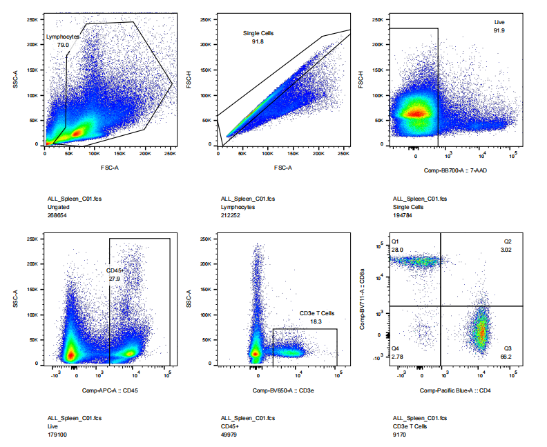
In this kit, mouse Spleen tissue was processed into a single-cell suspension and then flow-byme: cell viability, CD45, CD3, CD4, CD8 staining results
Note: This product is only used for scientific research experiments and does not support clinical research
5. Other product recommendations
|
Catalog number |
Product name |
specification |
|
abs9907 |
Flow cytometry absolute counting tubes |
50T |
|
abs9477 |
Mouse Fc receptor blocker |
200T/500T |
|
abs9476 |
Human Fc receptor blocker |
50T/200T |
|
abs9488 |
Sheath fluid |
20L |
|
abs9107 |
Phoboester |
1mg/5mg/25mg |
|
abs9108 |
Ionomycin calcium salt |
1mg/5mg |
|
abs970 |
D-PBS buffer (1 ×, no calcium magnesium) |
500mL |
|
abs810012 |
Brefeldin A |
10mg/25mg/50mg |
|
abs812975 |
Monensin sodium salt |
25mg/100mg |
|
abs9475 |
Cell staining buffer |
500mL |
|
abs9110 |
fixer |
50mL |
|
abs9111 |
Permeabilization agent |
150mL |
|
abs7303 |
5 mL flow cytometry tubes (sterile, enzyme-free, with caps) |
1 box |
|
abs1840002 |
APC Mouse anti-Human CD3 Antibody(3JI3-9) |
25T/100T |
|
PE Mouse anti-Human CD4 Antibody(5D4) |
25T/100T |
|
|
abs182430 |
FITC Mouse anti-Human CD8 Antibody(3C4) |
25T/100T |
|
abs180046 |
PE-Cy7 Mouse anti-Human CD25 Antibody(BC96) |
25T/100T |
|
abs1840696 |
FITC Mouse anti-Human CD80 Antibody(2D10.4) |
25T/100T |
|
abs182338 |
APC Rat anti-Mouse CD25 Antibody(4APC61.5) |
25T/100T |
|
More...... |
||
Absin provides antibodies, proteins, ELISA kits, cell culture, detection kits, and other research reagents. If you have any product needs, please contact us.
|
Absin Bioscience Inc. Email: worldwide@absin.cn |
 Follow us on Facebook: Absin Bio Follow us on Facebook: Absin Bio |


