WB result of Lamin B2 Rabbit mAb
Primary antibody: Lamin B2 Rabbit mAb at 1/1000 dilution
Lane 1: HeLa whole cell lysate 20 µg
Lane 2: Hep G2 whole cell lysate 20 µg
Lane 3: Raji whole cell lysate 20 µg
Lane 4: HCT 116 whole cell lysate 20 µg
Lane 5: Jurkat whole cell lysate 20 µg
Secondary antibody: Goat Anti-rabbit IgG, (H+L), HRP conjugated at 1/10000 dilution
Predicted MW: 68 kDa
Observed MW: 70 kDa
S-RMab®Lamin B2 Recombinant Rabbit mAb (S-951-70)
S-RMab®Lamin B2 Recombinant Rabbit mAb (S-951-70)
Price:
Regular price
$70 USD
Regular price
Sale price
$70 USD
Unit price
per
For shipping services or bulk orders, you may request a quotation.
Secure checkout with

View full details
Product Details
Product Details
Product Specification
| Host | Rabbit |
| Antigen | Lamin B2 |
| Synonyms | LMNB2, LMN2 |
| Immunogen | Synthetic Peptide |
| Location | Nucleus lamina |
| Accession | Q03252 |
| Clone Number | S-951-70 |
| Antibody Type | Recombinant mAb |
| Isotype | IgG |
| Application | WB, IHC-P, ICC, IP, IF, ICFCM |
| Reactivity | Hu, Ms, Rt |
| Predicted Reactivity | Xe |
| Purification | Protein A |
| Concentration | 0.5 mg/ml |
| Conjugation | Unconjugated |
| Physical Appearance | Liquid |
| Storage Buffer | PBS, 40% Glycerol, 0.05% BSA, 0.03% Proclin 300 |
| Stability & Storage | 12 months from date of receipt / reconstitution, -20 °C as supplied. |
Dilution
| application | dilution | species |
| WB | 1:1000-1:10000 | |
| IHC-P | 1:2000 | |
| ICC | 1:500 | |
| ICFCM | 1:500 | |
| IF | 1:500 | |
| IP | 1:50 |
Background
Lamins are components of the nuclear lamina, a fibrous layer on the nucleoplasmic side of the inner nuclear membrane, which is thought to provide a framework for the nuclear envelope and may also interact with chromatin. Lamin B2 is a protein that in humans is encoded by the LMNB2 gene. It is the second of two type B nuclear lamins, and it is associated with laminopathies.
Picture
Picture
Western Blot
WB result of Lamin B2 Rabbit mAb
Primary antibody: Lamin B2 Rabbit mAb at 1/1000 dilution
Lane 1: mouse spleen lysate 20 µg
Secondary antibody: Goat Anti-rabbit IgG, (H+L), HRP conjugated at 1/10000 dilution
Predicted MW: 68 kDa
Observed MW: 70 kDa
WB result of Lamin B2 Rabbit mAb
Primary antibody: Lamin B2 Rabbit mAb at 1/1000 dilution
Lane 1: rat spleen lysate 20 µg
Secondary antibody: Goat Anti-rabbit IgG, (H+L), HRP conjugated at 1/10000 dilution
Predicted MW: 68 kDa
Observed MW: 75 kDa
FC
Flow cytometric analysis of 4% PFA fixed 90% methanol permeabilized HeLa (Human cervix adenocarcinoma epithelial cell) labelling Lamin B2 antibody at 1/500 dilution (0.1 μg) / (Red) compared with a Rabbit monoclonal IgG (Black) isotype control and an unlabelled control (cells without incubation with primary antibody and secondary antibody) (Blue). Goat Anti - Rabbit IgG Alexa Fluor® 488 was used as the secondary antibody.
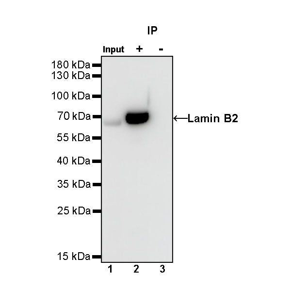
IP
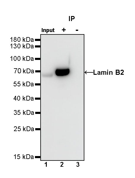
Lamin B2 Rabbit mAb at 1/50 dilution (1 µg) immunoprecipitating Lamin B2 in 0.4 mg HeLa whole cell lysate.
Western blot was performed on the immunoprecipitate using Lamin B2 Rabbit mAb at 1/1000 dilution.
Secondary antibody (HRP) for IP was used at 1/1000 dilution.
Lane 1: HeLa whole cell lysate 20 µg (Input)
Lane 2: Lamin B2 Rabbit mAb IP in HeLa whole cell lysate
Lane 3: Rabbit monoclonal IgG IP in HeLa whole cell lysate
Predicted MW: 68 kDa
Observed MW: 70 kDa
Immunohistochemistry
IHC shows positive staining in paraffin-embedded human cerebral cortex. Anti-Lamin B2 antibody was used at 1/2000 dilution, followed by a HRP Polymer for Mouse & Rabbit IgG (ready to use). Counterstained with hematoxylin. Heat mediated antigen retrieval with Tris/EDTA buffer pH9.0 was performed before commencing with IHC staining protocol.
IHC shows positive staining in paraffin-embedded human kidney. Anti-Lamin B2 antibody was used at 1/2000 dilution, followed by a HRP Polymer for Mouse & Rabbit IgG (ready to use). Counterstained with hematoxylin. Heat mediated antigen retrieval with Tris/EDTA buffer pH9.0 was performed before commencing with IHC staining protocol.
IHC shows positive staining in paraffin-embedded human testis. Anti-Lamin B2 antibody was used at 1/2000 dilution, followed by a HRP Polymer for Mouse & Rabbit IgG (ready to use). Counterstained with hematoxylin. Heat mediated antigen retrieval with Tris/EDTA buffer pH9.0 was performed before commencing with IHC staining protocol.
IHC shows positive staining in paraffin-embedded human prostatic cancer. Anti-Lamin B2 antibody was used at 1/2000 dilution, followed by a HRP Polymer for Mouse & Rabbit IgG (ready to use). Counterstained with hematoxylin. Heat mediated antigen retrieval with Tris/EDTA buffer pH9.0 was performed before commencing with IHC staining protocol.
IHC shows positive staining in paraffin-embedded human thyroid cancer. Anti-Lamin B2 antibody was used at 1/2000 dilution, followed by a HRP Polymer for Mouse & Rabbit IgG (ready to use). Counterstained with hematoxylin. Heat mediated antigen retrieval with Tris/EDTA buffer pH9.0 was performed before commencing with IHC staining protocol.
IHC shows positive staining in paraffin-embedded mouse cerebral cortex. Anti-Lamin B2 antibody was used at 1/2000 dilution, followed by a HRP Polymer for Mouse & Rabbit IgG (ready to use). Counterstained with hematoxylin. Heat mediated antigen retrieval with Tris/EDTA buffer pH9.0 was performed before commencing with IHC staining protocol.
IHC shows positive staining in paraffin-embedded mouse liver. Anti-Lamin B2 antibody was used at 1/2000 dilution, followed by a HRP Polymer for Mouse & Rabbit IgG (ready to use). Counterstained with hematoxylin. Heat mediated antigen retrieval with Tris/EDTA buffer pH9.0 was performed before commencing with IHC staining protocol.
IHC shows positive staining in paraffin-embedded rat spleen. Anti-Lamin B2 antibody was used at 1/2000 dilution, followed by a HRP Polymer for Mouse & Rabbit IgG (ready to use). Counterstained with hematoxylin. Heat mediated antigen retrieval with Tris/EDTA buffer pH9.0 was performed before commencing with IHC staining protocol.
IHC shows positive staining in paraffin-embedded rat testis. Anti-Lamin B2 antibody was used at 1/2000 dilution, followed by a HRP Polymer for Mouse & Rabbit IgG (ready to use). Counterstained with hematoxylin. Heat mediated antigen retrieval with Tris/EDTA buffer pH9.0 was performed before commencing with IHC staining protocol.
Immunocytochemistry
ICC shows positive staining in HeLa cells. Anti- Lamin B2 antibody was used at 1/500 dilution (Green) and incubated overnight at 4°C. Goat polyclonal Antibody to Rabbit IgG - H&L (Alexa Fluor® 488) was used as secondary antibody at 1/1000 dilution. The cells were fixed with 4% PFA and permeabilized with 0.1% PBS-Triton X-100. Nuclei were counterstained with DAPI (Blue). Counterstain with tubulin (Red).
ICC shows positive staining in NIH/3T3 cells. Anti-Lamin B2 antibody was used at 1/500 dilution (Green) and incubated overnight at 4°C. Goat polyclonal Antibody to Rabbit IgG - H&L (Alexa Fluor® 488) was used as secondary antibody at 1/1000 dilution. The cells were fixed with 4% PFA and permeabilized with 0.1% PBS-Triton X-100. Nuclei were counterstained with DAPI (Blue). Counterstain with tubulin (Red).
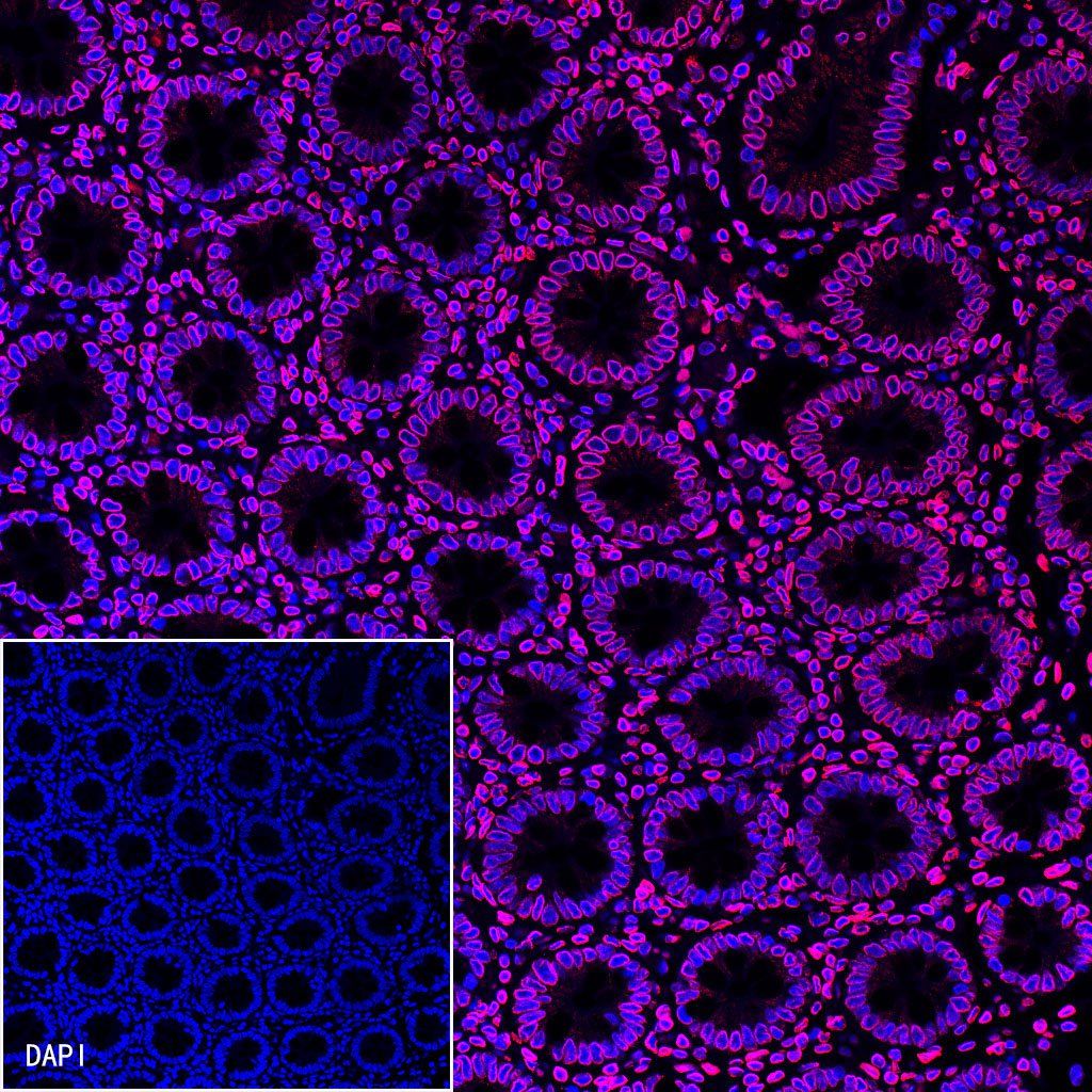
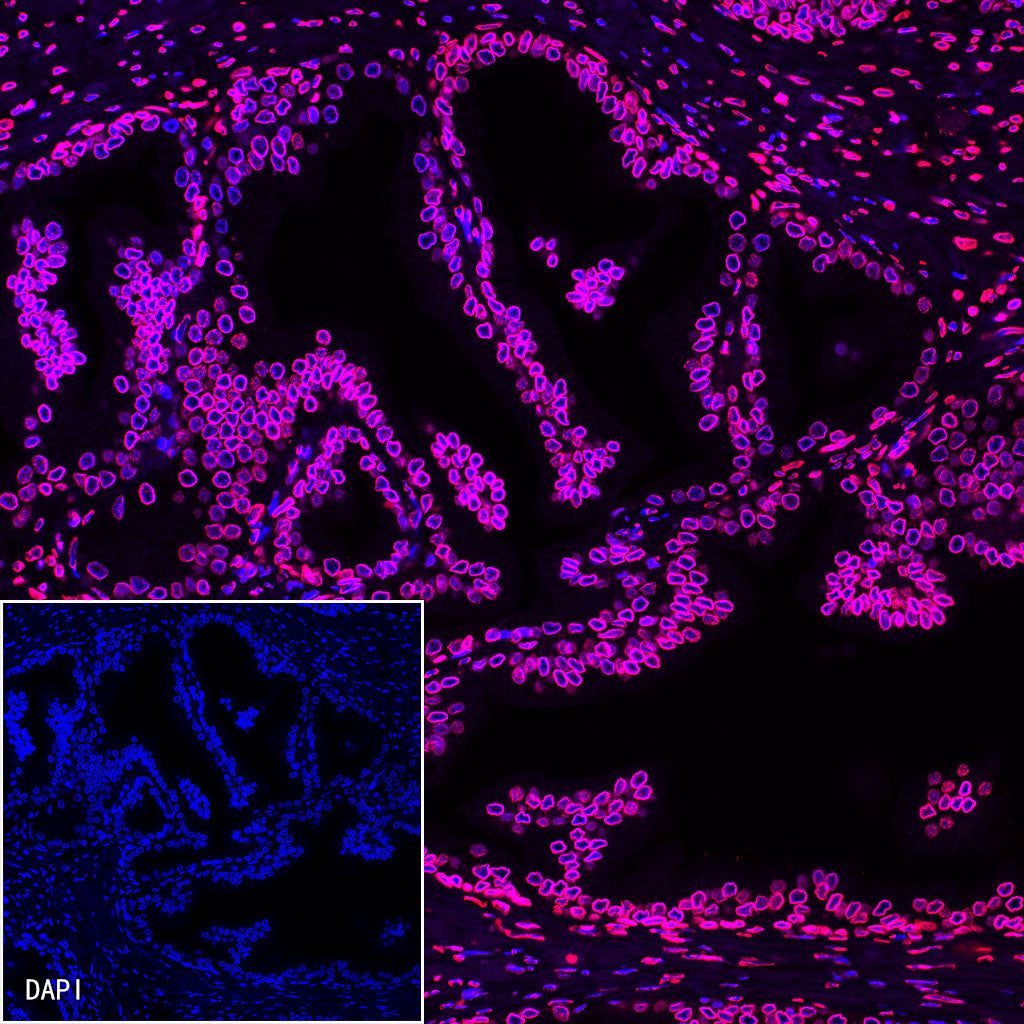
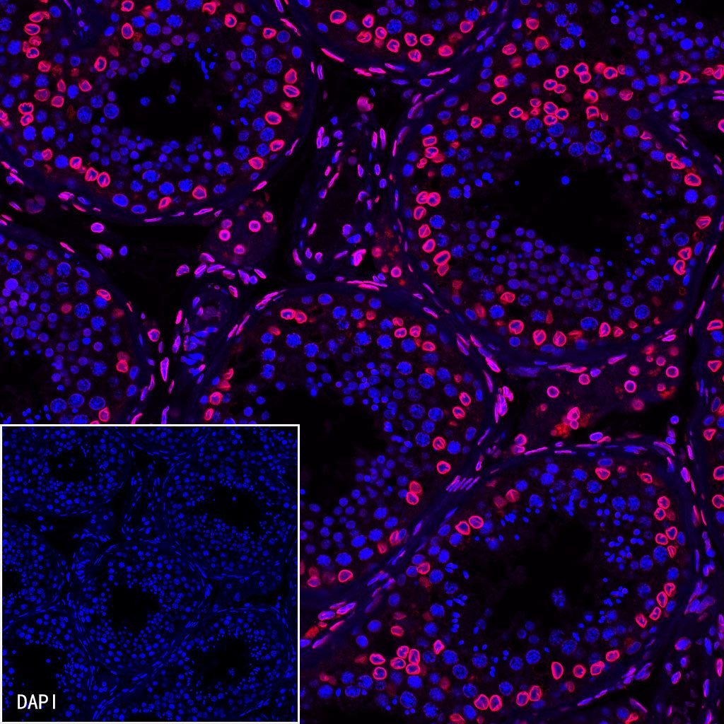
Immunofluorescence
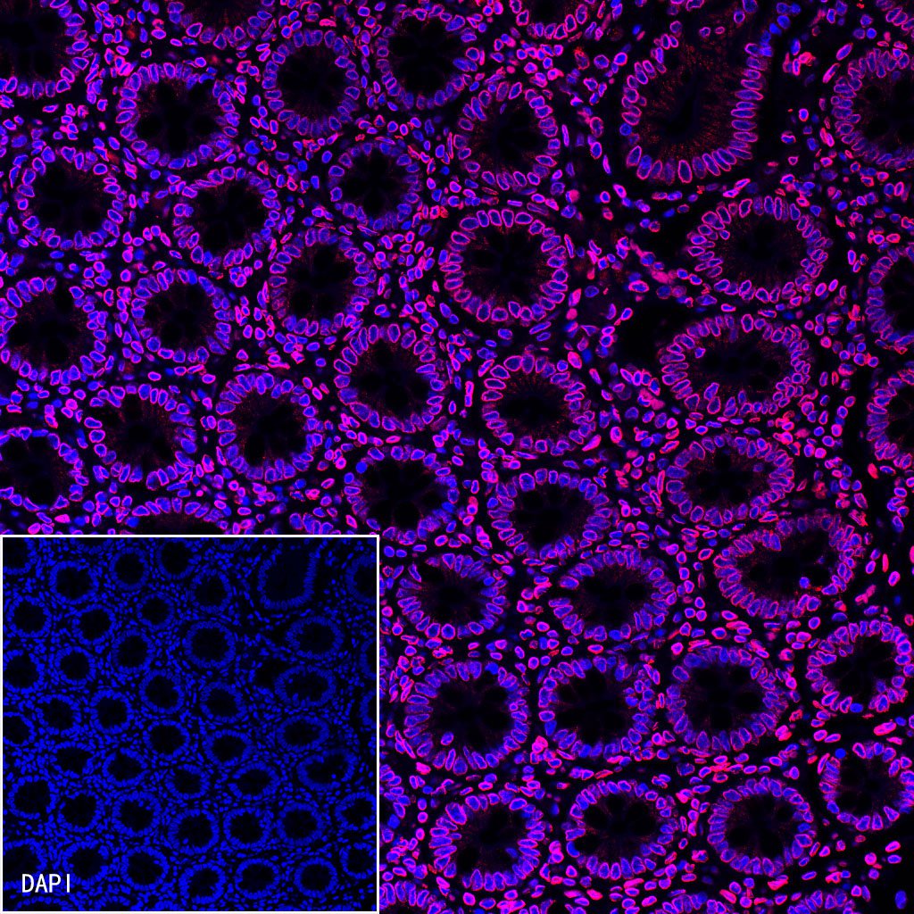
IF shows positive staining in paraffin-embedded human colon. Anti-Lamin B2 antibody was used at 1/500 dilution (Red) and incubated overnight at 4°C.Goat anti-Rabbit IgG(H+L) (Alexa Fluor® 594 Conjugate) (S0B4007) was used as secondary antibody at 1/1000 dilution.Counterstained with DAPI (Blue). Heat mediated antigen retrieval with EDTA buffer pH9.0 was performed before commencing with IF staining protocol.

IF shows positive staining in paraffin-embedded human prostate. Anti-Lamin B2 antibody was used at 1/500 dilution (Red) and incubated overnight at 4°C.Goat anti-Rabbit IgG(H+L) (Alexa Fluor® 594 Conjugate) (S0B4007) was used as secondary antibody at 1/1000 dilution.Counterstained with DAPI (Blue). Heat mediated antigen retrieval with EDTA buffer pH9.0 was performed before commencing with IF staining protocol.
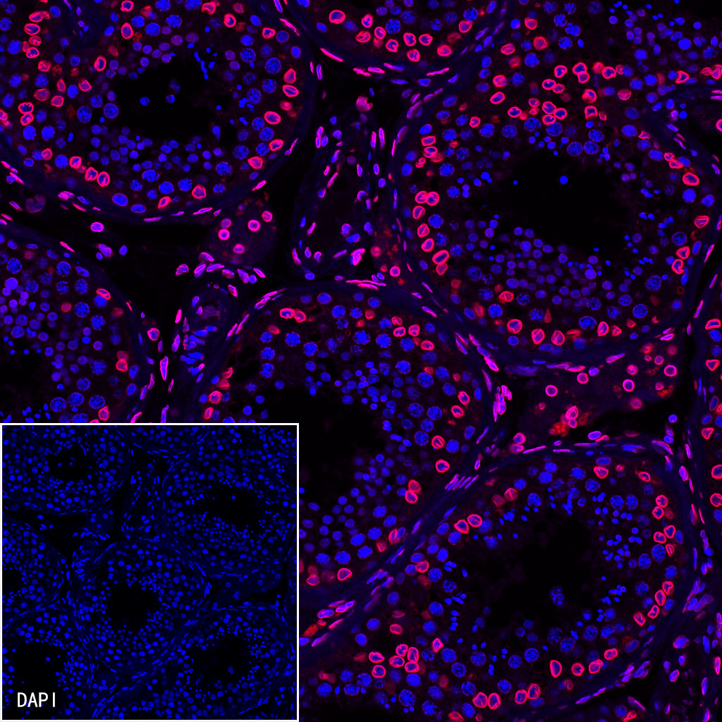
IF shows positive staining in paraffin-embedded human testis. Anti-Lamin B2 antibody was used at 1/500 dilution (Red) and incubated overnight at 4°C.Goat anti-Rabbit IgG(H+L) (Alexa Fluor® 594 Conjugate) (S0B4007) was used as secondary antibody at 1/1000 dilution.Counterstained with DAPI (Blue). Heat mediated antigen retrieval with EDTA buffer pH9.0 was performed before commencing with IF staining protocol.