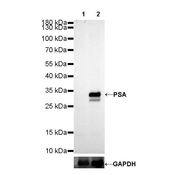
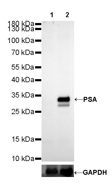
WB result of PSA Rabbit mAb
Primary antibody: PSA Rabbit mAb at 1/10000 dilution
Lane 1: HeLa whole cell lysate 20 µg
Lane 2: LNCaP whole cell lysate 20 µg
Negative control: HeLa whole cell lysate
Secondary antibody: Goat Anti-Rabbit IgG, (H+L), HRP conjugated at 1/10000 dilution
Predicted MW: 34 kDa
Observed MW: 34 kDa
S-RMab® PSA Recombinant Rabbit mAb (SDT-195-14)
S-RMab® PSA Recombinant Rabbit mAb (SDT-195-14)
Price:
Regular price
$100 USD
Regular price
Sale price
$100 USD
Unit price
per
For shipping services or bulk orders, you may request a quotation.
Secure checkout with

View full details
Product Details
Product Details
Product Specification
| Host | Rabbit |
| Antigen | PSA |
| Synonyms | Prostate-specific antigen, Seminin, Kallikrein-3, P-30 antigen, Semenogelase |
| Immunogen | Recombinant Protein |
| Location | Secreted |
| Accession | P07288 |
| Clone Number | SDT-195-14 |
| Antibody Type | Rabbit mAb |
| Application | WB, IHC-P, IP |
| Reactivity | Hu |
| Purification | Protein A |
| Concentration | 0.25 mg/ml |
| Physical Appearance | Liquid |
| Storage Buffer | PBS, 40% Glycerol, 0.05% BSA, 0.03% Proclin 300 |
| Stability & Storage | 12 months from date of receipt / reconstitution, -20 °C as supplied |
Dilution
| application | dilution | species |
| WB | 1:10000 | null |
| IHC-P | 1:1000 | null |
| IP | 1:25 | null |
Background
Prostate-specific antigen (PSA) is a biomarker for the diagnosis and management of prostate cancer and involved in the development of prostate cancer and/or its progression from the localized to the metastatic stage.
Picture
Picture
Western Blot
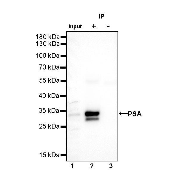
IP
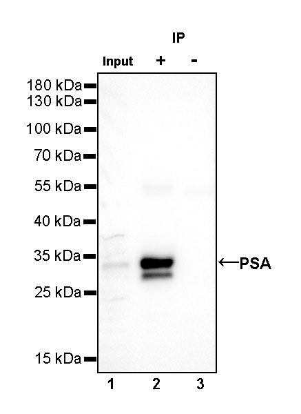
PSA Rabbit mAb at 1/25 dilution (1 µg) immunoprecipitating PSA in 0.4 mg LNCAP whole cell lysate.
Western blot was performed on the immunoprecipitate using PSA Rabbit mAb at 1/1000 dilution.
Secondary antibody (HRP) for IP was used at 1/400 dilution.
Lane 1: LNCAP whole cell lysate 10 µg (Input)
Lane 2: PSA Rabbit mAb IP in LNCAP whole cell lysate
Lane 3: Rabbit monoclonal IgG IP in LNCAP whole cell lysate
Predicted MW: 34 kDa
Observed MW: 34 kDa
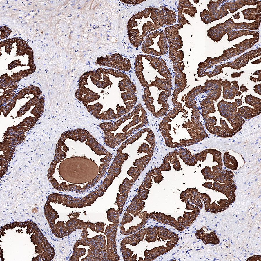
Immunohistochemistry
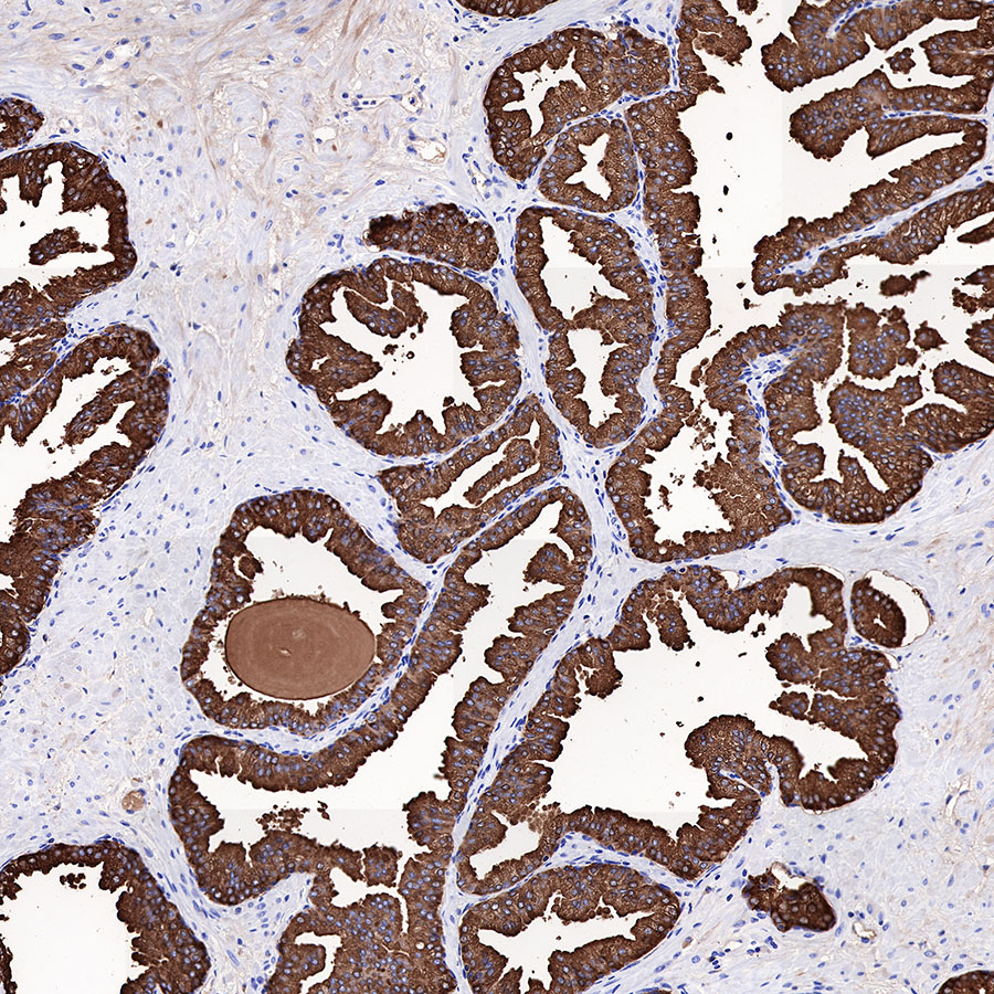
IHC shows positive staining in paraffin-embedded human prostatic hyperplasia. Anti-PSA antibody was used at 1/1000 dilution, followed by a HRP Polymer for Mouse & Rabbit IgG (ready to use). Counterstained with hematoxylin. Heat mediated antigen retrieval with Tris/EDTA buffer pH9.0 was performed before commencing with IHC staining protocol.
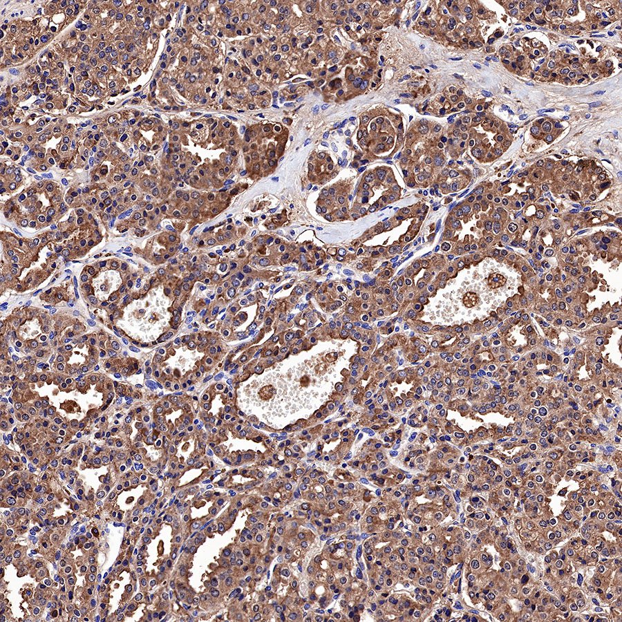
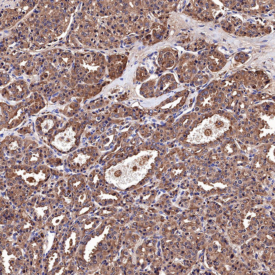
IHC shows positive staining in paraffin-embedded human prostate cancer. Anti-PSA antibody was used at 1/1000 dilution, followed by a HRP Polymer for Mouse & Rabbit IgG (ready to use). Counterstained with hematoxylin. Heat mediated antigen retrieval with Tris/EDTA buffer pH9.0 was performed before commencing with IHC staining protocol.
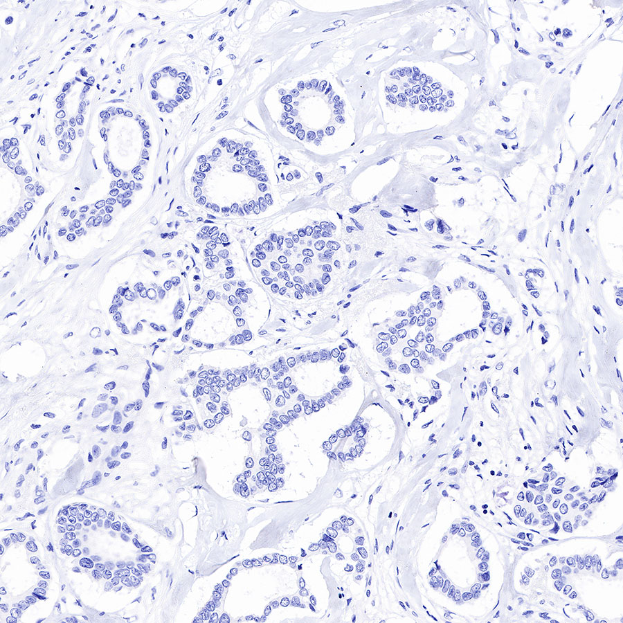
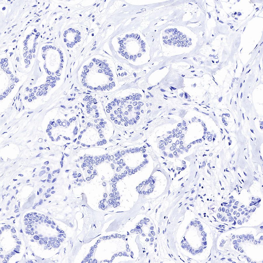
Negative control: IHC shows negative staining in paraffin-embedded human breast cancer. Anti-PSA antibody was used at 1/1000 dilution, followed by a HRP Polymer for Mouse & Rabbit IgG (ready to use). Counterstained with hematoxylin. Heat mediated antigen retrieval with Tris/EDTA buffer pH9.0 was performed before commencing with IHC staining protocol.
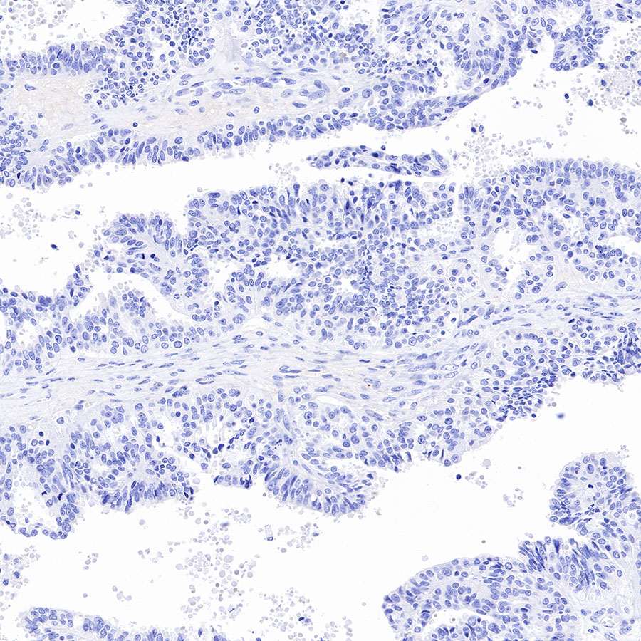
Negative control: IHC shows negative staining in paraffin-embedded human ovarian cancer. Anti-PSA antibody was used at 1/1000 dilution, followed by a HRP Polymer for Mouse & Rabbit IgG (ready to use). Counterstained with hematoxylin. Heat mediated antigen retrieval with Tris/EDTA buffer pH9.0 was performed before commencing with IHC staining protocol.