WB result of PGP9.5 Rabbit mAb
Primary antibody: PGP9.5 Rabbit mAb at 1/2000 dilution
Lane 1: LNCaP whole cell lysate 5 µg
Lane 2: DU 145 whole cell lysate 5 µg
Lane 3: SH-SY5Y whole cell lysate 5 µg
Lane 4: Ramos whole cell lysate 5 µg
Lane 5: HEK-293 whole cell lysate 5 µg
Negative control: LNCaP whole cell lysate
Secondary antibody: Goat Anti-rabbit IgG, (H+L), HRP conjugated at 1/10000 dilution
Predicted MW: 25 kDa
Observed MW: 23 kDa
PGP9.5 Recombinant Rabbit mAb (SDT-665-37)
PGP9.5 Recombinant Rabbit mAb (SDT-665-37)
Price:
Regular price
$100 USD
Regular price
Sale price
$100 USD
Unit price
per
For shipping services or bulk orders, you may request a quotation.
Secure checkout with

View full details
Product Details
Product Details
Product Specification
| Host | Rabbit |
| Antigen | PGP9.5 |
| Synonyms | Ubiquitin carboxyl-terminal hydrolase isozyme L1, UCH-L1, Neuron cytoplasmic protein 9.5, Ubiquitin thioesterase L1 |
| Immunogen | Synthetic Peptide |
| Location | Cytoplasm |
| Accession | P09936 |
| Clone Number | SDT-665-37 |
| Antibody Type | Recombinant mAb |
| Isotype | IgG |
| Application | WB, IHC-P, ICC, ICFCM |
| Reactivity | Hu, Ms, Rt |
| Predicted Reactivity | Mq, Pg, Hr, Dg |
| Purification | Protein A |
| Concentration | 0.5 mg/ml |
| Conjugation | Unconjugated |
| Physical Appearance | Liquid |
| Storage Buffer | PBS, 40% Glycerol, 0.05% BSA, 0.03% Proclin 300 |
| Stability & Storage | 12 months from date of receipt / reconstitution, -20 °C as supplied |
Dilution
| application | dilution | species |
| WB | 1:2000 | |
| IHC-P | 1:500-1:2000 | |
| ICC | 1:500 | |
| ICFCM | 1:5000 |
Background
Protein gene product 9.5 (PGP 9.5), also known as ubiquitin carboxyl-terminal hydrolase-1 (UCH-L1), is a 25 kDa protein originally isolated from whole brain extracts. In non-neoplastic tissues, PGP 9.5, a member of the ubiquitin hydrolase family of proteins, is confined to neural and neuroendocrine cells. In the field of diagnostic surgical pathology, PGP9.5 is used as a purportedly specific marker of putative neural and neuroectodermal tumors.
Picture
Picture
Western Blot
WB result of PGP9.5 Rabbit mAb
Primary antibody: PGP9.5 Rabbit mAb at 1/2000 dilution
Lane 1: mouse brain lysate 5 µg
Secondary antibody: Goat Anti-rabbit IgG, (H+L), HRP conjugated at 1/10000 dilution
Predicted MW: 25 kDa
Observed MW: 23 kDa
WB result of PGP9.5 Rabbit mAb
Primary antibody: PGP9.5 Rabbit mAb at 1/2000 dilution
Lane 1: rat brain lysate 5 µg
Secondary antibody: Goat Anti-rabbit IgG, (H+L), HRP conjugated at 1/10000 dilution
Predicted MW: 25 kDa
Observed MW: 23 kDa
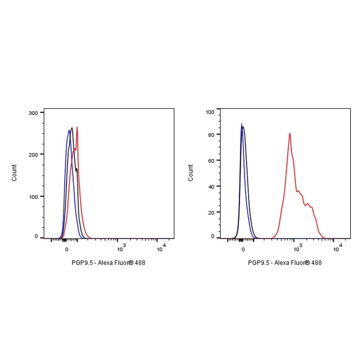
FC
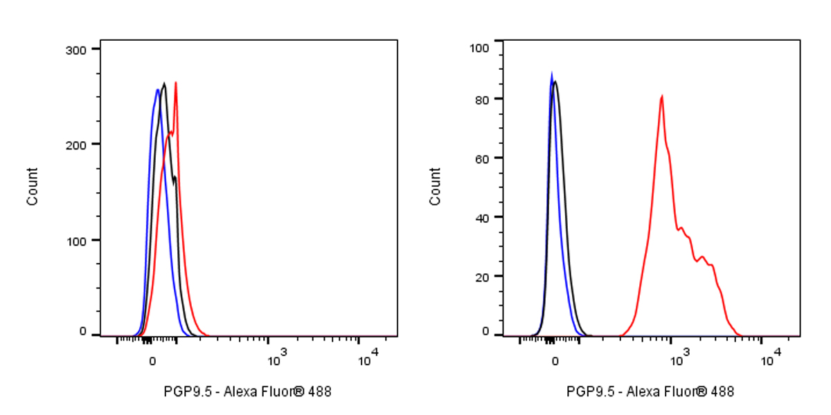
Flow cytometric analysis of 4% PFA fixed 90% methanol permeabilized Hela (Human cervix adenocarcinoma epithelial cell, left) / SH-SY5Y (Human neuroblastoma epithelial cell, Right) labelling PGP9.5 antibody at 1/5000 dilution (0.01 μg) / (Red) compared with a Rabbit monoclonal IgG (Black) isotype control and an unlabelled control (cells without incubation with primary antibody and secondary antibody) (Blue). Goat Anti - Rabbit IgG Alexa Fluor® 488 was used as the secondary antibody. Negative control: Hela
Immunohistochemistry
IHC shows positive staining in paraffin-embedded human cerebral cortex. Anti-PGP9.5 antibody was used at 1/2000 dilution, followed by a HRP Polymer for Mouse & Rabbit IgG (ready to use). Counterstained with hematoxylin. Heat mediated antigen retrieval with Tris/EDTA buffer pH9.0 was performed before commencing with IHC staining protocol.
IHC shows positive staining in paraffin-embedded human cerebellum. Anti-PGP9.5 antibody was used at 1/500 dilution, followed by a HRP Polymer for Mouse & Rabbit IgG (ready to use). Counterstained with hematoxylin. Heat mediated antigen retrieval with Tris/EDTA buffer pH9.0 was performed before commencing with IHC staining protocol.
IHC shows positive staining in paraffin-embedded human colon. Anti-PGP9.5 antibody was used at 1/500 dilution, followed by a HRP Polymer for Mouse & Rabbit IgG (ready to use). Counterstained with hematoxylin. Heat mediated antigen retrieval with Tris/EDTA buffer pH9.0 was performed before commencing with IHC staining protocol.
IHC shows positive staining in paraffin-embedded human kidney. Anti-PGP9.5 antibody was used at 1/2000 dilution, followed by a HRP Polymer for Mouse & Rabbit IgG (ready to use). Counterstained with hematoxylin. Heat mediated antigen retrieval with Tris/EDTA buffer pH9.0 was performed before commencing with IHC staining protocol.
IHC shows positive staining in paraffin-embedded human pancreas. Anti-PGP9.5 antibody was used at 1/2000 dilution, followed by a HRP Polymer for Mouse & Rabbit IgG (ready to use). Counterstained with hematoxylin. Heat mediated antigen retrieval with Tris/EDTA buffer pH9.0 was performed before commencing with IHC staining protocol.
IHC shows positive staining in paraffin-embedded human ovarian cancer. Anti-PGP9.5 antibody was used at 1/500 dilution, followed by a HRP Polymer for Mouse & Rabbit IgG (ready to use). Counterstained with hematoxylin. Heat mediated antigen retrieval with Tris/EDTA buffer pH9.0 was performed before commencing with IHC staining protocol.
IHC shows positive staining in paraffin-embedded human pancreatic cancer. Anti-PGP9.5 antibody was used at 1/500 dilution, followed by a HRP Polymer for Mouse & Rabbit IgG (ready to use). Counterstained with hematoxylin. Heat mediated antigen retrieval with Tris/EDTA buffer pH9.0 was performed before commencing with IHC staining protocol.
Negative control: IHC shows negative staining in paraffin-embedded human invasive ductal breast cancer. Anti-PGP9.5 antibody was used at 1/2000 dilution, followed by a HRP Polymer for Mouse & Rabbit IgG (ready to use). Counterstained with hematoxylin. Heat mediated antigen retrieval with Tris/EDTA buffer pH9.0 was performed before commencing with IHC staining protocol.
Immunocytochemistry
ICC shows positive staining in SH-SY5Y cells (top panel) and negative staining in HeLa cells (below panel). Anti-PGP9.5 antibody was used at 1/500 dilution (Green) and incubated overnight at 4°C. Goat polyclonal Antibody to Rabbit IgG - H&L (Alexa Fluor® 488) was used as secondary antibody at 1/1000 dilution. The cells were fixed with 100% ice-cold methanol and permeabilized with 0.1% PBS-Triton X-100. Nuclei were counterstained with DAPI (Blue). Counterstain with tubulin (Red).