Human IFN-α ELISA Kit
Human IFN-α ELISA Kit
Price:
Regular price
$450 USD
Regular price
Sale price
$450 USD
Unit price
per
For shipping services or bulk orders, you may request a quotation.
Secure checkout with

View full details
Product Details
Product Details
Product Specification
| protein | IFN-α | |||||||||||||||||||||||||||||||||||||||||||||||||||||||
| Usage |
Need to bring your own test equipment 1. Microplate reader (measurable 450nm Absorption value of detection wavelength and 540nm Or 570nm Absorption value of corrected wavelength) 2. High precision liquid dispenser and disposable tip 3. Distilled or deionized water 4. Bottle washer (spray bottle), multi-channel plate washer or automatic plate washer 5. 500mL Measuring cylinder 6. Microplate oscillator 1. Preparation before the experiment 1. Sample collection and storage ① Cell culture supernatant: particulate matter should be removed by centrifugation; Test the sample immediately. If the sample is not tested in time after collection, it is recommended to pack it according to the amount used once and store it frozen in -20℃ In the refrigerator, avoid repeated freezing and thawing. The sample may need to be used with a diluent ( 1× ) dilution. ② Serum: Use serum separation tubes ( SST ) Collect samples and place samples at room temperature 30 Minutes. Centrifugation 15 Minutes at a rotational speed of 1000g 。 The serum was removed immediately and tested immediately. If the sample is not tested in time after collection, it is recommended to pack it according to the amount used once and store it frozen in ≤-20℃ In the refrigerator, avoid repeated freezing and thawing. The sample may need to be used with a diluent ( 1× ) dilution. ③ Plasma: Use EDTA , heparin or citric acid as an anticoagulant to collect plasma, after collection 30 Centrifuge within minutes 15 Minutes, with a rotation speed of 1000g , and detect it immediately. If the sample is not tested in time after collection, it is recommended to pack it according to the amount used once and store it frozen in ≤-20℃ In the refrigerator, avoid repeated freezing and thawing. The sample may need to be used with a diluent ( 1× ) dilution. 2. Reagent Preparation ( Please place all reagents and samples at room temperature before use and let them stand 15 Minutes. All experimental samples and standards are recommended Do repeat hole detection ) ① Washing liquid preparation: The concentrated washing liquid taken out of the refrigerator may have crystals, which is a normal phenomenon; Place at room temperature, shake gently and mix well, and prepare washing liquid after the crystals are completely dissolved. The concentrated washing liquid can be diluted with distilled water or deionized water to obtain a washing liquid having a working concentration. The unused diluted wash liquid can be placed in 4℃ Stable Storage 30 Heaven. ② Assay buffer ( 1× ) Preparation: The amount of concentrated diluent used can be calculated according to the required dosage, and the concentrated diluent can be diluted with distilled water or deionized water to configure the diluent with working concentration. ③ Preparation of detection antibody working solution: Please fully mix the detection antibody before use, and calculate the amount of detection antibody used according to the required dosage. According to 1 : 100 Proportional use of 1× Dilute the test antibody with the test diluent and mix well. Please before use 30 Deploy detection antibodies within minutes. ④SA-HRP Preparation of working solution: Mix well before dilution. According to the number of standards and samples to be tested, use 1× Test buffer according to 1 : 100 Dilute streptavidin. ⑤ Standard: Please refer to the bottle label for the redissolved volume of the standard. Use distilled water or deionized water to redissolve the freeze-dried powder of the standard to obtain a concentration of 2000pg/mL Standard mother liquor of. Gently shake 5-15 Minutes, it is fully dissolved. Add in each dilution tube 230uL Standard dilution ( 1× ), refer to the figure below to make the above prepared standard mother liquor 2-fold Gradient dilution, each tube must be well mixed before pipetting to the next tube. Please use the standard solution in the first dilution tube as the highest point of the standard curve ( 1000pg/mL ), standard dilution ( 1× ) can be used as a standard curve zero ( 0pg/mL )。 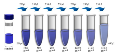 Reminder: In order to obtain more accurate experimental results, it is recommended to use the same as cell culture supernatant samples Media of supernatant sample replaced above 2-fold Standard diluents used during dilution ( 1× ), and using culture medium for It is a zero concentration standard control. ⑥ Sample dilution: If the sample needs to be diluted, use the test buffer provided in the kit ( 1× ) Perform the dilution operation of the sample. For cell culture supernatant samples, it is recommended that the same cell culture medium be used for sample dilution procedures. 2. Operation steps 1. Remove the unwanted slats, place them back in the aluminum foil bag containing the desiccant and re-seal the seal. In any case, avoid contact with the inner surface of the microplate. 2. To be used wells on each plate are added 300uL 1× Let the lotion stand and soak 30 Seconds. After discarding the wash, the plate was pat dry on absorbent paper. Use the plate immediately after the plate wash is completed and do not allow the plate to dry. 3. Add standard: Add standard well 100uL 2 Double-diluted standard. Blank hole addition 100uL Standard dilution ( Serum / Plasma samples ) Or medium ( Cell culture supernatant sample ) 。 4. Add sample: serum / Plasma: sample well addition 50uL 1× Assay buffer and 50uL Sample. Cell culture supernatant: sample well addition 100uL Cell culture supernatant. 5. Add detection antibody: add to each well to be tested 50uL Detect the antibody working solution. 6. Ensure that the loading of standards, samples and detection antibodies is in 15 Completed within minutes, It is continuous and cannot be interrupted. 7. Incubation: The plate was sealed using a sealing membrane. 100-300 turn / Shake for minutes (ensure that the solution in each well does not sprinkle and can be fully mixed), room temperature ( 25℃±3℃ ) Incubation 2 Hours. The sealing membrane should be sealed during incubation. 8. Wash: Discard the liquid and add per well 300uL Lotion washing plate, wash 6 Times. Pat dry on absorbent paper each time you wash the board. In order to achieve ideal experimental performance, it is necessary to completely move Remove residual liquid. 9. Enzyme addition: added per well 100uL SA-HRP Working fluid. 10. Incubation: The plate was sealed with a new plate sealing membrane. 100-300 turn / Shake for minutes (ensure that the solution in each well does not sprinkle and can be fully mixed), room temperature ( 25℃±3℃ ) Incubation 45 Minutes. 11. Repeat steps 8 。 12. Add substrate for color development: add per well 100uL Chromogenic substrate, protected from light, room temperature ( 25℃±3℃ ) Incubation 5-30 Minutes. 13. Add stop solution: add per well 100uL Stop liquid. The color changed from blue to yellow. If the color appears green or the color change is obviously uneven, it means that the stop solution and the color substrate are not fully mixed. Please gently tap the plate frame to fully mix. Addition of stop solution The addition order should be the same as the addition order of the chromogenic substrate. 14. Test readings: at 30 Within minutes, dual-wavelength detection was performed using a microplate reader to determine 450nm Absorption maximum wavelength and 570nm Or 630nm At reference wavelength OD Value. Quasi-post OD Value is 450nm Minus the measured value of 570nm Or 630nm The measured value of. Using 450nm The determination will result in OD The values are high and the accuracy is reduced. 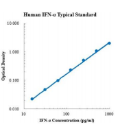 3. Kit parameters 1. Sensitivity: Human IFN-α The lowest measurable dose ( MDD ) is generally less than 1.04pg/mL 。 The lowest measurable value is determined according to 10 The corresponding concentration is calculated by adding two standard deviations to the mean value of the zero-point absorbance values of each standard curve. 2. Correction: This ELISA High purity recombinant human expressed by Escherichia coli IFN-α Corrected by protein. 3. Linearity: 5 Different samples were spiked with high concentrations of human IFN-α , followed by a diluent ( 1× ) Dilute the sample to the detection range and determine its linearity.
4. Recovery: Healthy human serum was spiked with different levels of human IFN-α The recovery rate was determined. The recoveries range from 97-118% , Average recovery Rates in 106% 。 5. Specificity: This ELISA Method can detect natural and recombinant human IFN-α Egg whites. The following factors were mixed with diluent ( 1× ) formulated into 50ng/mL Concentration to detect human IFN-α Cross-reactivity of. Will 50ng/mL Interference factors incorporated into the intermediate range of recombinant human IFN-α In the reference substance, to detect the effects of human IFN-α Of interference. No significant cross-reactivity or interference was observed.
|
|||||||||||||||||||||||||||||||||||||||||||||||||||||||
| Species Reactivity | Human | |||||||||||||||||||||||||||||||||||||||||||||||||||||||
| Theory | This kit uses double antibody sandwich ELISA technology. Specific anti-Human IFN-α capture antibody was pre-coated on a high affinity enzyme plate. The standard substance, the sample to be tested and the biotin-labeled detection antibody are sequentially added to the wells of the enzyme labeled plate, and then thoroughly shaken and mixed, and then placed at room temperature for an incubation process for 2 hours. The IFN-α present in the sample is combined with the solid phase antibody and the detection antibody. After thorough washing to remove free and unbound components, horseradish peroxidase-labeled Streptavidin-HRP (SA-HRP) was added. After washing again, TMB chromogenic substrate was added and incubated at room temperature in the dark from light to develop color. The depth of the color response is positively correlated with the concentration of IFN-α in the sample. The reaction was stopped by adding a stop solution, and the absorbance value was measured using a microplate reader at a detection wavelength of 450 nm (corrected wavelength of 570-630 nm). | |||||||||||||||||||||||||||||||||||||||||||||||||||||||
| Source | Human | |||||||||||||||||||||||||||||||||||||||||||||||||||||||
| Synonym | IFNA, IFNA2, IFNA2b, IFNalpha, IFN-alpha, IFN-alphaA, INFA2, interferon alpha 2, LeIF D | |||||||||||||||||||||||||||||||||||||||||||||||||||||||
| Composition |
Please use within the expiration date of the kit
|
|||||||||||||||||||||||||||||||||||||||||||||||||||||||
| Background | Interferons (IFNs) are a group of signaling proteins synthesized and released by host cells in response to pathogens (such as viruses, bacteria, parasites or tumor cells). Under normal circumstances, virus-infected cells will release interferon, which makes surrounding cells improve their antiviral defenses. Based on the receptor type, human interferons can be divided into three major types: type I interferons, including IFN-α, IFN-β, IFN-ε, IFN-κ, and IFN-ω, type II interferons (called IFN-γ in humans) and type III interferons. IFN-alpha protein is produced by leukocytes and is mainly involved in innate immunity in response to viral infection. Studies have shown that enhancing the expression of IFN-α in tumor-infiltrating macrophages can cause more effective dendritic cell activation and immune effector cytotoxicity. As a pyrogenic factor, IFN-α can cause fever by altering the activity of thermosensitive neurons in the hypothalamus. Using a similar mechanism, IFN-α can be used to alleviate pain, interacting with μ-opioid receptors as an analgesic. | |||||||||||||||||||||||||||||||||||||||||||||||||||||||
| General Notes | 1. Please use the kit within the validity period. 2. The components of different kits and different batch kits cannot be mixed. 3. If the sample value is greater than the highest value of the standard curve, the sample should be diluted with diluent (1 ×) and re-tested; If the cell culture supernatant sample needs to be distributed and diluted, cell culture medium can be used for other intermediate dilutions except dilution with diluent in the last step. 4. Differences in test results can be caused by a variety of factors, including the operation of the experimenter, the use of the pipette, the plate washing technique, the reaction time or temperature, the storage of the kit, etc. 5. The terminating solution in the kit is an acidic solution. Please protect your glasses, hands, face and clothes when using it. 6. For scientific research only, not for in vitro diagnosis. |
|||||||||||||||||||||||||||||||||||||||||||||||||||||||
| Storage Temp. | The unopened kit is stored at 2-8 ℃ and has a validity period of two years. | |||||||||||||||||||||||||||||||||||||||||||||||||||||||
| Test Range | 15.63 pg/mL-1000 pg/mL | |||||||||||||||||||||||||||||||||||||||||||||||||||||||