Product Specification
| Host |
Rabbit |
| Antigen |
CD79B |
| Synonyms |
B-cell-specific glycoprotein B29; Ig-beta; Immunoglobulin-associated B29 protein |
| Immunogen |
Synthetic Peptide |
| Location |
Membrane |
| Accession |
P40259 |
| Clone Number |
SDT-043-9 |
| Antibody Type |
Rabbit mAb |
| Application |
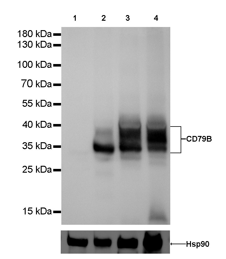
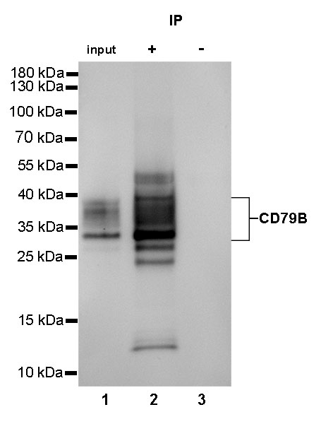
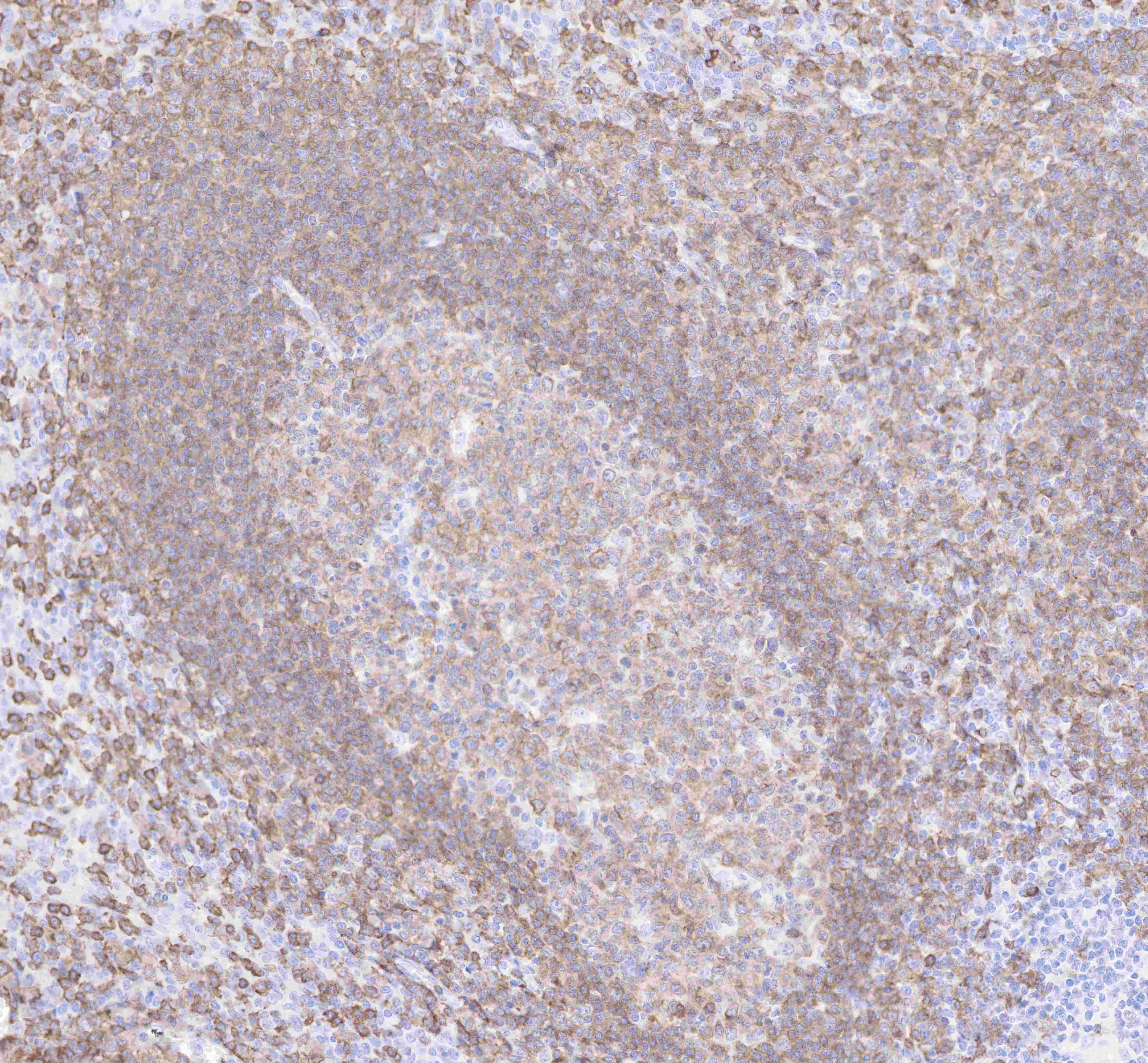
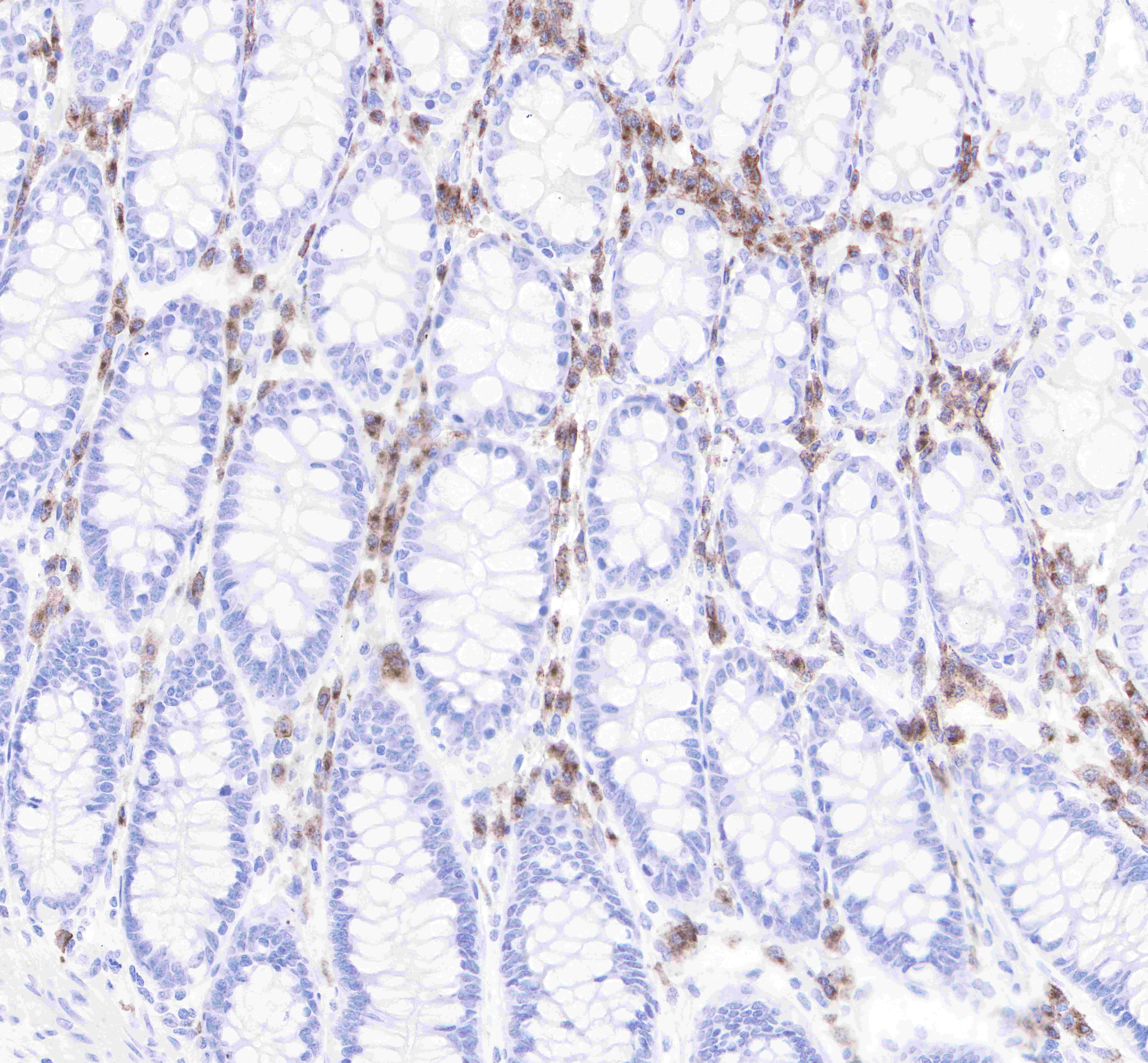
WB, IHC-P, ICC, IP |
| Reactivity |
Hu, Ms |
| Purification |
Protein A |
| Concentration |
0.5mg/ml |
| Molecular Weight |
30-50kDa |
| Conjugation |
Unconjugated |
| Physical Appearance |
Liquid |
| Storage Buffer |
PBS, 40% Glycerol, 0.05%BSA, 0.03% Proclin 300 |
| Stability & Storage |
12 months from date of receipt / reconstitution, -20 °C as supplied |
Dilution
| application |
dilution |
species |
| IP |
1:25 |
|
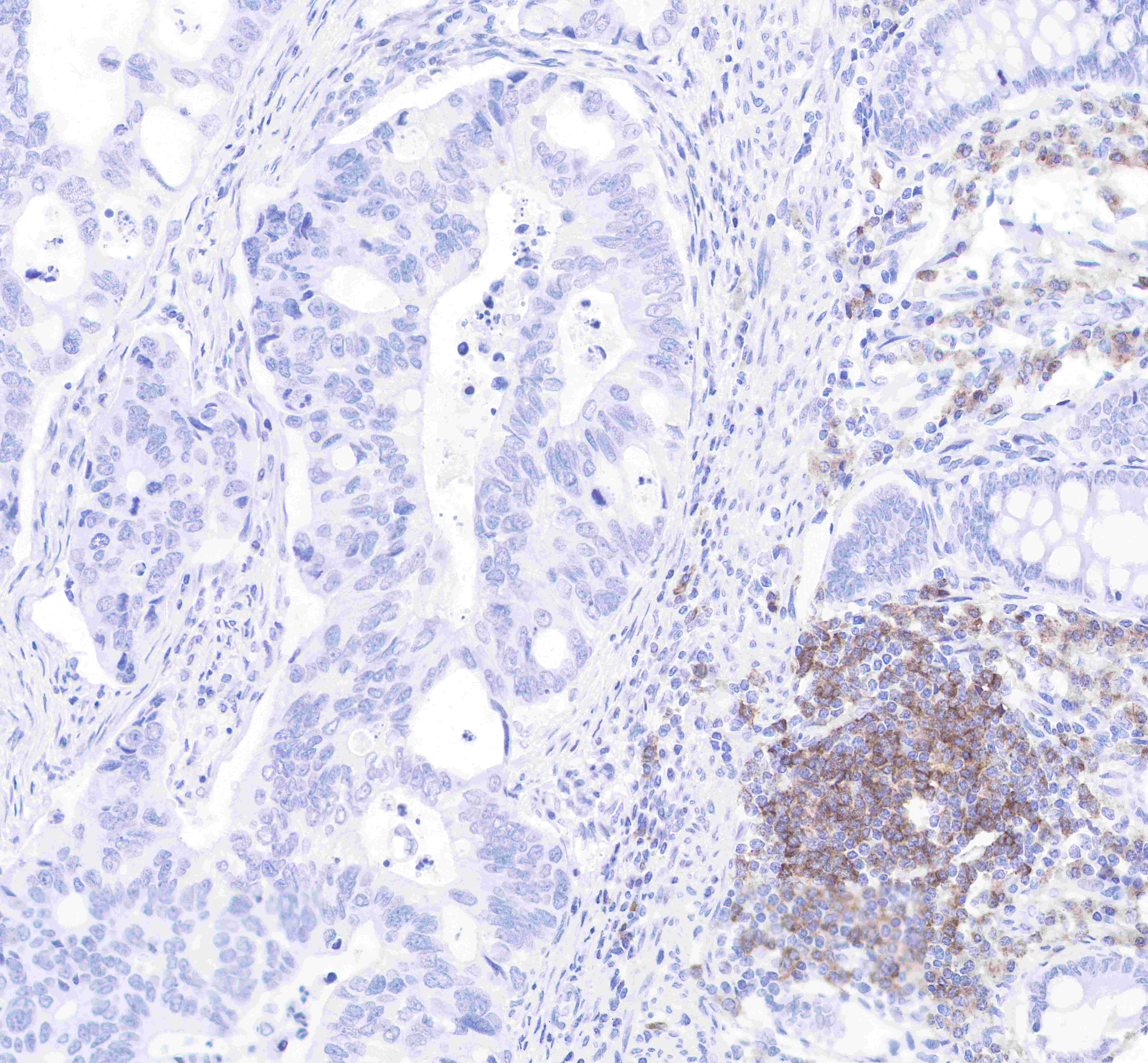
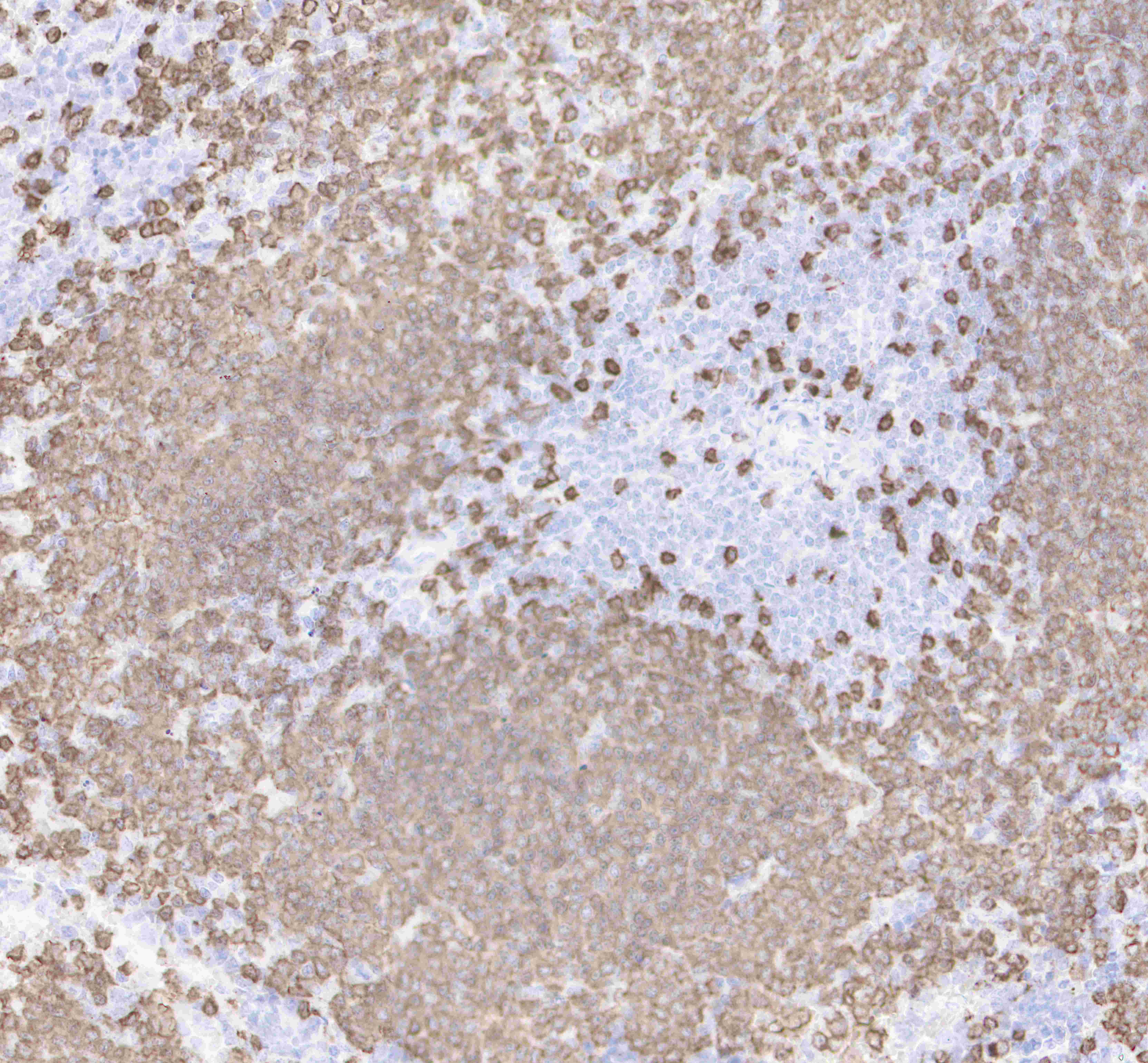
| IHC-P |
1:2000 |
|
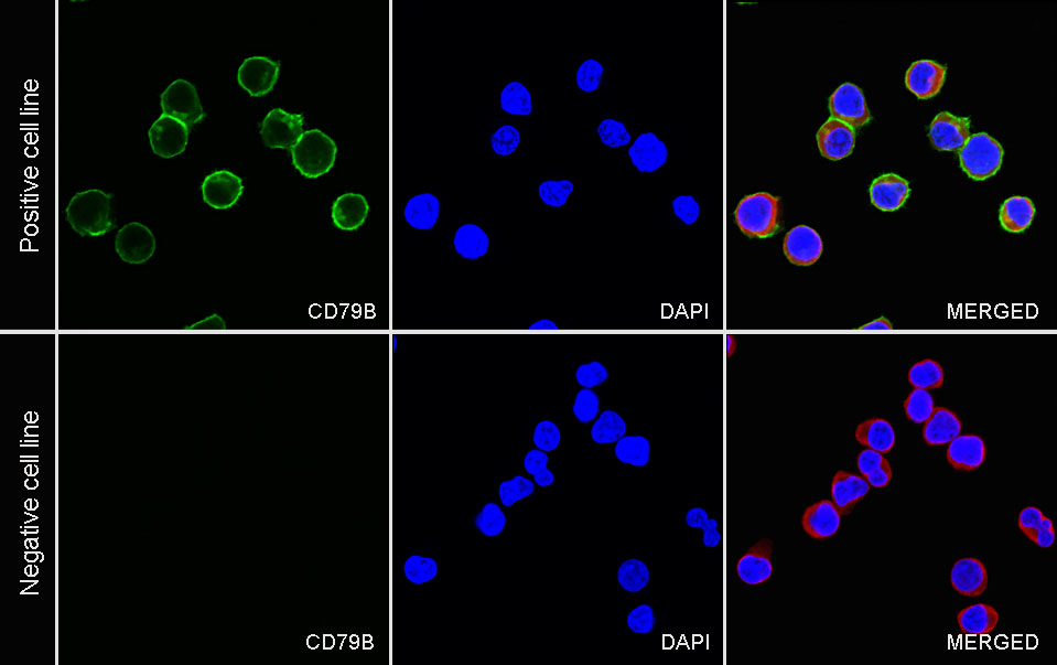
| ICC |
1:500 |
|
| WB |
1:1000 |
|
Background
CD79B is a multimeric complex that includes the antigen-specific component, surface immunoglobulin (Ig). Surface Ig non-covalently associates with two other proteins, Ig-alpha and Ig-beta, which are necessary for expression and function of the B-cell antigen receptor. This gene encodes the Ig-beta protein of the B-cell antigen component. CD79B is required in cooperation with CD79A for initiation of the signal transduction cascade activated by the B-cell antigen receptor complex (BCR) which leads to internalization of the complex, trafficking to late endosomes and antigen presentation. And it also enhances phosphorylation of CD79A, possibly by recruiting kinases which phosphorylate CD79A or by recruiting proteins which bind to CD79A and protect it from dephosphorylation.