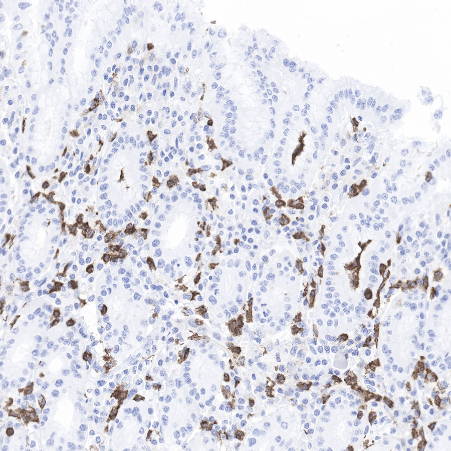
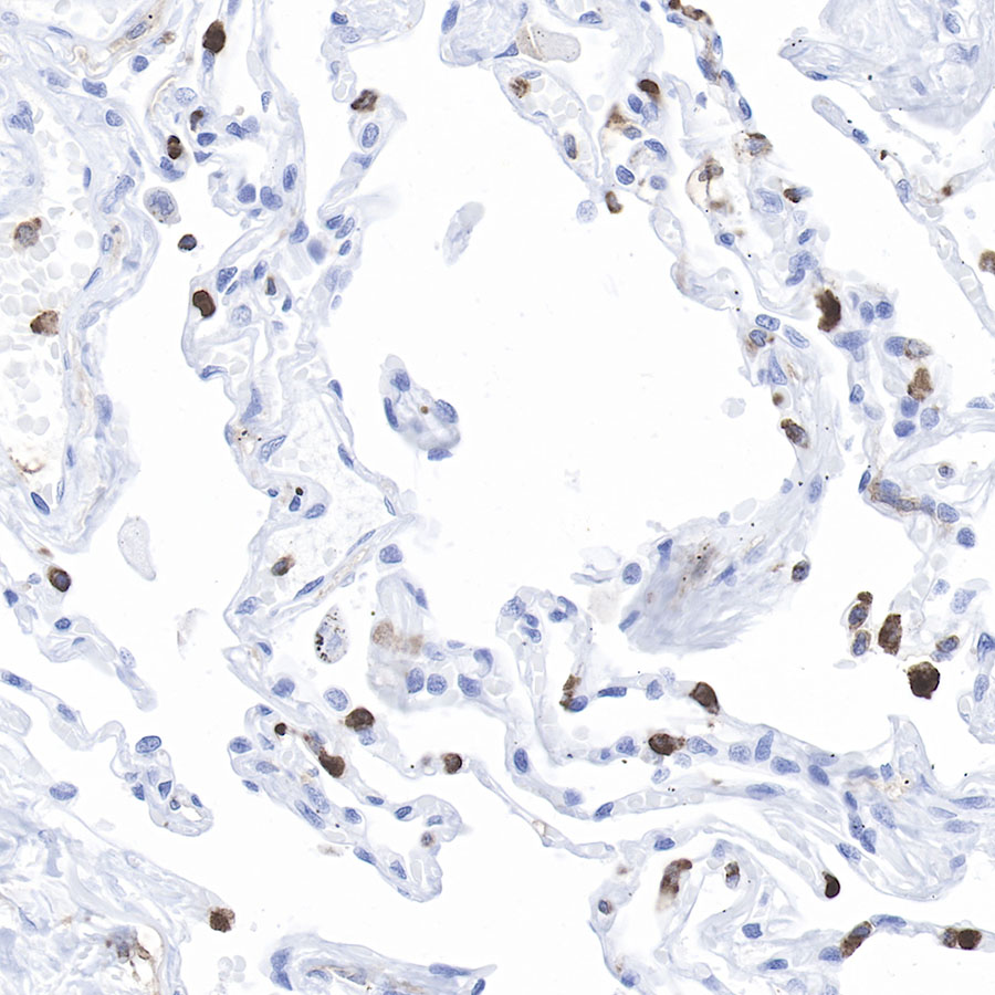
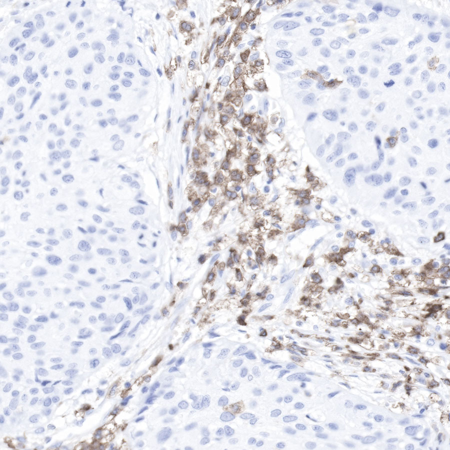
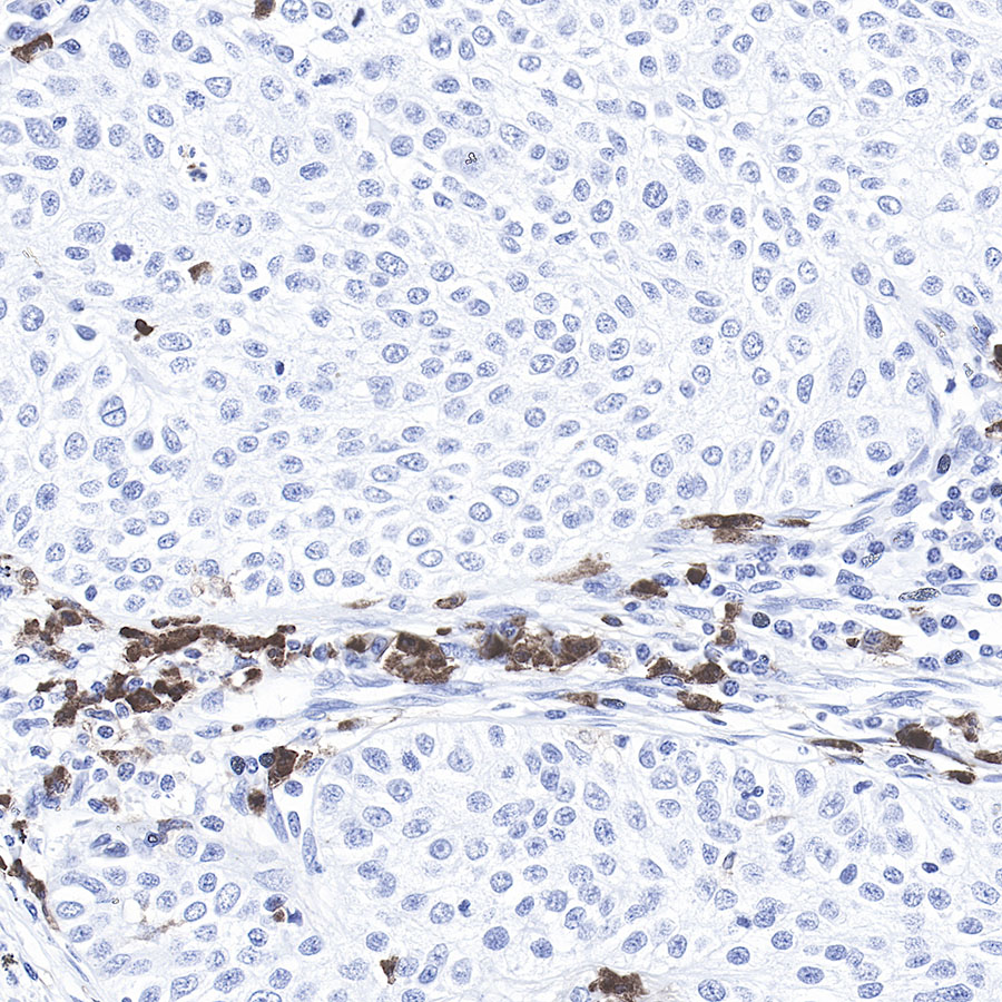
Product Specification
| Host |
Rabbit |
| Antigen |
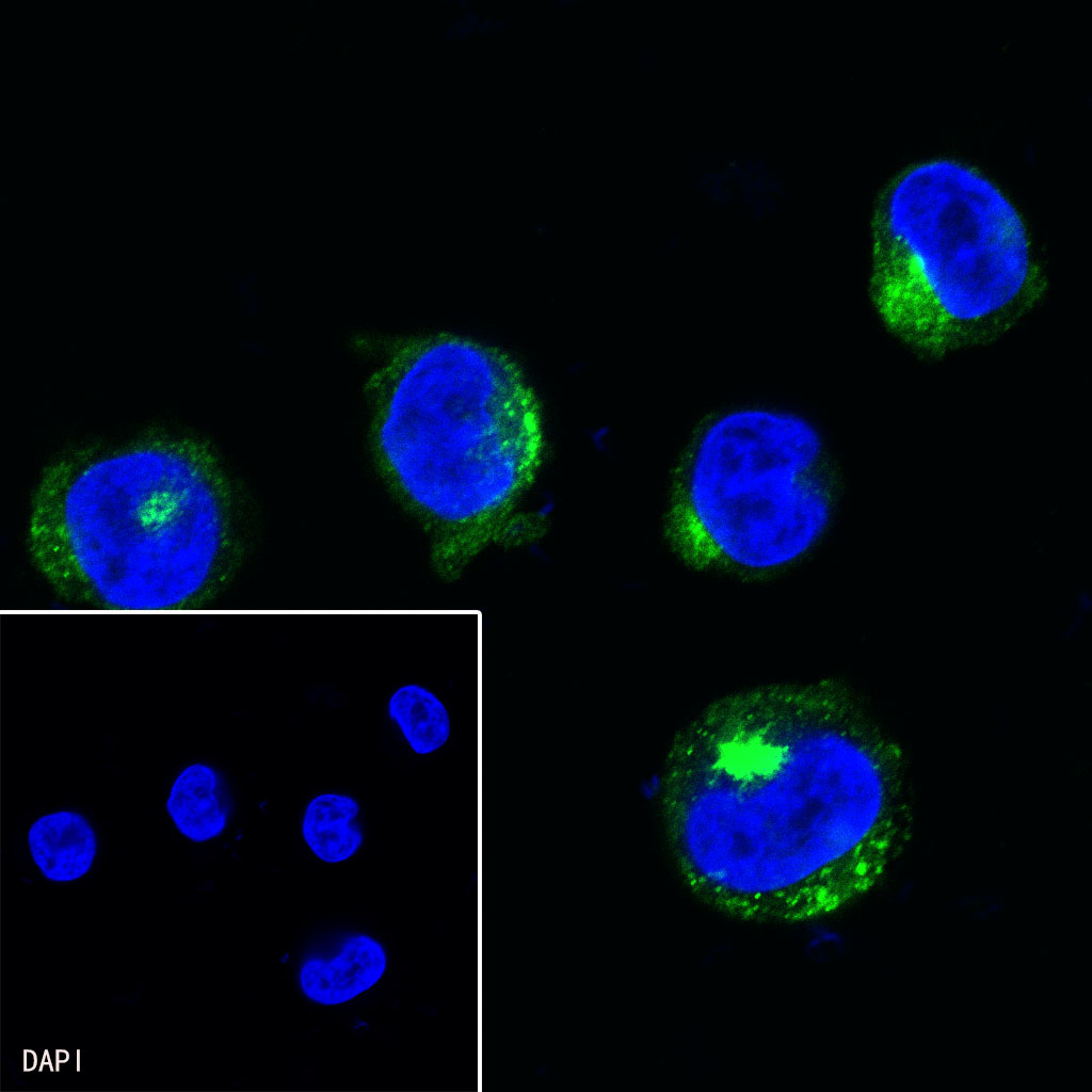
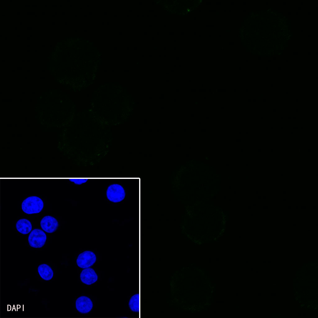
CD11b |
| Synonyms |
Integrin alpha-M |
| Immunogen |
Synthetic Peptide |
| Location |
Cell membrane |
| Accession |
P05555 |
| Clone Number |
SDT-058-44 |
| Antibody Type |
Rabbit mAb |
| Application |
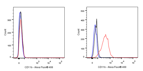
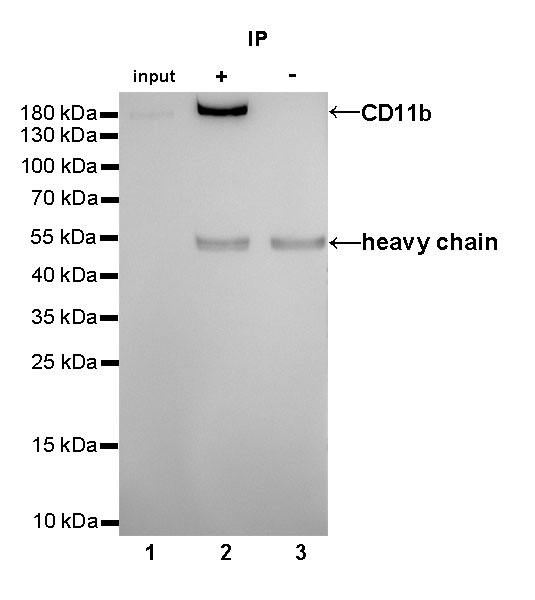
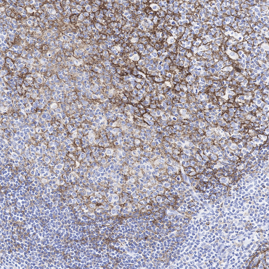
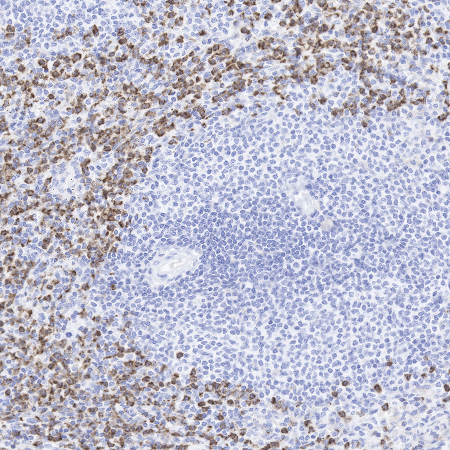
WB, IHC-P, ICC, IP, ICFCM |
| Reactivity |
Hu |
| Purification |
Protein A |
| Concentration |
1 mg/ml |
| Physical Appearance |
Liquid |
| Storage Buffer |
PBS |
| Stability & Storage |
12 months from date of receipt, 4°C as supplied |
Dilution
| application |
dilution |
species |
| WB |
1:1000 |
|
| IHC-P |
1:500 |
|
| IP |
1:25 |
|
| ICC |
1:500 |
|
| ICFCM |
1:500 |
|
Background
CD11b, also known as Integrated alpha-m, a transgender protein, can form an heterododerous composed of α and β subunit. It is a common bone marrow mark (neutral granulocyte, monocyte, macrophage, and small gel cells) and NK (natural kill cells) antigens. It can be used to distinguish between acute granulocyte deficiency (CD11B+, CD117--) and acute early early early elastic cell leukemia (CD11B-, CD117+).