WB result of TMPRSS2 Rabbit mAb
Primary antibody: TMPRSS2 Rabbit mAb at 1/1000 dilution
Lane 1: LNCaP whole cell lysate 20 µg
Secondary antibody: Goat Anti-Rabbit IgG, (H+L), HRP conjugated at 1/10000 dilution
Predicted MW: 54 kDa
Observed MW: 22, 55 kDa
(This blot was developed with high sensitivity substrate)
TMPRSS2 Recombinant Rabbit mAb (S-735-40)
TMPRSS2 Recombinant Rabbit mAb (S-735-40)
Price:
Regular price
$100 USD
Regular price
Sale price
$100 USD
Unit price
per
For shipping services or bulk orders, you may request a quotation.
Secure checkout with

View full details
Product Details
Product Details
Product Specification
| Host | Rabbit |
| Synonyms | Transmembrane protease serine 2, Serine protease 10, PRSS10 |
| Immunogen | Synthetic Peptide |
| Location | Cell membrane |
| Accession | O15393 |
| Clone Number | S-735-40 |
| Antibody Type | Recombinant mAb |
| Isotype | IgG |
| Application | WB, IHC-P, IP |
| Reactivity | Hu |
| Purification | Protein A |
| Concentration | 0.5 mg/ml |
| Conjugation | Unconjugated |
| Physical Appearance | Liquid |
| Storage Buffer | PBS, 40% Glycerol, 0.05% BSA, 0.03% Proclin 300 |
| Stability & Storage | 12 months from date of receipt / reconstitution, -20 °C as supplied |
Dilution
| application | dilution | species |
| WB | 1:1000 | |
| IHC-P | 1:500 | |
| IP | 1:50 |
Background
TMPRSS2 is a protein that belongs to the serine protease family. The protein contains a type II transmembrane domain, a low density lipoprotein receptor class A domain, a scavenger receptor cysteine-rich domain and a protease domain. Serine proteases are known to be involved in many physiological and pathological processes. This protein is up-regulated by androgenic hormones in prostate cancer cells and down-regulated in androgen-independent prostate cancer tissue. The protease domain of this protein is thought to be cleaved and secreted into cell media after autocleavage. TMPRSS2 participates in proteolytic cascades necessary for normal physiological function of the prostate.
Picture
Picture
Western Blot
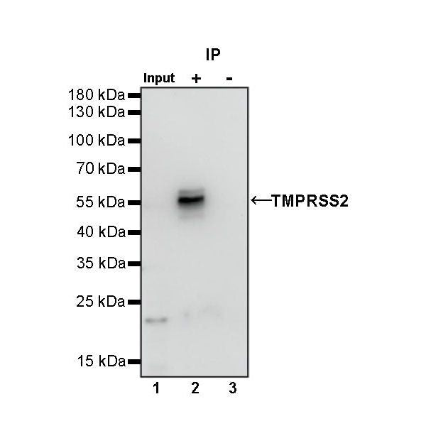
IP
TMPRSS2 Rabbit mAb at 1/50 dilution (1 µg) immunoprecipitating TMPRSS2 in 0.4 mg LnCap whole cell lysate.
Western blot was performed on the immunoprecipitate using TMPRSS2 Rabbit mAb at 1/1000 dilution.
Secondary antibody (HRP) for IP was used at 1/400 dilution.
Lane 1: LnCap whole cell lysate 20 µg (Input)
Lane 2: TMPRSS2 Rabbit mAb IP in LnCap whole cell lysate
Lane 3: Rabbit monoclonal IgG IP in LnCap whole cell lysate
Predicted MW: 54 kDa
Observed MW: 22, 55 kDa
This blot was developed with high sensitivity substrate
Immunohistochemistry
IHC shows positive staining in paraffin-embedded human colon. Anti-TMPRSS2 antibody was used at 1/500 dilution, followed by a HRP Polymer for Mouse & Rabbit IgG (ready to use). Counterstained with hematoxylin. Heat mediated antigen retrieval with Tris/EDTA buffer pH9.0 was performed before commencing with IHC staining protocol.
IHC shows positive staining in paraffin-embedded human colon cancer. Anti-TMPRSS2 antibody was used at 1/500 dilution, followed by a HRP Polymer for Mouse & Rabbit IgG (ready to use). Counterstained with hematoxylin. Heat mediated antigen retrieval with Tris/EDTA buffer pH9.0 was performed before commencing with IHC staining protocol.
IHC shows positive staining in paraffin-embedded human prostatic cancer. Anti-TMPRSS2 antibody was used at 1/500 dilution, followed by a HRP Polymer for Mouse & Rabbit IgG (ready to use). Counterstained with hematoxylin. Heat mediated antigen retrieval with Tris/EDTA buffer pH9.0 was performed before commencing with IHC staining protocol.