WB result of Runx1 Rabbit mAb
Primary antibody: Runx1 Rabbit mAb at 1/1000 dilution
Lane 1: HEK-293 whole cell lysate 20 µg
Lane 2: Jurkat whole cell lysate 20 µg
Lane 3: THP-1 whole cell lysate 20 µg
Lane 4: MOLT-4 whole cell lysate 20 µg
Lane 5: SW620 whole cell lysate 20 µg
Weak expression: HEK-293 whole cell lysate
Secondary antibody: Goat Anti-rabbit IgG, (H+L), HRP conjugated at 1/10000 dilution
Predicted MW: 48 kDa
Observed MW: 55 kDa
Runx1 Recombinant Rabbit mAb (S-959-64)
Runx1 Recombinant Rabbit mAb (S-959-64)
Price:
Regular price
$100 USD
Regular price
Sale price
$100 USD
Unit price
per
For shipping services or bulk orders, you may request a quotation.
Secure checkout with

View full details
Product Details
Product Details
Product Specification
| Host | Rabbit |
| Synonyms | Runt-related transcription factor 1, Acute myeloid leukemia 1 protein, Core-binding factor subunit alpha-2 (CBF-alpha-2), Oncogene AML-1, Polyomavirus enhancer-binding protein 2 alpha B subunit (PEA2-alpha B; PEBP2-alpha B), SL3-3 enhancer factor 1 alpha B subunit, SL3/AKV core-binding factor alpha B subunit |
| Immunogen | Synthetic Peptide |
| Location | Nucleus |
| Accession | Q01196 |
| Clone Number | S-959-64 |
| Antibody Type | Recombinant mAb |
| Isotype | IgG |
| Application | WB, ICC, ICFCM, IP |
| Reactivity | Hu, Ms, Rt |
| Purification | Protein A |
| Concentration | 0.5 mg/ml |
| Conjugation | Unconjugated |
| Physical Appearance | Liquid |
| Storage Buffer | PBS, 40% Glycerol, 0.05% BSA, 0.03% Proclin 300 |
| Stability & Storage | 12 months from date of receipt / reconstitution, -20 °C as supplied |
Dilution
| application | dilution | species |
| WB | 1:1000 | |
| ICC | 1:500 | |
| ICFCM | 1:5000 | |
| IP | 1:50 |
Background
Runx1 is a crucial transcription factor that binds to the core elements of many enhancers and promoters, regulating the expression of specific genes. This protein plays a pivotal role in cellular development, particularly in the development of hematopoietic stem cells. RUNX1 interacts with CBFβ (core-binding factor β) to form a set of transcription factors, and this interaction is essential in the development of neurons and hematopoietic stem cells. However, when CBFβ undergoes translocation, it can form a fusion protein CBFβ-SMMHC that abnormally binds tightly to RUNX1, participating in the pathogenesis of acute lymphocytic leukemia (ALL). Therefore, the RUNX1−CBFβ interaction is also considered an important target for cancer treatment. Furthermore, research on RUNX1 protein has revealed its association with a range of cancers, particularly leukemia. Chromosome translocations involving the RUNX1 gene have been clearly linked to several types of leukemia.
Picture
Picture
Western Blot
WB result of Runx1 Rabbit mAb
Primary antibody: Runx1 Rabbit mAb at 1/1000 dilution
Lane 1: mouse thymus lysate 20 µg
Secondary antibody: Goat Anti-rabbit IgG, (H+L), HRP conjugated at 1/10000 dilution
Predicted MW: 48 kDa
Observed MW: 55 kDa
WB result of Runx1 Rabbit mAb
Primary antibody: Runx1 Rabbit mAb at 1/1000 dilution
Lane 1: rat thymus lysate 20 µg
Secondary antibody: Goat Anti-rabbit IgG, (H+L), HRP conjugated at 1/10000 dilution
Predicted MW: 48 kDa
Observed MW: 55 kDa
FC
Flow cytometric analysis of 4% PFA fixed 90% methanol permeabilized Jurkat (Human T cell leukemia T lymphocyte) labelling Runx1 antibody at 1/5000 dilution (0.01 μg) / (Red) compared with a Rabbit monoclonal IgG (Black) isotype control and an unlabelled control (cells without incubation with primary antibody and secondary antibody) (Blue). Goat Anti - Rabbit IgG Alexa Fluor® 488 was used as the secondary antibody.
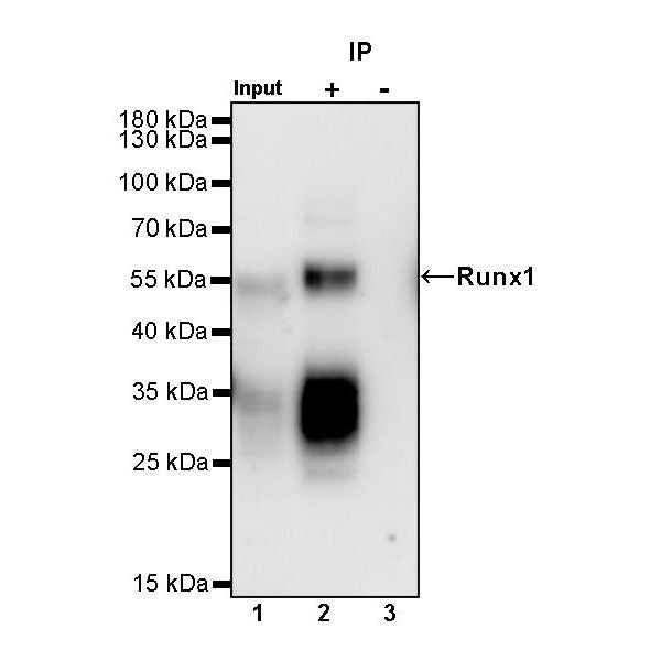
IP
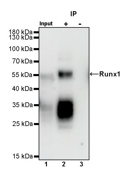
Runx1 Rabbit mAb at 1/50 dilution (1 µg) immunoprecipitating Runx1 in 0.4 mg Jurkat whole cell lysate.
Western blot was performed on the immunoprecipitate using Runx1 Rabbit mAb at 1/1000 dilution.
Secondary antibody (HRP) for IP was used at 1/1000 dilution.
Lane 1: Jurkat whole cell lysate 10 µg (Input)
Lane 2: Runx1 Rabbit mAb IP in Jurkat whole cell lysate
Lane 3: Rabbit monoclonal IgG IP in Jurkat whole cell lysate
Predicted MW: 48 kDa
Observed MW: 55 kDa
This blot was developed with high sensitivity substrate
Immunocytochemistry
ICC shows positive staining in Jurkat cells (top panel) and weak staining in HEK293 cells (below panel). Anti-RUNX1 antibody was used at 1/500 dilution (Green) and incubated overnight at 4°C. Goat polyclonal Antibody to Rabbit IgG - H&L (Alexa Fluor® 488) was used as secondary antibody at 1/1000 dilution. The cells were fixed with 100% ice-cold methanol and permeabilized with 0.1% PBS-Triton X-100. Nuclei were counterstained with DAPI (Blue). Counterstain with tubulin (Red).