Rat FSH ELISA Kit
Rat FSH ELISA Kit
Product Details
Product Details
Product Specification
| protein | FSH | ||||||||||||||||||||||||||||||||||||
| Usage |
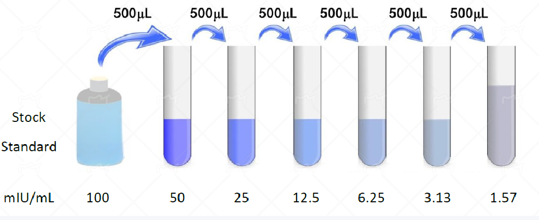
Sample collection preparation and preservation1. Serum: Whole blood sample placed at room temperature 2 Hour or 4°C Overnight after 1000×g Centrifugation 20 Minutes, take the supernatant to detect. 2. Plasma: Sample after collection 30 Within minutes 2-8°C 、 1000×g Centrifugation 15 Minutes, take the supernatant to detect. 3. Tissue homogenate: Take an appropriate amount of tissue block and add it to the pre-cooled PBS ( 0.01M , pH7.0-7.2 ) to remove blood (lysed red blood cells in the homogenate will affect the measurement result), cut the tissue into pieces after weighing, and then mix it with the corresponding volume of PBS (generally according to 1:9 The mass-to-volume ratio, the specific volume can be appropriately adjusted according to the needs of the experiment, and recorded. 4. Cell culture supernatant: Take the cell supernatant from 1000×g Centrifugation 20 Minutes, impurities and cell debris were removed. 5. Urine: Please collect the first urine in the morning (mid-section urine), or 24 Hourly urine, 2000×g Centrifugation 15 The supernatant was collected after minutes, And save the sample At -20°C And repeated freezing and thawing should be avoided. 6. Saliva: A sample is collected with a saliva sample collection tube, and then 2-8°C, 1000×g Centrifugation 15 Minutes, take the supernatant to detect, Or after sub-packaging -20°C Save. 7. Other biological samples: Please 1000×g Centrifugation 20 Minutes, take the supernatant to detect. Notes1. The sample should be clear and transparent, and the suspended solids should be removed by centrifugation. 2. After sample collection, if 1 Testing within weeks can be stored at 4°C , if it cannot be detected in time, please pack it according to the one-time usage amount and freeze it in -20°C ( 1 Within months), or -80°C ( 3-6 Test within a month) to avoid repeated freezing and thawing. Principles of sample dilutionIf your test sample needs to be diluted, refer to the general dilution principles below: 1. Dilution 50 Times: One-step dilution. 2. Dilution 100 Times: One-step dilution. 3. Dilution 1000 Times: Two-step dilution. 4. Dilution 100000 Times: Three-step dilution. 5. The amount of liquid taken during each dilution step is not less than 3 μL , the dilution factor is not more than 100 Times. 6. When the dilution factor is very high, you can use it first PBS Dilution, last step using standard in kit & Sample dilution. Sample dilution recommendations1. Normal fresh serum / Plasma Sample Recommendation (Original solution-1:2) Testing. 2. Due to individual variations, the recommended dilution factor is for informational purposes only. Preparation for testing1. Please advance 30 Minutes remove the kit from the refrigerator and equilibrate to room temperature. 2. Use double distilled water 25× The concentrated wash liquid is diluted to 1× Working fluid, put back unused 4°C 。 3. Standard: Add standard & Sample Universal Diluent 1.0 mL Into the lyophilized standard, screw the tube cap tightly and let stand 10 Minutes, and after it is fully dissolved, gently mix (concentration of 100 mIU/mL )。
4. Biotinylated antibody working solution: calculate the dosage required for the current experiment before the experiment (according to 100 μL/ Hole meter, should be configured more in actual configuration 100-200 μL ), before use 15 Min, concentrated biotinylated antibody was diluted with biotinylated antibody diluent ( 1:100 ) into working concentration, use on the same day. 5. Enzyme conjugate working solution: calculate the dosage required for the current experiment before the experiment (according to 100 μL/ Hole meter, should be configured more in actual configuration 100-200 μL )。 6.TMB Substrate —— Pipette the desired dose of solution and do not pour the residual solution back into the reagent vial again. Notes1. Please make sure that all components are dissolved and mixed before use of the kit. 2. Concentrated biotinylated antibody, the volume of concentrated enzyme conjugate is small, may be dispersed in various parts of the tube during transportation, please 1000×g Centrifugation 1 Minutes to allow the liquid of the tube wall or cap to deposit to the bottom of the tube. |
||||||||||||||||||||||||||||||||||||
| Species Reactivity | Rat | ||||||||||||||||||||||||||||||||||||
| Theory | This kit adopts the principle of sandwich method. The specific anti-rat FSH antibody was coated in a 96-well microplate, and rat FSH standards or samples were added to the microwells respectively, so that the rat FSH protein in the standard or the rat FSH protein in the sample was bound to the anti-rat FSH antibody solid on the microplate, then biotinylated anti-rat FSH antibody was added, the unbound biotinylated antibody was washed, HRP-labeled streptavidin was added, and TMB substrate was added to develop color. TMB is converted to blue under peroxidase catalysis and to final yellow under the action of acid. There is a positive correlation between the depth of color and rat FSH protein in the sample. The absorbance (OD value) was measured with a microplate reader at a wavelength of 450 nm, and the sample concentration was calculated by drawing a standard curve. | ||||||||||||||||||||||||||||||||||||
| Source | Rat | ||||||||||||||||||||||||||||||||||||
| Synonym | FSH; Follicle Stimulating Hormone | ||||||||||||||||||||||||||||||||||||
| Detection Type | Rat FSH can be detected in samples and does not cross-react with other related proteins | ||||||||||||||||||||||||||||||||||||
| Composition |
|
||||||||||||||||||||||||||||||||||||
| General Notes |
1. Incubate in strict accordance with the specified time and temperature to ensure accurate results. All reagents must reach room temperature 20-25 °C prior to use. Store reagents in refrigeration immediately after use. 2. Incorrect plate washing can lead to inaccurate results. Make sure to drain the liquid from the wells as much as possible before adding the substrate. Do not allow the wells to dry out during incubation. 3. Eliminate the residual liquid and fingerprints at the bottom of the plate, otherwise it will affect the OD value. 4. The substrate color development solution should be colorless or very light in color, and the substrate solution that has turned blue cannot be used. 5. Avoid cross-contamination of reagents and specimens to avoid wrong results. 6. Avoid direct exposure to strong light during storage and incubation. 7. After equilibrating to room temperature, open the sealed bag to prevent water droplets from condensing on the cold slats. 8. Any reaction reagent cannot come into contact with the bleaching solvent or the strong gas emitted by the bleaching solvent. Any bleaching component will destroy the biological activity of the reaction reagents in the kit. 9. Expired products cannot be used. 10. If it is possible to spread diseases, all samples should be managed, and the samples and testing devices should be handled according to the prescribed procedures. |
||||||||||||||||||||||||||||||||||||
| Storage Temp. | Unopened kit, stored at 4 °C, shelf life 6 months. | ||||||||||||||||||||||||||||||||||||
| Test Range | 1.57-100 mIU/mL; Sensitivity: 0.72 mIU/mL |