WB result of Phospho-Akt (Ser473) Rabbit mAb
Primary antibody: Phospho-Akt (Ser473) Rabbit mAb at 1/1000 dilution
Lane 1: untreated Jurkat whole cell lysate 20 µg
Lane 2: Jurkat treated with 100 nM Calyculin A for 30 minutes whole cell lysate 20 µg
Secondary antibody: Goat Anti-rabbit IgG, (H+L), HRP conjugated at 1/10000 dilution
Predicted MW: 56 kDa
Observed MW: 60 kDa
Phospho-Akt (Ser473) Recombinant Rabbit mAb (S-622-64)
Phospho-Akt (Ser473) Recombinant Rabbit mAb (S-622-64)
Price:
Regular price
$100 USD
Regular price
Sale price
$100 USD
Unit price
per
For shipping services or bulk orders, you may request a quotation.
Secure checkout with

View full details
Product Details
Product Details
Product Specification
| Host | Rabbit |
| Antigen | Phospho-Akt (Ser473) |
| Synonyms | RAC-alpha serine/threonine-protein kinase, Protein kinase B (PKB), Protein kinase B alpha (PKB alpha), Proto-oncogene c-Akt, RAC-PK-alpha, PKB, RAC, AKT1 |
| Immunogen | Synthetic Peptide |
| Location | Cytoplasm, Nucleus, Cell membrane |
| Accession | P31749 |
| Clone Number | S-622-64 |
| Antibody Type | Recombinant mAb |
| Isotype | IgG |
| Application | WB, ICC, ICFCM, IP |
| Reactivity | Hu |
| Predicted Reactivity | Rt, Ms |
| Purification | Protein A |
| Concentration | 0.5 mg/ml |
| Conjugation | Unconjugated |
| Physical Appearance | Liquid |
| Storage Buffer | PBS, 40% Glycerol, 0.05% BSA, 0.03% Proclin 300 |
| Stability & Storage | 12 months from date of receipt / reconstitution, -20 °C as supplied |
Dilution
| application | dilution | species |
| WB | 1:1000 | |
| ICC | 1:500 | |
| ICFCM | 1:500 | |
| Dot Blot | 1:1000 | |
| IP | 1:50 |
Background
Akt or Protein kinase B, is a serine/threonine kinase that plays an important role in regulating a number of cellular processes such as growth, metabolism and survival. The importance of the Akt pathway is highlighted by the mutation of various components of the pathway in human cancers such as the PTEN and PI3-kinase (P110α), which occur in more than 30% of human tumors. For Akt to achieve full activation, phosphorylation is needed at both serine 473 (ser473) of the hydrophobic tail and threonine 308 (thr308) of the activation motif, upon growth factor ligation to the receptor tyrosine kinases.
Picture
Picture
Western Blot
FC
Flow cytometric analysis of 4% PFA fixed 90% methanol permeabilized Jurkat (Human T cell leukemia T lymphocyte) cells, treated with 100nM Calyculin A for 30 min (Red) or untreated (Green), labeling Phospho-Akt (Ser473) at 1/500 dilution (0.1 μg) compared with a rabbit monoclonal IgG isotype control (Black) and an unlabeled control (cells without incubation with primary antibody and secondary antibody) (Blue). Goat Anti - Rabbit IgG Alexa Fluor® 488 was used as the secondary antibody.
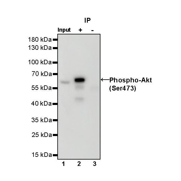
IP
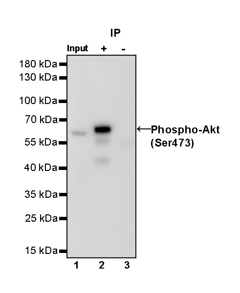
Phospho-Akt (Ser473) Rabbit mAb at 1/50 dilution (1 µg) immunoprecipitating Phospho-Akt (Ser473) in 0.4 mg Jurkat treated with 100 nM Calyculin A for 30 minutes whole cell lysate.
Western blot was performed on the immunoprecipitate using Phospho-Akt (Ser473) Rabbit mAb at 1/1000 dilution.
Secondary antibody (HRP) for IP was used at 1/1000 dilution.
Lane 1: Jurkat treated with 100 nM Calyculin A for 30 minutes whole cell lysate 20 µg (Input)
Lane 2: Phospho-Akt (Ser473) Rabbit mAb IP in Jurkat treated with 100 nM Calyculin A for 30 minutes whole cell lysate
Lane 3: Rabbit monoclonal IgG IP in Jurkat treated with 100 nM Calyculin A for 30 minutes whole cell lysate
Predicted MW: 56 kDa
Observed MW: 60 kDa
Dot Blot
Dot blot result of Phospho-Akt (Ser473) Rabbit mAb
Lane1: Phospho-Akt (Ser473) peptide
Lane2: Akt unmodified peptide Primary antibody: Phospho-Akt (Ser473) Rabbit mAb at 1/1000 dilution
Secondary antibody: Goat Anti-Rabbit IgG, (H+L), HRP conjugated at 1/10000 dilution
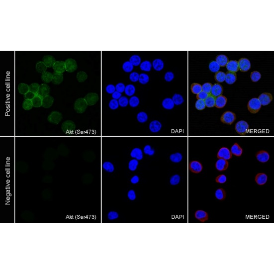
Immunocytochemistry
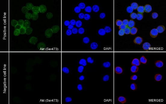
ICC analysis of Jurkat cells treated with Calyculin A (100nM, 30 min) (top panel) and Jurkat cells untreated with Calyculin A (100nM, 30 min) (below panel). Anti-Phospho-Akt (Ser473) antibody was used at 1/500 dilution (Green) and incubated overnight at 4°C. Goat polyclonal Antibody to Rabbit IgG - H&L (Alexa Fluor® 488) was used as secondary antibody at 1/1000 dilution. The cells were fixed with 4% PFA and permeabilized with 0.1% PBS-Triton X-100. Nuclei were counterstained with DAPI (Blue). Counterstain with tubulin (Red).