WB result of PDCD4 Rabbit polyclonal antibody
Primary antibody: PDCD4 Rabbit polyclonal antibody at 1/1000 dilution
Lane 1: HeLa whole cell lysate 20 µg
Lane 2: HEK-293 whole cell lysate 20 µg
Lane 3: 293T whole cell lysate 20 µg
Lane 4: Jurkat whole cell lysate 20 µg
Secondary antibody: Goat Anti-Rabbit IgG, (H+L), HRP conjugated at 1/10000 dilution
Predicted MW: 52 kDa
Observed MW: 60 kDa
PDCD4 Rabbit polyclonal antibody
PDCD4 Rabbit polyclonal antibody
Price:
Regular price
$80 USD
Regular price
Sale price
$80 USD
Unit price
per
For shipping services or bulk orders, you may request a quotation.
Secure checkout with

View full details
Product Details
Product Details
Product Specification
| Host | Rabbit |
| Antigen | PDCD4 |
| Synonyms | Programmed cell death protein 4, Neoplastic transformation inhibitor protein, Nuclear antigen H731-like, Protein 197/15a, H731 |
| Immunogen | Synthetic Peptide |
| Location | Cytoplasm, Nucleus |
| Accession | Q53EL6 |
| Antibody Type | Polyclonal antibody |
| Isotype | IgG |
| Application | WB, IHC-P, ICC, ICFCM, IP |
| Reactivity | Hu, Ms, Rt |
| Predicted Reactivity | Or |
| Purification | Immunogen Affinity |
| Concentration | Lot specific* (generally 0.5 to 5 mg/ml)* |
| Conjugation | Unconjugated |
| Physical Appearance | Liquid |
| Storage Buffer | PBS, 40% Glycerol, 0.05%BSA, 0.03% Proclin 300 |
| Stability & Storage | 12 months from date of receipt / reconstitution, -20 °C as supplied |
Dilution
| application | dilution | species |
| WB | 1:1000 | |
| IP | 1:50 | |
| IHC | 1:400-1:800 | |
| ICC | 1:500 | |
| ICFCM | 1:500 |
Background
PDCD4 was considered to encode a nuclear antigen and located on chromosome 10. The expression of PDCD4 is regulated by interleukins IL-2, IL-12, and IL-15. The PDCD4 acts as a tumor-suppressor gene and performs essential functions in many biological events, including apoptosis, protein translation, signal transduction, and stimulation of inflammation mediators. The expression of PDCD4 is tightly regulated at the level of transcription, translation, and protein degradation. The loss of expression of PDCD4 is diagnostic indicator for different human cancers, and is prognostic indicator for survival in cancers of the breast, liver, colon, lung, glioma, and esophagus.
Picture
Picture
Western Blot
WB result of PDCD4 Rabbit polyclonal antibody
Primary antibody: PDCD4 Rabbit polyclonal antibody at 1/1000 dilution
Lane 1: NIH/3T3 whole cell lysate 20 µg
Secondary antibody: Goat Anti-Rabbit IgG, (H+L), HRP conjugated at 1/10000 dilution
Predicted MW: 52 kDa
Observed MW: 60 kDa
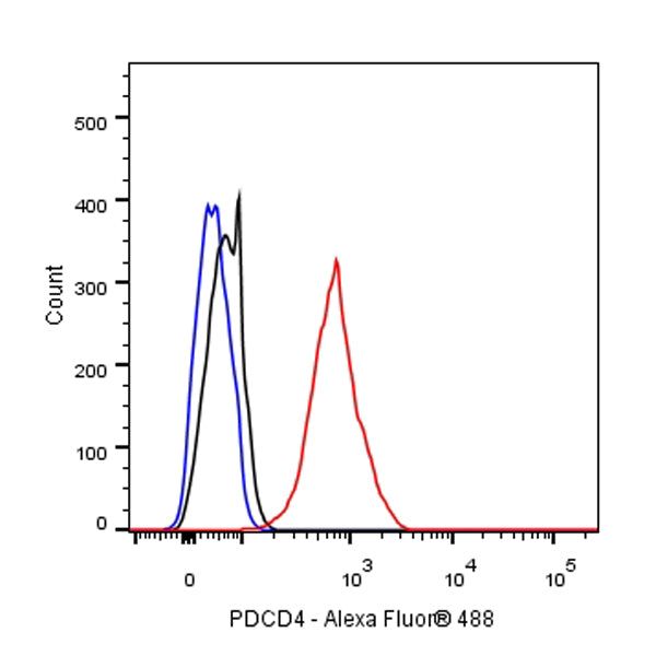
FC
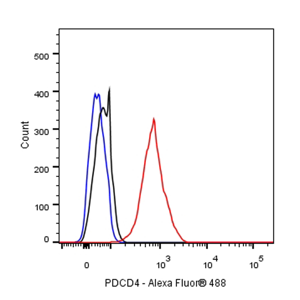
Flow cytometric analysis of 4% PFA fixed 90% methanol permeabilized HeLa (Human cervix adenocarcinoma epithelial cell) cells labelling PDCD4 antibody at 1/500 dilution (0.1 μg)/ (Red) compared with a Rabbit monoclonal IgG (Black) isotype control and an unlabelled control (cells without incubation with primary antibody and secondary antibody) (Blue). Goat Anti - Rabbit IgG Alexa Fluor® 488 was used as the secondary antibody.
IP
PDCD4 Rabbit pAb at 1/50 dilution (1 µg) immunoprecipitating PDCD4 in 0.4 mg HeLa whole cell lysate.
Western blot was performed on the immunoprecipitate using PDCD4 Rabbit pAb at 1/1000 dilution.
Secondary antibody (HRP) for IP was used at 1/400 dilution.
Lane 1: HeLa whole cell lysate 20 µg (Input)
Lane 2: PDCD4 Rabbit pAb IP in HeLa whole cell lysate
Lane 3: Rabbit monoclonal IgG IP in HeLa whole cell lysate
Predicted MW: 52 kDa
Observed MW: 60 kDa
Immunohistochemistry
IHC shows positive staining in paraffin-embedded human colon. Anti-PDCD4 antibody was used at 1/400 dilution, followed by a HRP Polymer for Mouse & Rabbit IgG (ready to use). Counterstained with hematoxylin. Heat mediated antigen retrieval with Tris/EDTA buffer pH9.0 was performed before commencing with IHC staining protocol.
IHC shows positive staining in paraffin-embedded human stomach. Anti-PDCD4 antibody was used at 1/400 dilution, followed by a HRP Polymer for Mouse & Rabbit IgG (ready to use). Counterstained with hematoxylin. Heat mediated antigen retrieval with Tris/EDTA buffer pH9.0 was performed before commencing with IHC staining protocol.
IHC shows positive staining in paraffin-embedded human tonsil. Anti-PDCD4 antibody was used at 1/400 dilution, followed by a HRP Polymer for Mouse & Rabbit IgG (ready to use). Counterstained with hematoxylin. Heat mediated antigen retrieval with Tris/EDTA buffer pH9.0 was performed before commencing with IHC staining protocol.
IHC shows positive staining in paraffin-embedded human breast. Anti-PDCD4 antibody was used at 1/800 dilution, followed by a HRP Polymer for Mouse & Rabbit IgG (ready to use). Counterstained with hematoxylin. Heat mediated antigen retrieval with Tris/EDTA buffer pH9.0 was performed before commencing with IHC staining protocol.
IHC shows positive staining in paraffin-embedded human breast cancer. Anti-PDCD4 antibody was used at 1/400 dilution, followed by a HRP Polymer for Mouse & Rabbit IgG (ready to use). Counterstained with hematoxylin. Heat mediated antigen retrieval with Tris/EDTA buffer pH9.0 was performed before commencing with IHC staining protocol.
IHC shows positive staining in paraffin-embedded mouse esophagus. Anti-PDCD4 antibody was used at 1/400 dilution, followed by a HRP Polymer for Mouse & Rabbit IgG (ready to use). Counterstained with hematoxylin. Heat mediated antigen retrieval with Tris/EDTA buffer pH9.0 was performed before commencing with IHC staining protocol.
IHC shows positive staining in paraffin-embedded rat stomach. Anti-PDCD4 antibody was used at 1/400 dilution, followed by a HRP Polymer for Mouse & Rabbit IgG (ready to use). Counterstained with hematoxylin. Heat mediated antigen retrieval with Tris/EDTA buffer pH9.0 was performed before commencing with IHC staining protocol.
Immunocytochemistry
ICC shows positive staining in HeLa cells. Anti-PDCD4 antibody was used at 1/500 dilution (Green) and incubated overnight at 4°C. Goat polyclonal Antibody to Rabbit IgG - H&L (Alexa Fluor® 488) was used as secondary antibody at 1/1000 dilution. The cells were fixed with 4% PFA and permeabilized with 0.1% PBS-Triton X-100. Nuclei were counterstained with DAPI (Blue). Counterstain with tubulin (red).