WB result of IMP-3 Rabbit mAb
Primary antibody: IMP-3 Rabbit mAb at 1/1000 dilution
Lane 1: MCF7 whole cell lysate 20 µg
Lane 2: HeLa whole cell lysate 20 µg
Lane 3: HepG2 whole cell lysate 20 µg
Negative control: MCF7 whole cell lysate
Secondary antibody: Goat Anti-Rabbit IgG, (H+L), HRP conjugated at 1/10000 dilution
Predicted MW: 63 kDa
Observed MW: 63 kDa
IMP-3 Recombinant Rabbit mAb (S-439-10)
IMP-3 Recombinant Rabbit mAb (S-439-10)
Price:
Regular price
$100 USD
Regular price
Sale price
$100 USD
Unit price
per
For shipping services or bulk orders, you may request a quotation.
Secure checkout with

View full details
Product Details
Product Details
Product Specification
| Host | Rabbit |
| Antigen | IMP-3 |
| Synonyms | Insulin-like growth factor 2 mRNA-binding protein 3, IGF2 mRNA-binding protein 3, IGF-II mRNA-binding protein 3, KH domain-containing protein overexpressed in cancer (hKOC), VICKZ family member 3, IGF2BP3, KOC1, VICKZ3 |
| Immunogen | Recombinant Protein |
| Location | Cytoplasm, Nucleus |
| Accession | O00425 |
| Clone Number | S-439-10 |
| Antibody Type | Recombinant mAb |
| Isotype | IgG |
| Application | WB, IHC-P, ICC, ICFCM |
| Reactivity | Hu, Ms, Rt |
| Purification | Protein A |
| Concentration | 0.5 mg/ml |
| Conjugation | Unconjugated |
| Physical Appearance | Liquid |
| Storage Buffer | PBS, 40% Glycerol, 0.05%BSA, 0.03% Proclin 300 |
| Stability & Storage | 12 months from date of receipt / reconstitution, -20 °C as supplied |
Dilution
| application | dilution | species |
| WB | 1:1000 | null |
| IHC | 1:200 | null |
| ICC | 1:500 | null |
| ICFCM | 1:500 | null |
Background
Insulin-like growth factor-II (IGF-II) messenger RNA (mRNA)-binding protein-3 (IMP-3), also known as K homology domain-containing protein overexpressed in cancer (KOC) and L523S, is a member of the IGF-II mRNA-binding protein (IMP) family, which also includes IMP-1 and IMP-2. IMP-3 is a 580 amino-acid protein encoded by a 4350-bp mRNA transcript produced by a gene located on chromosome 7p11.5. As a translational activator of IGF-II leader-3 mRNA, it is associated with cell proliferation and is considered an oncofetal protein due to its expression during embryogenesis and in some malignancies. Normal adult tissues that express IMP-3 include term placenta, ovary, testis, brain, lymph node germinal centers, and intestinal mucosa. Increased levels of IMP-3 have been identified in pancreatic carcinoma, renal cell carcinoma, germ cell neoplasms, ovarian carcinoma, and extrapulmonary small-cell carcinoma, as well as high-grade neuroendocrine carcinoma, squamous cell carcinoma, and adenocarcinoma of the lung. Additionally, IMP-3 was shown to be a prognostic marker in patients with renal cell carcinoma, with lack of expression in the primary tumor predicting longer metastases-free survival.
Picture
Picture
Western Blot
WB result of IMP-3 Rabbit mAb
Primary antibody: IMP-3 Rabbit mAb at 1/1000 dilution
Lane 1: NIH/3T3 whole cell lysate 20 µg
Lane 2: mouse placenta lysate 20 µg
Secondary antibody: Goat Anti-Rabbit IgG, (H+L), HRP conjugated at 1/10000 dilution
Predicted MW: 63 kDa
Observed MW: 63 kDa
WB result of IMP-3 Rabbit mAb
Primary antibody: IMP-3 Rabbit mAb at 1/1000 dilution
Lane 1: rat placenta lysate 20 µg
Secondary antibody: Goat Anti-Rabbit IgG, (H+L), HRP conjugated at 1/10000 dilution
Predicted MW: 63 kDa
Observed MW: 63 kDa
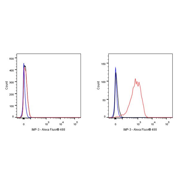
FC
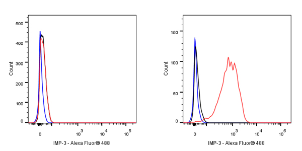
Flow cytometric analysis of 4% PFA fixed 90% methanol permeabilized MCF7 (Human breast adenocarcinoma epithelial cell, left) / HepG2 (Human hepatocellular carcinoma epithelial cell, Right) labelling IMP-3 antibody at 1/500 dilution (0.1 μg) / (Red) compared with a Rabbit monoclonal IgG (Black) isotype control and an unlabelled control (cells without incubation with primary antibody and secondary antibody) (Blue). Goat Anti - Rabbit IgG Alexa Fluor® 488 was used as the secondary antibody. Negative control: MCF7
Immunohistochemistry
IHC shows positive staining in paraffin-embedded human placenta. Anti-IMP-3 antibody was used at 1/200 dilution, followed by a HRP Polymer for Mouse & Rabbit IgG (ready to use). Counterstained with hematoxylin. Heat mediated antigen retrieval with Tris/EDTA buffer pH9.0 was performed before commencing with IHC staining protocol.
IHC shows positive staining in paraffin-embedded human tonsil. Anti-IMP-3 antibody was used at 1/200 dilution, followed by a HRP Polymer for Mouse & Rabbit IgG (ready to use). Counterstained with hematoxylin. Heat mediated antigen retrieval with Tris/EDTA buffer pH9.0 was performed before commencing with IHC staining protocol.
IHC shows positive staining in paraffin-embedded human testis. Anti-IMP-3 antibody was used at 1/200 dilution, followed by a HRP Polymer for Mouse & Rabbit IgG (ready to use). Counterstained with hematoxylin. Heat mediated antigen retrieval with Tris/EDTA buffer pH9.0 was performed before commencing with IHC staining protocol.
Negative control: IHC shows negative staining in paraffin-embedded human cerebral cortex. Anti-IMP-3 antibody was used at 1/200 dilution, followed by a HRP Polymer for Mouse & Rabbit IgG (ready to use). Counterstained with hematoxylin. Heat mediated antigen retrieval with Tris/EDTA buffer pH9.0 was performed before commencing with IHC staining protocol.
IHC shows positive staining in paraffin-embedded human colon cancer. Anti-IMP-3 antibody was used at 1/200 dilution, followed by a HRP Polymer for Mouse & Rabbit IgG (ready to use). Counterstained with hematoxylin. Heat mediated antigen retrieval with Tris/EDTA buffer pH9.0 was performed before commencing with IHC staining protocol.
IHC shows positive staining in paraffin-embedded human lung squamous cell carcinoma. Anti-IMP-3 antibody was used at 1/200 dilution, followed by a HRP Polymer for Mouse & Rabbit IgG (ready to use). Counterstained with hematoxylin. Heat mediated antigen retrieval with Tris/EDTA buffer pH9.0 was performed before commencing with IHC staining protocol.
Negative control: IHC shows negative staining in paraffin-embedded human hepatocellular carcinoma. Anti-IMP-3 antibody was used at 1/200 dilution, followed by a HRP Polymer for Mouse & Rabbit IgG (ready to use). Counterstained with hematoxylin. Heat mediated antigen retrieval with Tris/EDTA buffer pH9.0 was performed before commencing with IHC staining protocol.
IHC shows positive staining in paraffin-embedded mouse testis. Anti-IMP-3 antibody was used at 1/200 dilution, followed by a HRP Polymer for Mouse & Rabbit IgG (ready to use). Counterstained with hematoxylin. Heat mediated antigen retrieval with Tris/EDTA buffer pH9.0 was performed before commencing with IHC staining protocol.
IHC shows positive staining in paraffin-embedded rat testis. Anti-IMP-3 antibody was used at 1/200 dilution, followed by a HRP Polymer for Mouse & Rabbit IgG (ready to use). Counterstained with hematoxylin. Heat mediated antigen retrieval with Tris/EDTA buffer pH9.0 was performed before commencing with IHC staining protocol.
Immunocytochemistry
ICC shows positive staining in HeLa cells. Anti-IMP-3 antibody was used at 1/500 dilution (Green) and incubated overnight at 4°C. Goat polyclonal Antibody to Rabbit IgG - H&L (Alexa Fluor® 488) was used as secondary antibody at 1/1000 dilution. The cells were fixed with 4% PFA and permeabilized with 0.1% PBS-Triton X-100. Nuclei were counterstained with DAPI (Blue). Counterstain with tubulin (red).