Flow cytometric analysis of HeLa (Human cervix adenocarcinoma epithelial cell, left) / Molt-4 (Human lymphoblastic leukemia T lymphoblast, Right) cells labelling CD5 antibody at 1/2000 dilution (0.1 μg) / (Red) compared with a Mouse monoclonal IgG (Black) isotype control and an unlabelled control (cells without incubation with primary antibody and secondary antibody) (Blue). Goat Anti - Mouse IgG Alexa Fluor® 488 was used as the secondary antibody.
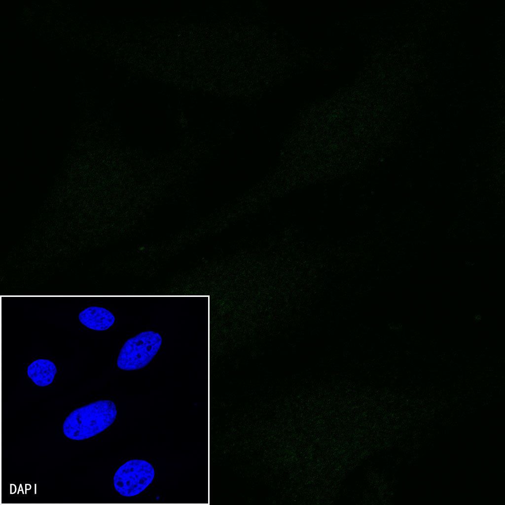
Negative control: HeLa
CD5 Mouse mAb (S-576-35)
CD5 Mouse mAb (S-576-35)
Price:
Regular price
$100 USD
Regular price
Sale price
$100 USD
Unit price
per
For shipping services or bulk orders, you may request a quotation.
Secure checkout with

View full details
Product Details
Product Details
Product Specification
| Host | Mouse |
| Antigen | CD5 |
| Synonyms | T-cell surface glycoprotein CD5, Lymphocyte antigen T1/Leu-1, LEU1 |
| Immunogen | Recombinant Protein |
| Location | Cell membrane |
| Accession | P06127 |
| Clone Number | S-576-35 |
| Antibody Type | Mouse mAb |
| Isotype | IgG1,k |
| Application | ICC, FCM |
| Reactivity | Hu |
| Purification | Protein G |
| Concentration | 2 mg/ml |
| Conjugation | Unconjugated |
| Physical Appearance | Liquid |
| Storage Buffer | PBS, 40% Glycerol, 0.05%BSA, 0.03% Proclin 300 |
| Stability & Storage | 12 months from date of receipt / reconstitution, -20 °C as supplied |
Dilution
| application | dilution | species |
| FCM | 1:2000 | null |
| ICC | 1:100 | null |
Background
CD5 is a cluster of differentiation expressed on the surface of T cells (various species) and in a subset of murine B cells known as B-1a. CD5 serves to mitigate activating signals from the BCR so that the B-1 cells can only be activated by very strong stimuli (such as bacterial proteins) and not by normal tissue proteins. CD5 was used as a T-cell marker until monoclonal antibodies against CD3 were developed. There is no confirmed ligand for CD5 but there is evidence that CD72, a C-type lectin, may be a ligand or that CD5 may be homophilic, binding CD5 on the surface of other cells.
Picture
Picture
FC
Immunocytochemistry

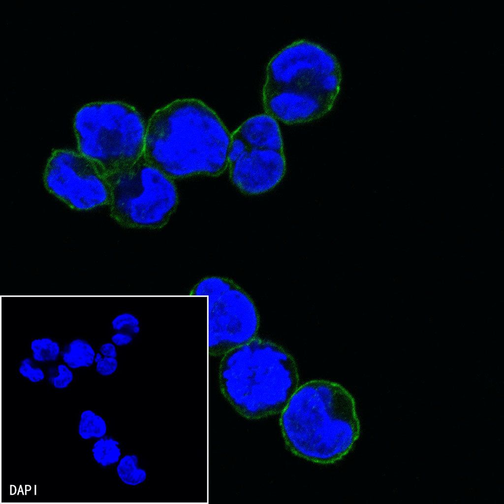
ICC shows positive staining in Molt-4 cells. Anti-CD5 antibody was used at 1/100 dilution (Green) and incubated overnight at 4°C. Goat polyclonal Antibody to Mouse IgG - H&L (Alexa Fluor® 488) was used as secondary antibody at 1/1000 dilution. The cells were fixed with 4%PFA and permeabilized with 0.1% PBS-Triton X-100. Nuclei were counterstained with DAPI (Blue).
Negative control:ICC shows negative staining in HeLa cells. Anti-CD5 antibody was used at 1/100 dilution and incubated overnight at 4°C. Goat polyclonal Antibody to Mouse IgG - H&L (Alexa Fluor® 488) was used as secondary antibody at 1/1000 dilution. The cells were fixed with 4%PFA and permeabilized with 0.1% PBS-Triton X-100. Nuclei were counterstained with DAPI (Blue).