WB result of ATF-4 Rabbit mAb
Primary antibody: ATF-4 Rabbit mAb at 1/1000 dilution
Lane 1: HeLa whole cell lysate 40 µg
Lane 2: HeLa treated with Tunicamycin (2µg/mL, 8hr) whole cell lysate 40 µg
Lane 3: HEK-293 whole cell lysate 40 µg
Lane 4: HEK-293 treated with Tunicamycin (2µg/mL, 8hr) whole cell lysate 40 µg
Secondary antibody: Goat Anti-Rabbit IgG, (H+L), HRP conjugated at 1/10000 dilution
Predicted MW: 38 kDa
Observed MW: 50 kDa
ATF-4 Recombinant Rabbit mAb (S-471-111)
ATF-4 Recombinant Rabbit mAb (S-471-111)
Price:
Regular price
$100 USD
Regular price
Sale price
$100 USD
Unit price
per
For shipping services or bulk orders, you may request a quotation.
Secure checkout with

View full details
Product Details
Product Details
Product Specification
| Host | Rabbit |
| Antigen | ATF-4 |
| Synonyms | cAMP-dependent transcription factor ATF-4; Activating transcription factor 4; Cyclic AMP-responsive element-binding protein 2; cAMP-responsive element-binding protein 2; Tax-responsive enhancer element-binding protein 67; CREB2; TXREB; TaxREB67 |
| Immunogen | Recombinant Protein |
| Location | Nucleus, Cytoplasm, Cell membrane |
| Accession | P18848 |
| Clone Number | S-471-111 |
| Antibody Type | Recombinant mAb |
| Isotype | IgG |
| Application | WB, ICC |
| Reactivity | Hu, Ms |
| Purification | Protein A |
| Concentration | 0.5 mg/ml |
| Conjugation | Unconjugated |
| Physical Appearance | Liquid |
| Storage Buffer | PBS, 40% Glycerol, 0.05%BSA, 0.03% Proclin 300 |
| Stability & Storage | 12 months from date of receipt / reconstitution, -20 °C as supplied |
Dilution
| application | dilution | species |
| WB | 1:1000 | |
| ICC | 1:100 |
Background
ATF-4 is a transcription factor that was originally identified as a widely expressed mammalian DNA binding protein that could bind a tax-responsive enhancer element in the LTR of HTLV-1. It belongs to a family of DNA-binding proteins that includes the AP-1 family of transcription factors, cAMP-response element binding proteins (CREBs) and CREB-like proteins. ATF4 transcription factor is also known to play role in osteoblast differentiation along with RUNX2 and osterix. ATF4 is also involved in the cannabinoid Δ9-tetrahydrocannabinol–induced apoptosis in cancer cells, by the proapoptotic role of the stress protein p8 via its upregulation of the endoplasmic reticulum stress-related genes ATF4, CHOP, and TRB3.
Picture
Picture
Western Blot
WB result of ATF-4 Rabbit mAb
Primary antibody: ATF-4 Rabbit mAb at 1/1000 dilution
Lane 1: NIH/3T3 whole cell lysate 20 µg
Lane 2: NIH/3T3 treated with Tunicamycin (2µg/mL, 8hr) whole cell lysate 20 µg
Secondary antibody: Goat Anti-Rabbit IgG, (H+L), HRP conjugated at 1/10000 dilution
Predicted MW: 38 kDa
Observed MW: 50 kDa
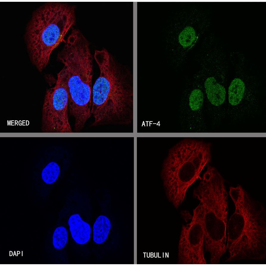
Immunocytochemistry
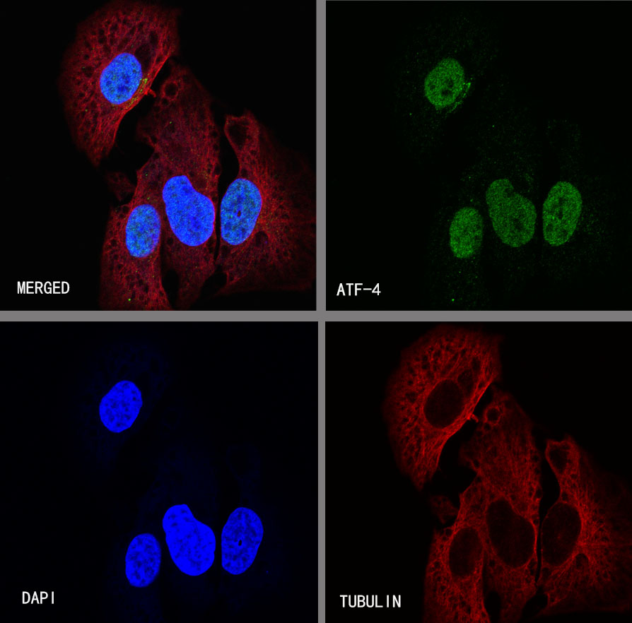
ICC shows positive staining in HeLa cells treated with Tunicamycin (2µg/mL, 8hr). Anti- ATF-4 antibody was used at 1/100 dilution (Green) and incubated overnight at 4°C. Goat polyclonal Antibody to Rabbit IgG - H&L (Alexa Fluor® 488) was used as secondary antibody at 1/1000 dilution. The cells were fixed with 4% PFA and permeabilized with 0.1% PBS-Triton X-100. Nuclei were counterstained with DAPI (Blue). Counterstain with tubulin (Red).
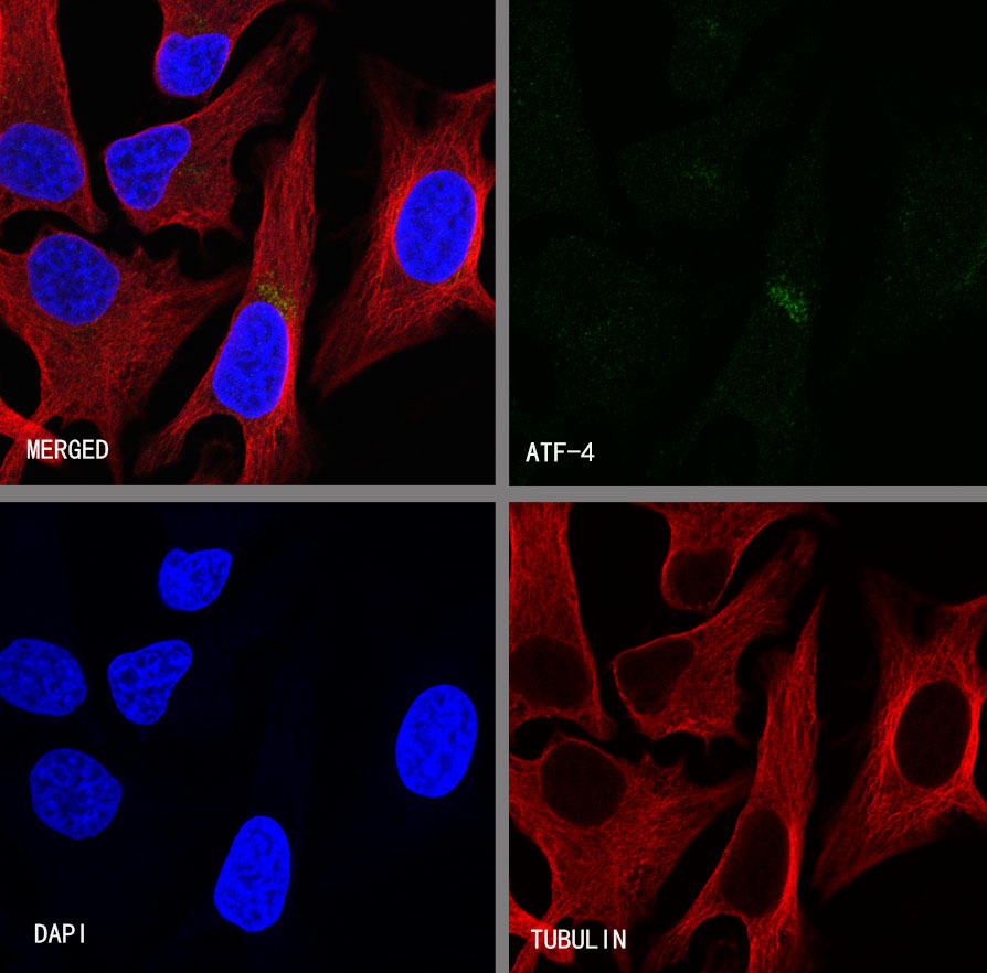
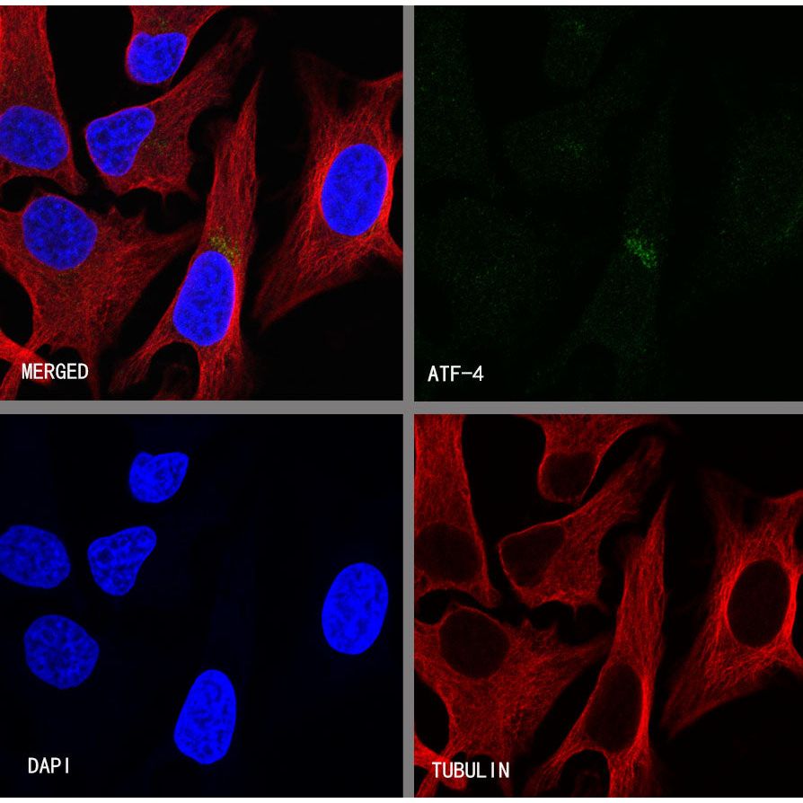
Negative control:ICC shows weakly positive staining in HeLa cells untreated with Tunicamycin (2μg/mL, 8hr). Anti- ATF-4 antibody was used at 1/100 dilution and incubated overnight at 4°C. Goat polyclonal Antibody to Rabbit IgG - H&L (Alexa Fluor® 488) was used as secondary antibody at 1/1000 dilution. The cells were fixed with 4% PFA and permeabilized with 0.1% PBS-Triton X-100. Nuclei were counterstained with DAPI (Blue). Counterstain with tubulin (Red).