
UniOne® TR-FRET Human IgG Sandwich Assay Kit
UniOne® TR-FRET Human IgG Sandwich Assay Kit
Product Details
Product Details
Product Specification
| Host | Human |
| Stability & Storage | -80℃ |
Background
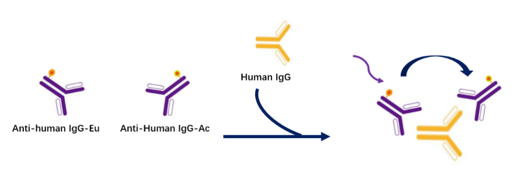
The kit employs homogeneous time-resolved fluorescence (HTRF) technology for the precise measurement of human IgG antibody or hFc-tagged protein concentrations in cell culture supernatants. The kit includes an antibody-labeled donor Eu (Anti-human IgG-Eu, Donor) and another antibody-labeled acceptor Ac (Anti-Human IgG-Ac, Acceptor) targeting a different epitope. When Anti-human IgG-Eu and Anti-human IgG-Ac bind to Human IgG, the Donor releases energy under 320 nm excitation and transfers it to the Acceptor, which then emits light at a specific wavelength (665 nm).
In the kit, Anti-human IgG-Eu and Anti-human IgG-Ac form a sandwich complex with Human IgG in samples or standards, leading to an increase in the 665 nm signal. This signal is proportional to the concentration of Human IgG in the standards, enabling accurate quantification of Human IgG in samples via a standard curve. The homogeneous assay is simple to perform, requiring no washing steps, and can be completed in a single step.
Components
Table 1. Kit Components
Component |
Concentration |
100T |
500T |
2500T |
10000T |
Storage Temperature |
Anti-Human IgG-Eu |
50× |
10μL |
50μL |
250μL |
1000μL |
-80℃ |
Anti-Human IgG-Ac |
50× |
10μL |
50μL |
250μL |
1000μL |
-80℃ |
IgG Standard |
40μg/ml |
10μL |
50μL |
200μL |
400μL |
-80℃ |
Detection Buffer |
1× |
1.5mL |
6ml |
30mL |
120mL |
-80℃ |
Diluent Buffer |
1× |
1.5mL极速翻译 |
6ml |
30mL |
120mL |
-80℃ |
Note: Aliquot immediately after first thaw and store at recommended temperature. Avoid storing after dilution or repeated freeze-thaw cycles.
Protocol
1.Reagent Preparation
Thaw all reagents at room temperature before use (equilibrate at room temperature for at least 30 min). The reaction volume for the 384-well shallow plate is 20 μL (reagent volumes for the reaction system are shown in the table). Calculate the required volume for the experiment before preparation and prepare as needed; the following preparation is for reference only, using 500 assays as an example.
1.1 Reagent Preparation:
Table 2. Reagent Preparation and Volumes
Reagent Name |
Preparation |
Volume (μL) |
IgG Standard or Test Sample |
Dilute the compound to the desired concentration using Diluent Buffer according to the reaction system. |
10 |
Antibody Mix |
Take 50 μL of Anti-Human IgG-Eu and dilute to 2500 μL with 1× Detection Buffer, mix well; take 50 μL of Anti-Human IgG-Ac and dilute to 2500 μL with 1× Detection Buffer, mix well, then mix 1:1 to prepare Antibody Mix. |
10 |
1.2Standard or Sample Dilution:
Use 1× Diluent Buffer to prepare the test samples and standards. If the sample stock is in DMSO, it is recommended to keep the DMSO concentration consistent in the system (the Detection Buffer does not contain DMSO; please add DMSO to the 1× Detection Buffer according to the dilution of the standard or test sample), with a maximum of 2%. Prepare the total volume of standards according to experimental needs; the following content is for reference only.
Table 3. Serial Dilution of Standards
|
Standard Preparation Concentration (ng/mL) |
Preparation Method |
① |
4000 |
5 μL of 40 μg/mL standard stock solution + 45 μL of 1× Diluent Buffer |
② |
2000 |
25 μL of ① + 25 μL of 1× Diluent Buffer |
③ |
1000 |
25 μL of ② + 25 μL of 1× Diluent Buffer |
④ |
500 |
25 μL of ③ + 25 μL of 1× Diluent Buffer |
⑤ |
250 |
25 μL of ④ + 25 μL of 1× Diluent Buffer |
⑥ |
125 |
25 μL of ⑤ + 25 μL of 1× Diluent Buffer |
⑦ |
62.5 |
25 μL of ⑥ + 25 μL of 1× Diluent Buffer |
⑧ |
31.25 |
25 μL of ⑦ + 25 μL of 1× Diluent Buffer |
⑨ |
15.63 |
25 μL of ⑧ + 25 μL of 1× Diluent Buffer |
⑩ |
7.81 |
25 μL of ⑨ + 25 μL of 1× Diluent Buffer |
⑪ |
3.91 |
25 μL of ⑩ + 25 μL of 1× Diluent Buffer |
Blank |
0 |
25 μL of 1× Diluent Buffer |
*The Blank normally receives Anti-human IgG-Eu and Anti-human IgG-Ac. The NC group receives only Anti-human IgG-Eu, used for calculatingNet Signal.
Dilute all test samples with dilution buffer or culture medium. Serial dilutions should be performed within the range of 2000-3.9 ng/mL (working solution); if the test sample concentration is within the 2000-3.9 ng/mL range, directly take 10 μL of supernatant for detection.
2. Sample Addition and Controls
2.1 Sample addition order for experimental wells: 10 μL of well-mixedAntibody Mix, 10 μL of standard or test sample, added sequentially into the 384-well shallow plate;
2.2 Blank control well (Blank): 1× Detection Buffer;
2.3 NC: 15 μL of 1× Detection Buffer plus 5 μL of dilutedAnti-Human IgG-Eu.
Table 4. Sample Addition and Controls
|
Blank |
Sample |
NC |
Step 1 |
10 μL 1× Detection Buffer |
10 μL Serially diluted test sample |
15 μL 1× Detection Buffer plus 5 μL 1× Anti-Human IgG-Eu |
10 μL Antibody Mix | |||
Step 2 |
Seal the plate wells with a plate sealer, and incubate at room temperature for 2 h. |
||
*For negative controls and quality control, it is recommended to use a solution with the same matrix as the test sample to replace 1× Detection Buffer.
*Keep DMSO or other matrices consistent across all detection wells.
3.Detection
Seal the plate wells with a plate sealer, centrifuge at 1000 rpm for 1 min, and incubate at room temperature for 2 h. Detect on a TR-FRET compatible microplate reader (excitation wavelength 320 nm, emission wavelengths 620 nm and 665 nm).
[Result Calculation]
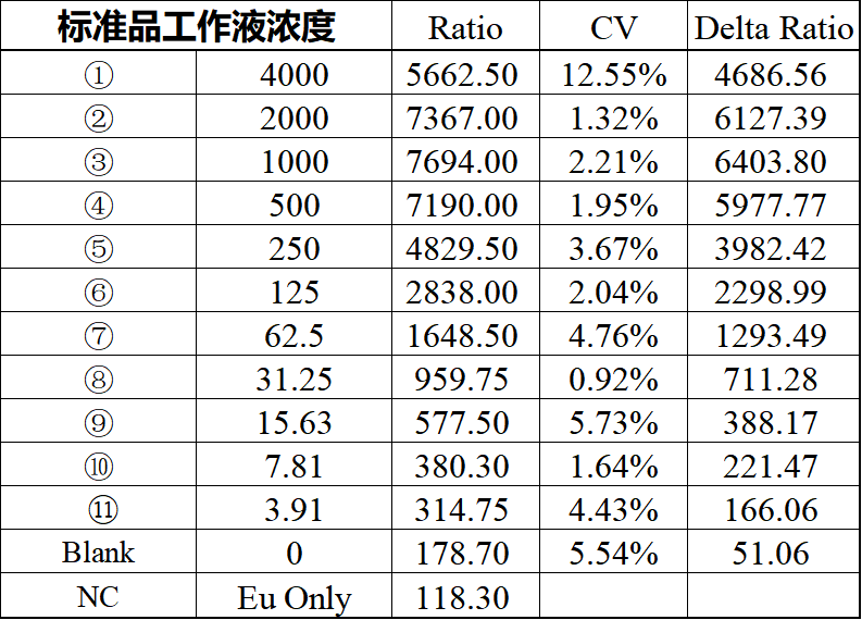
1. Calculate the signal value: Divide the 665 nm fluorescence signal by the 620 nm fluorescence signal and multiply by 10000, i.e., Signal Value = (665/620) * 10000
2. Calculate Net Signal based on the signal value: Net Signal = (Std - NC) / NC * 100
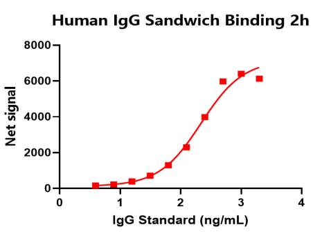
[Data Example]
Please note that the following data cannot replace the data obtained in experiments and is provided only as an example. Results may vary depending on the TR-FRET compatible instrument.
Note: Recommended microplate (384-well plate, white, shallow)
