WB result of E-Cadherin Rabbit mAb Primary antibody: E-Cadherin Rabbit mAb at 1/1000 dilution Lane 1: MDA-MB-231 whole cell lysate 20 µg Lane 2: MCF7 whole cell lysate 20 µg Lane 3: HT-29 whole cell lysate 20 µg Negative control: MDA-MB-231 whole cell lysate Secondary antibody: Goat Anti-Rabbit IgG, (H+L), HRP conjugated at 1/10000 dilution Predicted MW: 97 kDa Observed MW: 80~125 kDa
S-RMab® E-Cadherin Recombinant Rabbit mAb (SDT-438-5)
S-RMab® E-Cadherin Recombinant Rabbit mAb (SDT-438-5)
Price:
Regular price
$100 USD
Regular price
Sale price
$100 USD
Unit price
per
For shipping services or bulk orders, you may request a quotation.
Secure checkout with

View full details
Product Details
Product Details
Product Specification
| Host | Rabbit |
| Antigen | E-Cadherin |
| Synonyms | Cadherin-1, CAM 120/80, Epithelial cadherin, Uvomorulin, CD324, Uvomorulin, L-CAM |
| Immunogen | Recombinant Protein |
| Location | Cell membrane |
| Accession | P12830 |
| Clone Number | SDT-438-5 |
| Antibody Type | Rabbit mAb |
| Application | WB, IHC-P, ICC, ICFCM |
| Reactivity | Hu, Ms, Rt |
| Predicted Reactivity | Orangutan (95%) |
| Purification | Protein A |
| Concentration | 0.5 mg/ml |
| Conjugation | Unconjugated |
| Physical Appearance | Liquid |
| Storage Buffer | PBS, 40% Glycerol, 0.05% BSA, 0.03% Proclin 300 |
| Stability & Storage | 12 months from date of receipt / reconstitution, -20 °C as supplied |
Dilution
| application | dilution | species |
| WB | 1:1000 | |
| IHC | 1:1000 | |
| ICC | 1:500 | |
| ICFCM | 1:50 |
Background
E-cadherin is a transmembrane glycoprotein which connects epithelial cells together at adherens junctions. In normal cells, E-cadherin exerts its tumour suppressing role mainly by sequestering β-catenin from its binding to LEF (Lymphoid enhancer factor)/TCF (T cell factor) which serves the function of transcribing genes of the proliferative Wnt signaling pathway. Despite the ongoing debate on whether the loss of E-cadherin is the cause or effect of epithelial-mesenchymal transition (EMT), E-cadherin functional loss has frequently been associated with poor prognosis and survival in patients of various cancers. The dysregulation of E-cadherin expression that leads to carcinogenesis happens mostly at the epigenetic level but there are cases of genetic alterations as well. E-cadherin expression has been linked to the cellular functions of invasiveness reduction, growth inhibition, apoptosis, cell cycle arrest and differentiation.
Picture
Picture
Western Blot
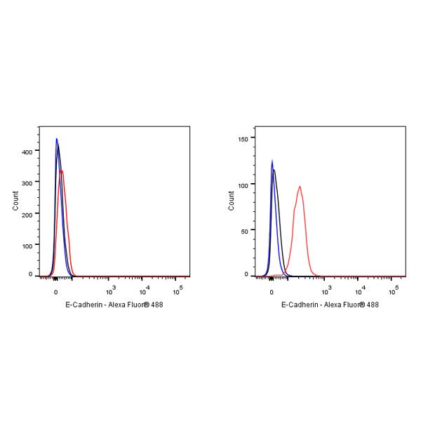
FC
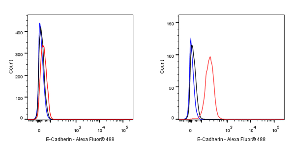
Flow cytometric analysis of 4% PFA fixed 90% methanol permeabilized MDA-MB-231 (Human breast adenocarcinoma epithelial cell, left) / HT29 (Human colorectal adenocarcinoma epithelial cell, Right) cells labelling E-Cadherin antibody at 1/500 dilution (0.1 μg) / (red) compared with a Rabbit monoclonal IgG (Black) isotype control and an unlabelled control (cells without incubation with primary antibody and secondary antibody) (Blue). Goat Anti - Rabbit IgG Alexa Fluor® 488 was used as the secondary antibody. Negative control: MDA-MB-231
Immunohistochemistry
IHC shows positive staining in paraffin-embedded human colon. Anti-E-Cadherin antibody was used at 1/1000 dilution, followed by a HRP Polymer for Mouse & Rabbit IgG (ready to use). Counterstained with hematoxylin. Heat mediated antigen retrieval with Tris/EDTA buffer pH9.0 was performed before commencing with IHC staining protocol.
IHC shows positive staining in paraffin-embedded human stomach. Anti-E-Cadherin antibody was used at 1/1000 dilution, followed by a HRP Polymer for Mouse & Rabbit IgG (ready to use). Counterstained with hematoxylin. Heat mediated antigen retrieval with Tris/EDTA buffer pH9.0 was performed before commencing with IHC staining protocol.
Negative control: IHC shows negative staining in paraffin-embedded human spleen. Anti-E-Cadherin antibody was used at 1/1000 dilution, followed by a HRP Polymer for Mouse & Rabbit IgG (ready to use). Counterstained with hematoxylin. Heat mediated antigen retrieval with Tris/EDTA buffer pH9.0 was performed before commencing with IHC staining protocol.
IHC shows positive staining in paraffin-embedded human breast cancer. Anti-E-Cadherin antibody was used at 1/1000 dilution, followed by a HRP Polymer for Mouse & Rabbit IgG (ready to use). Counterstained with hematoxylin. Heat mediated antigen retrieval with Tris/EDTA buffer pH9.0 was performed before commencing with IHC staining protocol.
IHC shows positive staining in paraffin-embedded human colon cancer. Anti-E-Cadherin antibody was used at 1/1000 dilution, followed by a HRP Polymer for Mouse & Rabbit IgG (ready to use). Counterstained with hematoxylin. Heat mediated antigen retrieval with Tris/EDTA buffer pH9.0 was performed before commencing with IHC staining protocol.
IHC shows positive staining in paraffin-embedded human lung squamous cell carcinoma. Anti-E-Cadherin antibody was used at 1/1000 dilution, followed by a HRP Polymer for Mouse & Rabbit IgG (ready to use). Counterstained with hematoxylin. Heat mediated antigen retrieval with Tris/EDTA buffer pH9.0 was performed before commencing with IHC staining protocol.
IHC shows positive staining in paraffin-embedded human pancreatic carcinoma. Anti-E-Cadherin antibody was used at 1/1000 dilution, followed by a HRP Polymer for Mouse & Rabbit IgG (ready to use). Counterstained with hematoxylin. Heat mediated antigen retrieval with Tris/EDTA buffer pH9.0 was performed before commencing with IHC staining protocol.
IHC shows positive staining in paraffin-embedded mouse colon. Anti-E-Cadherin antibody was used at 1/1000 dilution, followed by a HRP Polymer for Mouse & Rabbit IgG (ready to use). Counterstained with hematoxylin. Heat mediated antigen retrieval with Tris/EDTA buffer pH9.0 was performed before commencing with IHC staining protocol.
IHC shows positive staining in paraffin-embedded rat stomach. Anti-E-Cadherin antibody was used at 1/1000 dilution, followed by a HRP Polymer for Mouse & Rabbit IgG (ready to use). Counterstained with hematoxylin. Heat mediated antigen retrieval with Tris/EDTA buffer pH9.0 was performed before commencing with IHC staining protocol.
Immunocytochemistry
ICC shows positive staining in HT-29 cells. Anti-E-Cadherin antibody was used at 1/500 dilution (Green) and incubated overnight at 4°C. Goat polyclonal Antibody to Rabbit IgG - H&L (Alexa Fluor® 488) was used as secondary antibody at 1/1000 dilution. The cells were fixed with 100% ice-cold methanol and permeabilized with 0.1% PBS-Triton X-100. Nuclei were counterstained with DAPI (Blue).
ICC shows positive staining in MCF7 cells. Anti-E-Cadherin antibody was used at 1/500 dilution (Green) and incubated overnight at 4°C. Goat polyclonal Antibody to Rabbit IgG - H&L (Alexa Fluor® 488) was used as secondary antibody at 1/1000 dilution. The cells were fixed with 100% ice-cold methanol and permeabilized with 0.1% PBS-Triton X-100. Nuclei were counterstained with DAPI (Blue).
Negative control: ICC shows negative staining in MDA-MB-231 cells. Anti-E-Cadherin antibody was used at 1/500 dilution and incubated overnight at 4°C. Goat polyclonal Antibody to Rabbit IgG - H&L (Alexa Fluor® 488) was used as secondary antibody at 1/1000 dilution. The cells were fixed with 100% ice-cold methanol and permeabilized with 0.1% PBS-Triton X-100. Nuclei were counterstained with DAPI (Blue).