Flow cytometric analysis of C2C12 (Mouse myoblasts myoblast, left) / RAW 264.7 (Mouse Abelson murine leukemia virus-induced tumor macrophage, right) labelling Mouse F4/80 antibody at 1/2000 dilution (0.1 μg) / (Red) compared with a Rat monoclonal IgG (Black) isotype control and an unlabelled control (cells without incubation with primary antibody and secondary antibody) (Blue). Goat Anti - Rat IgG Alexa Fluor® 647 was used as the secondary antibody.
Negative control: C2C12
Rat Anti-Mouse F4/80 Antibody (S-R537)
Rat Anti-Mouse F4/80 Antibody (S-R537)
Price:
Regular price
$100 USD
Regular price
Sale price
$100 USD
Unit price
per
For shipping services or bulk orders, you may request a quotation.
Secure checkout with

View full details
Product Details
Product Details
Product Specification
| Host | Rat |
| Antigen | F4/80 |
| Synonyms | Adhesion G protein-coupled receptor E1; Cell surface glycoprotein F4/80; EGF-like module receptor 1; EGF-like module-containing mucin-like hormone receptor-like 1; EMR1 hormone receptor; Emr1; Gpf480; Adgre1 |
| Location | Cell membrane |
| Accession | Q61549 |
| Clone Number | S-R537 |
| Antibody Type | Rat mAb |
| Isotype | IgG2a,k |
| Application | ICC, FCM |
| Reactivity | Ms |
| Positive Sample | RAW 264.7 |
| Purification | Protein G |
| Concentration | 2 mg/ml |
| Conjugation | Unconjugated |
| Physical Appearance | Liquid |
| Storage Buffer | PBS pH7.4 |
| Stability & Storage | 12 months from date of receipt / reconstitution, 2 to 8 °C as supplied |
Dilution
| application | dilution | species |
| ICC | 1:500 | Ms |
| FCM | 1:2000 | Ms |
Background
F4/80, also known as EGF-like module-containing mucin-like hormone receptor-like 1 (EMR1), is a cell surface glycoprotein that is a member of the EGF-TM7 protein family. It is primarily used as a marker for mature macrophages in mice and shows significant expression changes during the maturation and activation of these cells. F4/80 is expressed on various mature macrophage populations, including Kupffer cells, Langerhans cells, microglia, and macrophages located in the peritoneum, intestinal lamina propria, splenic red pulp, lung, thymus, and bone marrow stroma. This antigen is encoded by the Adgre1 gene, which is a seven transmembrane G protein-coupled receptor with an extracellular domain containing repeated Epidermal Growth Factor (EGF)-like calcium binding domains. The F4/80 antigen is not only crucial for the phenotypic characterization of macrophages but also plays a role in immune regulation, as indicated by studies showing its necessity for inducing CD8+ regulatory T cells responsible for peripheral immune tolerance.
Picture
Picture
FC
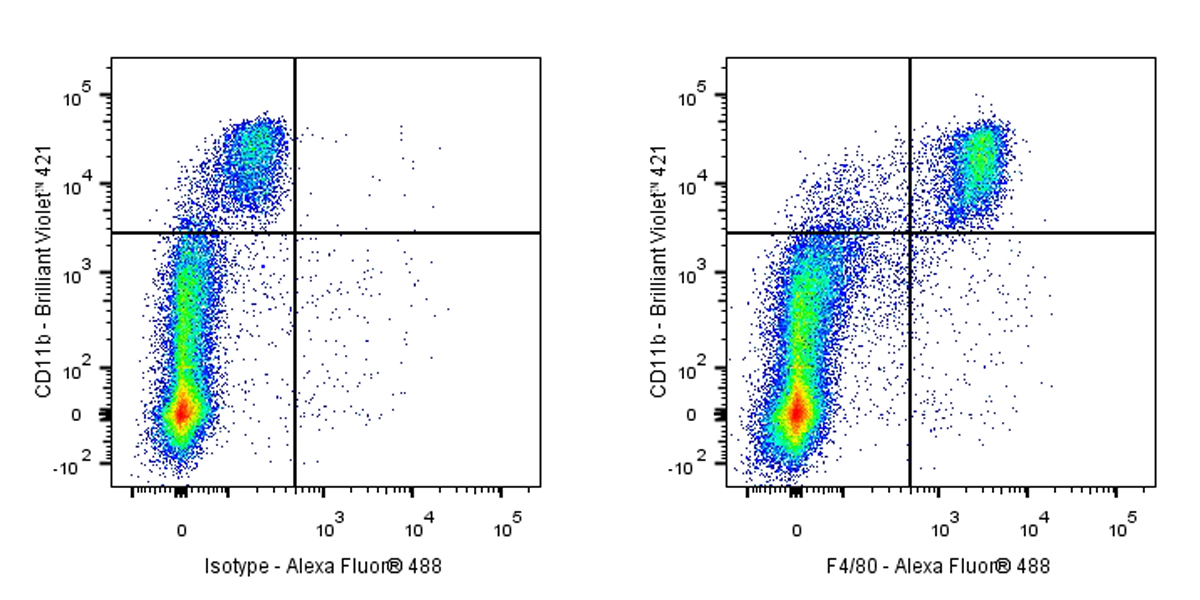
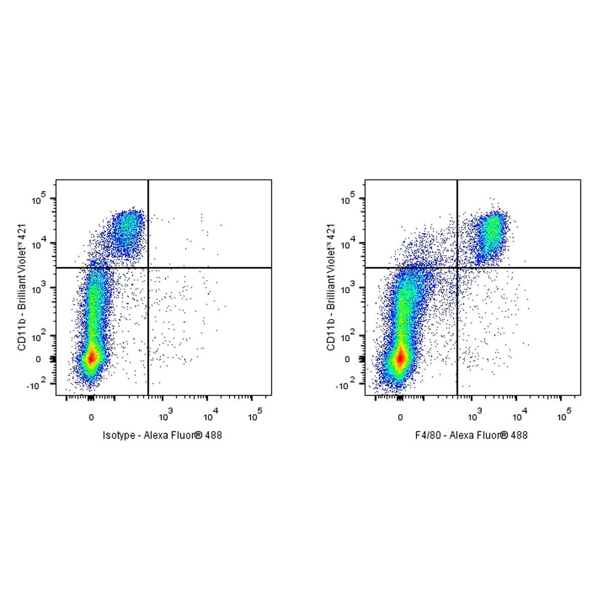
Flow cytometric analysis of mouse peritoneal exudates cells labelling Mouse F4/80 antibody at 1/2000 (0.1 μg) dilution/ (Right panel) compared with a Rat IgG2a, κ Isotype Control / (Left panel). Goat Anti-Rat IgG Alexa Fluor® 488 was used as the secondary antibody. Then cells were stained with CD11b - Brilliant Violet 421™ Antibody separately.
Immunocytochemistry
ICC shows positive staining in RAW 264.7 cells (top panel) and negative staining in C2C12 cells (below panel). Rat Anti-Mouse F4/80 Antibody was used at 1/500 dilution (Green) and incubated overnight at 4°C. Goat polyclonal Antibody to Rat IgG - H&L (Alexa Fluor® 488) was used as secondary antibody at 1/1000 dilution. The cells were fixed with 100% ice-cold methanol and permeabilized with 0.1% PBS-Triton X-100. Nuclei were counterstained with DAPI (Blue). Counterstain with tubulin (Red).