WB result of NeuN Rabbit mAb Primary antibody: NeuN Rabbit mAb at 1/1000 dilution Lane 1: mouse brain lysate 20 µg Secondary antibody: Goat Anti-Rabbit IgG, (H+L), HRP conjugated at 1/10000 dilution Predicted MW: 34 kDa Observed MW: 46~55 kDa
NeuN Recombinant Rabbit mAb (SDT-432-1)
NeuN Recombinant Rabbit mAb (SDT-432-1)
Price:
Regular price
$100 USD
Regular price
Sale price
$100 USD
Unit price
per
For shipping services or bulk orders, you may request a quotation.
Secure checkout with

View full details
Product Details
Product Details
Product Specification
| Host | Rabbit |
| Antigen | NeuN |
| Synonyms | RNA binding protein fox-1 homolog 3, Fox-3, Rbfox3, Hexaribonucleotide Binding Protein-3, Fox-1 homolog C Neuronal nuclei antigen, NeuN antigen |
| Immunogen | Synthetic Peptide |
| Location | Cytoplasm, Nucleus |
| Accession | A6NFN3 |
| Clone Number | SDT-432-1 |
| Antibody Type | Rabbit mAb |
| Application | WB, IHC-P, ICFCM, IP, IF |
| Reactivity | Hu, Ms, Rt |
| Purification | Protein A |
| Concentration | 0.5 mg/ml |
| Conjugation | Unconjugated |
| Physical Appearance | Liquid |
| Storage Buffer | PBS, 40% Glycerol, 0.05% BSA, 0.03% Proclin 300 |
| Stability & Storage | 12 months from date of receipt / reconstitution, -20 °C as supplied |
Dilution
| application | dilution | species |
| WB | 1:1000 | |
| IHC | 1:500 | |
| ICFCM | 1:500 | |
| IP | 1:50 | |
| IF | 1:100 |
Background
NeuN (Fox-3, Rbfox3, or Hexaribonucleotide Binding Protein-3), a protein which is a homologue to the protein product of a sex-determining gene in Caenorhabditis elegans, is a neuronal nuclear antigen that is commonly used as a biomarker for neurons. A few neuronal cell types are not recognized by NeuN antibodies, such as Purkinje cells, stellate and Golgi cells of the cerebellum, olfactory Mitral cells, retinal photoreceptors and spinal cord gamma motor neurons. However the vast majority of neurons are strongly NeuN positive, and NeuN immunoreactivity has been widely used to identify neurons in tissue culture and in sections and to measure the neuron/glia ratio in brain regions [PMID: 15758160].
Picture
Picture
Western Blot
WB result of NeuN Rabbit mAb Primary antibody: NeuN Rabbit mAb at 1/1000 dilution Lane 1: rat brain lysate 20 µg Secondary antibody: Goat Anti-Rabbit IgG, (H+L), HRP conjugated at 1/10000 dilution Predicted MW: 34 kDa Observed MW: 46~55 kDa
FC
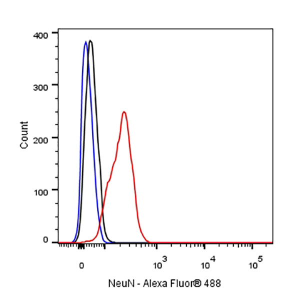
Flow cytometric analysis of 4% PFA fixed 90% methanol permeabilized SH-SY5Y (Human neuroblastoma epithelial cell) cells labelling NeuN antibody at 1/500 (0.1 μg) dilution / (red) compared with a Rabbit monoclonal IgG (Black) isotype control and an unlabelled control (cells without incubation with primary antibody and secondary antibody) (Blue). Goat Anti - Rabbit IgG Alexa Fluor® 488 was used as the secondary antibody.
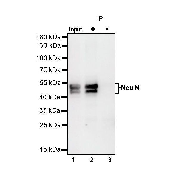
IP
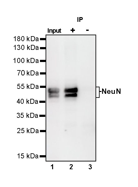
NeuN Rabbit mAb at 1/50 dilution (1 µg) immunoprecipitating NeuN in 0.4 mg mouse brain lysate.
Western blot was performed on the immunoprecipitate using NeuN Rabbit mAb at 1/1000 dilution.
Secondary antibody (HRP) for IP was used at 1/400 dilution.
Lane 1: mouse brain lysate 20 µg (Input)
Lane 2: S100B Rabbit mAb IP in mouse brain lysate
Lane 3: Rabbit monoclonal IgG IP in mouse brain lysate
Predicted MW: 34 kDa
Observed MW: 46, 48 kDa
Immunohistochemistry
IHC shows positive staining in paraffin-embedded human cerebellum. Anti-NeuN antibody was used at 1/500 dilution, followed by a HRP Polymer for Mouse & Rabbit IgG (ready to use). Counterstained with hematoxylin. Heat mediated antigen retrieval with Tris/EDTA buffer pH9.0 was performed before commencing with IHC staining protocol.
Negative control: IHC shows negative staining in paraffin-embedded human cardiac muscle. Anti-NeuN antibody was used at 1/500 dilution, followed by a HRP Polymer for Mouse & Rabbit IgG (ready to use). Counterstained with hematoxylin. Heat mediated antigen retrieval with Tris/EDTA buffer pH9.0 was performed before commencing with IHC staining protocol.
IHC shows positive staining in paraffin-embedded human glioma. Anti-NeuN antibody was used at 1/500 dilution, followed by a HRP Polymer for Mouse & Rabbit IgG (ready to use). Counterstained with hematoxylin. Heat mediated antigen retrieval with Tris/EDTA buffer pH9.0 was performed before commencing with IHC staining protocol.
Negative control: IHC shows negative staining in paraffin-embedded human baldder cancer. Anti-NeuN antibody was used at 1/500 dilution, followed by a HRP Polymer for Mouse & Rabbit IgG (ready to use). Counterstained with hematoxylin. Heat mediated antigen retrieval with Tris/EDTA buffer pH9.0 was performed before commencing with IHC staining protocol.
IHC shows positive staining in paraffin-embedded mouse cerebral cortex. Anti-NeuN antibody was used at 1/500 dilution, followed by a HRP Polymer for Mouse & Rabbit IgG (ready to use). Counterstained with hematoxylin. Heat mediated antigen retrieval with Tris/EDTA buffer pH9.0 was performed before commencing with IHC staining protocol.
Negative control: IHC shows negative staining in paraffin-embedded mouse liver. Anti-NeuN antibody was used at 1/500 dilution, followed by a HRP Polymer for Mouse & Rabbit IgG (ready to use). Counterstained with hematoxylin. Heat mediated antigen retrieval with Tris/EDTA buffer pH9.0 was performed before commencing with IHC staining protocol.
IHC shows positive staining in paraffin-embedded rat cerebral cortex. Anti-NeuN antibody was used at 1/500 dilution, followed by a HRP Polymer for Mouse & Rabbit IgG (ready to use). Counterstained with hematoxylin. Heat mediated antigen retrieval with Tris/EDTA buffer pH9.0 was performed before commencing with IHC staining protocol.
Negative control: IHC shows negative staining in paraffin-embedded rat liver. Anti-NeuN antibody was used at 1/500 dilution, followed by a HRP Polymer for Mouse & Rabbit IgG (ready to use). Counterstained with hematoxylin. Heat mediated antigen retrieval with Tris/EDTA buffer pH9.0 was performed before commencing with IHC staining protocol.
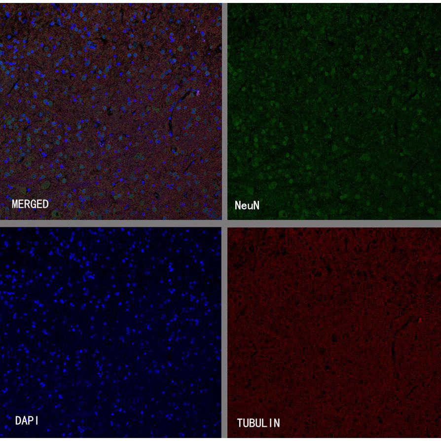
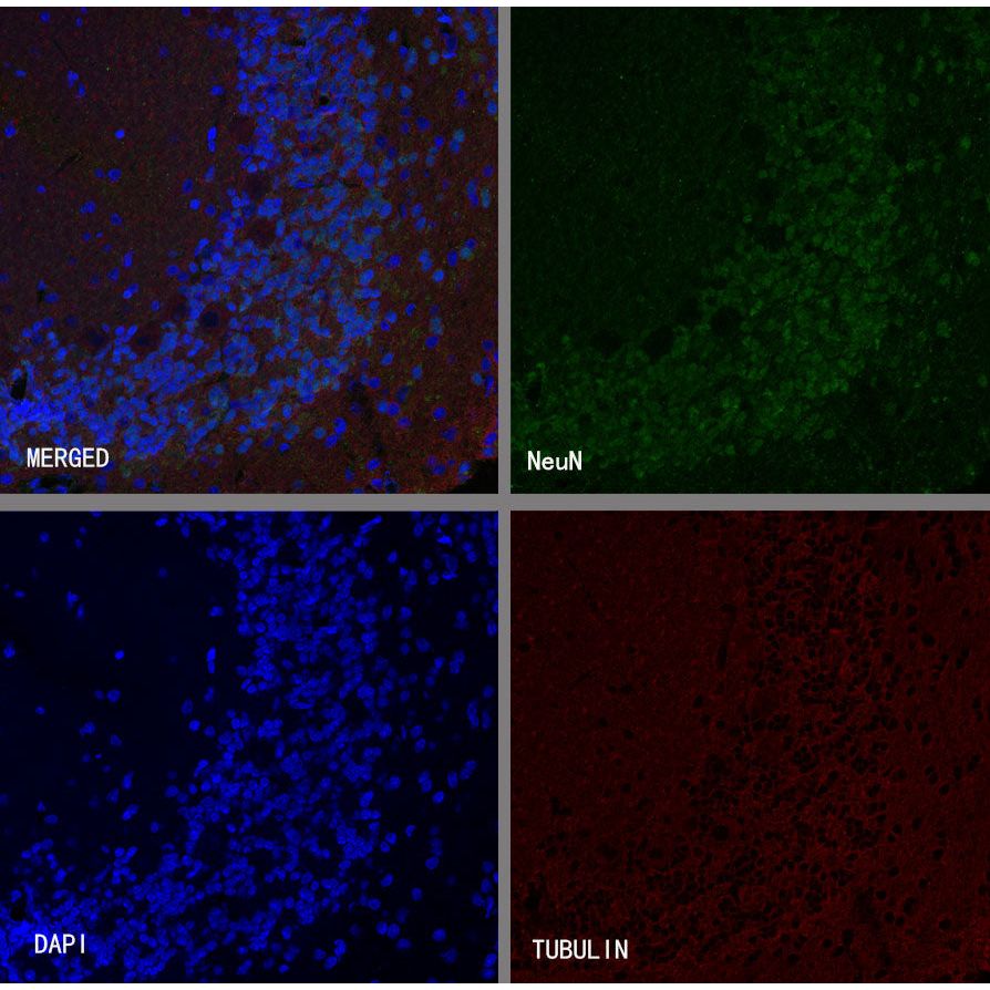
Immunofluorescence
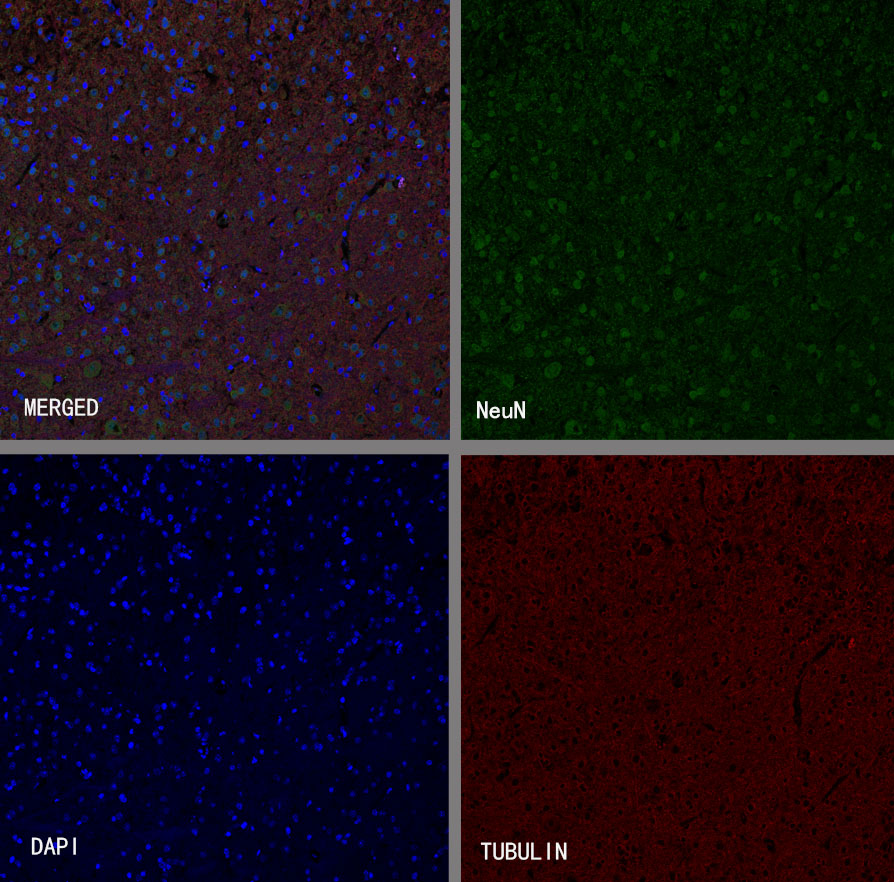
IF shows positive staining in paraffin-embedded mouse cerebellum. Anti-NeuN antibody was used at 1/100 dilution (Green) and incubated overnight at 4°C. Goat polyclonal Antibody to Rabbit IgG - H&L (Alexa Fluor® 488) was used as secondary antibody at 1/1000 dilution. Counterstained with DAPI (Blue). Counterstain with tubulin (Red).Heat mediated antigen retrieval with EDTA buffer pH9.0 was performed before commencing with IF staining protocol.
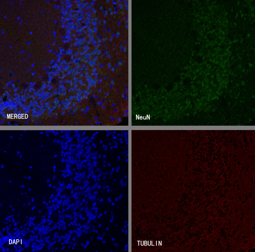
IF shows positive staining in paraffin-embedded rat cerebellum. Anti-NeuN antibody was used at 1/100 dilution (Green) and incubated overnight at 4°C. Goat polyclonal Antibody to Rabbit IgG - H&L (Alexa Fluor® 488) was used as secondary antibody at 1/1000 dilution. Counterstained with DAPI (Blue).Counterstain with tubulin (Red). Heat mediated antigen retrieval with EDTA buffer pH9.0 was performed before commencing with IF staining protocol.