
Mouse TGF-β ELISA Kit
Mouse TGF-β ELISA Kit

Product Details
Product Details
Product Specification
| protein | TGF-β | |||||||||||||||||||||||||||||||||||||||
| Usage |
Self-prepared test equipment required for the experiment: 1 , plate reader ( 450nm ) 2 , high-precision sampler and gun head: 0.5-10uL 、 5-50uL 、 20-200uL 、 200-1000uL 3 、 37℃℃ Incubator 4 Distilled water or deionized water, Sample handling and requirements: The detection range of the kit is not equivalent to the concentration range of the test substance in the sample. It is recommended to estimate the concentration of the test substance in the sample through relevant literature before the experiment and determine the actual concentration of the sample through pre-experiment. If the concentration of the test substance in the sample is too high or too low, please dilute or concentrate the sample appropriately. If the tested sample is not among the samples listed in the instructions, it is recommended to conduct pre-experiments to verify its detection effectiveness. Serum: Whole blood samples collected in serum separation tubes were placed at room temperature 2 Hour or 4℃ Overnight, then 1000×g Centrifugation 20 Minutes, take the supernatant, or place the supernatant in -20℃ Or -80℃ Store, but repeated freezing and thawing should be avoided. Plasma: with EDTA Or heparin as an anticoagulant to collect specimens, and collect the specimens after collection 30 Within minutes 2-8℃1000×g Centrifugation 15 Minutes, take the supernatant to detect, or place the supernatant in -20℃ Or -80℃℃ Store, but repeated freezing and thawing should be avoided. Tissue homogenate:: with pre-cooled PBS(0.01M,pH=7.4) The tissue is flushed to remove residual blood (lysed red blood cells in the homogenate can affect the measurement), and the tissue is weighed and cut into pieces. Combining the shredded tissue with the corresponding volume PBS (Generally according to 1:9 Weight to volume ratio, such as 1g The tissue samples correspond to 9mL Of PBS The specific volume can be appropriately adjusted according to the experimental needs and recorded. Recommended in PBS Add protease inhibitor) into a glass homogenizer and ground thoroughly on ice. For further lysis of tissue cells, the homogenate can be sonicated, or freeze-thawed repeatedly. Finally, the homogenate was mixed in 5000×g Centrifugation 5~10 Minutes, take the supernatant for detection. Cell culture supernatant: please 1000×g Centrifugation 20 Minutes, take the supernatant to detect, or place the supernatant in -20℃ Or -80℃ Store, but repeated freezing and thawing should be avoided. Other biological specimens: 1000×g Centrifugation 20 Minutes, take the supernatant to detect. Sample appearance: The sample should be clear and transparent, and the suspended solids should be removed by centrifugation. Sample storage: after sample collection, if 1 Those tested within weeks can be stored in 4℃ , if it cannot be detected in time, please pack it according to the one-time usage amount and freeze it in -20℃ ( 1 Within months), or -80℃ ( 6 Test within months) to avoid repeated freezing and thawing. Hemolysis of the specimen will affect the final test result, so hemolyzed specimens are not suitable for this test. Sample Activation: 1 , please advance 10 Minutes remove the kit from the refrigerator and equilibrate to room temperature. 2 , Standard gradient working solution preparation: add 1mL Universal diluent into lyophilized standard and let stand 15 Minutes until it is completely dissolved and then gently mix (the concentration is 2000pg/mL ) And then according to the following concentrations: 2000pg/mL 、 1000pg/mL 、 500pg/mL 、 250pg/mL 、 125pg/mL 、 62.5pg/mL 、 31.25pg/mL 、 0pg/mL The dilution was performed. Double dilution method : Take 7 branch EP Tube , added to each tube 500uL Universal diluent ,2000pg/mL Pipette from the standard working solution 500uL To the first EP Mix evenly in a tube 1000pg/mL Standard Working Solution , according to this step, absorb and mix evenly in turn. The last tube is directly used as a blank hole , there is no need to suck liquid from the penultimate tube, as shown in the figure below. 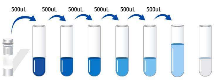 3 Preparation of biotinylated antibody detection working solution: before use 15 Min. The concentrated biotinylated antibody was concentrated in 1000×g Centrifugation 1 Minutes, with a universal diluent 100× The concentrated biotinylated antibody was diluted into 1× Working concentration (ex: 10uL Concentrate +990uL Universal Diluent) , used on the same day. 4 Preparation of enzyme conjugate working solution: before use 15 Minutes will 100× Concentrated enzyme conjugate in 1000×g Centrifugation 1 Minutes, with a universal diluent 100× concentrate HRP The enzyme conjugate is diluted into 1× Working concentration (ex: 10uL Concentrate +990uL Universal Diluent) , used on the same day. 5 、 1× Wash liquid preparation: Take 10mL 20× Wash liquid to 190mL In distilled water (the concentrated washing liquid taken out of the refrigerator may have crystals, which is a normal phenomenon. It can be left at room temperature and prepared after the crystals are completely dissolved). Operation steps: 1 Equilibration from room temperature 10 After minutes, remove the required slats from the aluminum foil bag, and seal the remaining slats with ziplock bag and put them back 4℃ 。 2 , Adding samples: respectively add samples or different concentration standards according to 100μl Each well is added to the corresponding well, and the blank well is added 100μL Universal diluent. After covering the sealing film 37℃ Incubation 1 Hours. (Recommendation: minimum dilution of sample to be tested with universal diluent 1 Times later, add the enzyme labeled plate for testing. So as to reduce the error influence of matrix effect on test results, and finally, the sample concentration needs to be multiplied by the corresponding dilution factor when calculating. It is recommended to set up double wells for all samples and standards to be tested during testing). 3 Add biotinylated antibody: take out the enzyme plate, discard the liquid without washing. Directly add biotinylated antibody working solution to each well 100μL , after covering the sealing film 37℃ Incubation 1 Hours. 4 Plate washing: discard the liquid and add to each well 300μL1x Wash liquid, stand 1 Minutes, throw off the washing liquid, pat dry on absorbent paper, and repeat washing the plate 3 Times (the plate can also be washed with a plate washing machine). 5 Adding enzyme conjugate working solution: adding enzyme conjugate working solution to each well 100μL , after covering the sealing film 37℃ Incubation 30 Minutes. 6 Plate washing: discard the liquid according to the steps 4 Washing method, wash plate 5 Times. 7 Add substrate: add substrate per well (TMB)90μL Covered with a sealing film, 37℃ Incubation protected from light 15 Minutes. 8 Add stop solution: take out the enzyme label plate and directly add stop solution to each well 50μL , immediately in 450nm Wavelength measurement of each well OD Value. Calculation of experimental results: Result judgment: 1 , calculate the average of the standard and sample replica well OD Value and subtract the blank hole's OD Values as correction values. Taking concentration as the abscissa, OD Value is ordinate , draw the standard curve of the four-parameter logic function on the double logarithmic coordinate paper. 2 If the sample OD If the value is higher than the upper limit of the standard curve, the test should be retested after appropriate dilution and multiplied by the corresponding dilution factor when calculating the sample concentration.
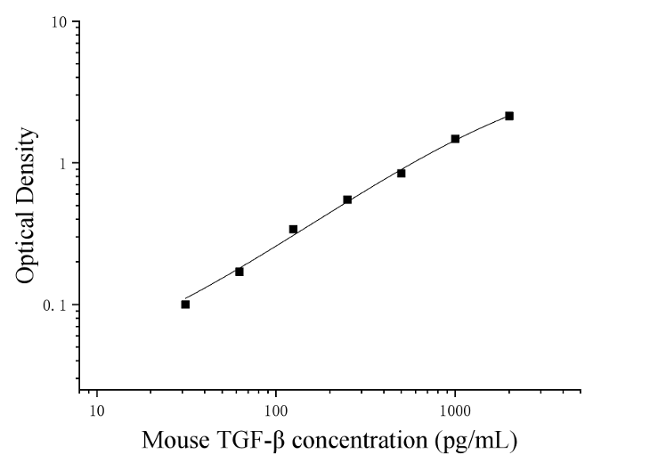 Kit Performance: 1 , repeatability: the coefficient of variation in the plate is less than 10% , the interplate coefficient of variation is less than 10% 。 2 Recovery rate: added to serum, plasma and cell culture supernatant of selected healthy mice 3 Mice at different concentration levels TGF-β , calculate the recovery.
3 , linear dilution: respectively in the selected 4 Serum, plasma and cell culture supernatant of healthy mice were added with high concentration of mice TGF-β , linearity was assessed by dilution within the standard curve kinetic range.
|
|||||||||||||||||||||||||||||||||||||||
| Sensitivity | 15.3pg/mL | |||||||||||||||||||||||||||||||||||||||
| Theory | This kit uses double antibody sandwich enzyme-linked immunosorbent assay (ELISA). To the microwells pre-coated with mouse transforming growth factor β (TGF-β) capture antibody, sample, standard product, biotin-labeled detection antibody, and HRP enzyme conjugate were sequentially added, incubated and washed in the middle, and colored with substrate TMB. TMB turned blue under the catalysis of peroxidase (HRP), and turned final yellow under the action of acid. The depth of color was positively correlated with mouse transforming growth factor β (TGF-β) in the sample. The absorbance (OD value) was measured with a microplate reader at 450nm wavelength, and the sample concentration was calculated. | |||||||||||||||||||||||||||||||||||||||
| Source | Mouse | |||||||||||||||||||||||||||||||||||||||
| Synonym | Mouse TGF-β ELISA Kit | |||||||||||||||||||||||||||||||||||||||
| Detection Type | Double antibody sandwich method | |||||||||||||||||||||||||||||||||||||||
| Composition |
|
|||||||||||||||||||||||||||||||||||||||
| Background | Transforming growth factor beta (TGF-beta) is a multifunctional cytokine belonging to the transforming growth factor superfamily that includes three different mammalian isoforms (TGF-β1 to 3, HGNC symbols TGFB1, TGFB2, TGFB3) and many other signaling proteins. TGFB protein is produced by all leukocyte lines. The activated TGF-β complexes with other factors to form a serine/threonine kinase complex, which binds to the TGF-β receptor. The TGF-beta receptor consists of type 1 and type 2 receptor subunits. Following TGF-beta binding, type 2 receptor kinases phosphorylate and activate type 1 receptor kinases, thereby activating a signaling cascade. This leads to the activation of different downstream substrates and regulatory proteins, inducing the transcription of different target genes, playing a role in differentiation, chemotaxis, proliferation, and the activation of many immune cells. TGF-β is secreted by many cell types, including macrophages, in a latent state, complexing with two other polypeptides, latent TGF-β binding protein (LTBP) and latent related peptide (LAP). Serum proteases such as plasma proteases catalyze the release of active TGF-β from the complex. This usually occurs on the surface of macrophages, where the latent TGF-β complex binds to CD36 through its ligand thrombochondroitin-1 (TSP-1). Inflammatory stimuli that activate macrophages enhance the release of active TGF-β by promoting the activation of plasma proteins. Macrophages can also endocytose latent TGF-β complexes bound by IgG secreted by plasma cells and then release active TGF-β into the extracellular fluid. One of its main functions is to regulate inflammatory processes, especially in the gut. TGF-β also plays a key role in stem cell differentiation and T cell regulation and differentiation. Due to its role in immunity and stem cell regulation and differentiation, it is a cytokine widely studied in the fields of cancer, autoimmune diseases and infectious diseases. The TGF-β superfamily includes endogenous growth inhibitory proteins; Increased expression of TGF-β is often associated with the degree of malignancy of many cancers and defects in the growth-inhibitory response of cells to TGF-β. Dysregulation of its immunosuppressive function is also implicated in the pathogenesis of autoimmune diseases, although its action is mediated by the environment of other cytokines present. | |||||||||||||||||||||||||||||||||||||||
| General Notes | 1. Carry out incubation in strict accordance with the specified time and temperature to ensure accurate results. All reagents must reach room temperature 20-25 °C prior to use. Store reagents in refrigeration immediately after use. 2. Incorrect plate washing may lead to inaccurate results. Make sure to drain the liquid from the wells as much as possible before adding the substrate. Do not allow the wells to dry out during incubation. 3. Eliminate the residual liquid and fingerprints at the bottom of the plate, otherwise it will affect the OD value. 4. The substrate color development solution should be colorless or very light in color, and the substrate solution that has turned blue cannot be used. 5. Avoid cross-contamination of reagents and specimens to avoid wrong results. 6. Avoid direct exposure to strong light during storage and incubation. 7. Any reaction reagent cannot come into contact with the bleaching solvent or the strong gas emitted by the bleaching solvent. Any bleaching component will destroy the biological activity of the reaction reagents in the kit. 8. Expired products cannot be used, and components with different item numbers and batch numbers cannot be mixed. 9. Recombinant proteins from sources other than the kit may not match the antibodies in this kit and are not recognized. 10. If the disease may be spread, all samples should be managed well, and the samples and testing devices should be handled according to the prescribed procedures. |
|||||||||||||||||||||||||||||||||||||||
| Storage Temp. | Unopened kit, stored at 4 °C, shelf life 6 months. | |||||||||||||||||||||||||||||||||||||||
| Test Range | 31.25-2000pg/mL |