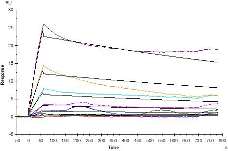
Anti-His antibody Immobilized on CM5 Chip captured PD1 His Tag, Mouse, can bind PD-1 Recombinant Rat mAb (
RMP1-14
) with an affinity constant of 0.238 μM as determined in SPR assay.
Invivo anti-mouse PD-1 Recombinant mAb (RMP1-14)
Invivo anti-mouse PD-1 Recombinant mAb (RMP1-14)
Price:
Regular price
$120 USD
Regular price
Sale price
$120 USD
Unit price
per
For shipping services or bulk orders, you may request a quotation.
Secure checkout with

View full details
Product Details
Product Details
Product Specification
| Host | Rat |
| Antigen | PD-1 |
| Synonyms | Programmed cell death protein 1, mPD-1, CD279 |
| Immunogen | N/A |
| Location | Cell membrane |
| Accession | Q02242 |
| Clone Number | RMP1-14 |
| Antibody Type | Recombinant mAb |
| Isotype | Rat IgG2a, κ |
| Isotype Control | S0B0932 |
| Application | WB, ICC, SPR, in vivo blocking of PD-1/PD-L signaling |
| Reactivity | Ms |
| Purification | Protein G |
| Concentration | 5 mg/ml |
| Purity | >95% (Determined by SDS-PAGE) |
| Endotoxin | <1EU/mg |
| Tag | N/A |
| Physical Appearance | Liquid |
| Storage Buffer | PBS pH7.4, containing no preservative |
| Stability & Storage |
2 to 8 °C for 2 weeks under sterile conditions; -20 °C for 3 months under sterile conditions; -80 °C for 24 months under sterile conditions. Please avoid repeated freeze-thaw cycles. |
Dilution
| application | dilution | species |
| WB | 1:250 | |
| SPR | 2000nM-31.25nM | |
| ICC | 1:500 |
Background
Programmed cell death protein 1, also known as PD-1 and CD279 (cluster of differentiation 279), is a protein on the surface of T and B cells that has a role in regulating the immune system's response to the cells of the human body by down-regulating the immune system and promoting self-tolerance by suppressing T cell inflammatory activity. This prevents autoimmune diseases, but it can also prevent the immune system from killing cancer cells. PD-1 is an immune checkpoint and guards against autoimmunity through two mechanisms. First, it promotes apoptosis (programmed cell death) of antigen-specific T-cells in lymph nodes. Second, it reduces apoptosis in regulatory T cells (anti-inflammatory, suppressive T cells).
Picture
Picture
Validation Data
Western Blot
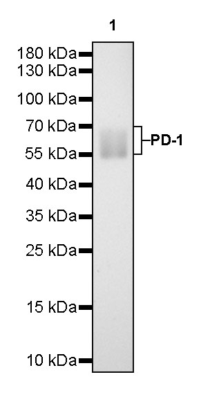
WB result of PD-1 Rat mAb
Primary antibody: PD-1 Rat mAb at 1/250 dilution
Lane 1:PD-1 Fc Chimera, Mouse (recombinant protein) lysate 1 μg
Secondary antibody: Goat Anti-Rat IgG, (H+L), HRP conjugated at 1/10000 dilution
Predicted MW: 58~70 kDa
Observed MW: 58~70 kDa
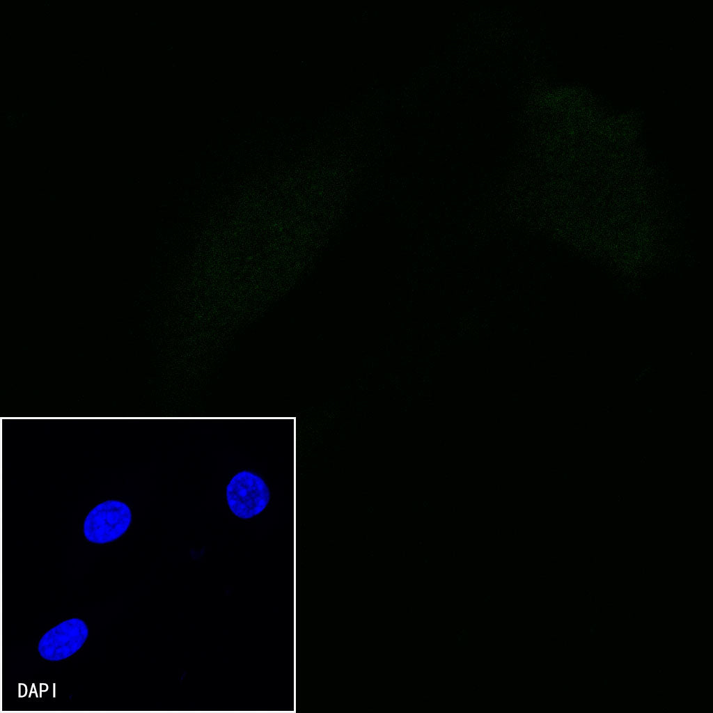
Immunocytochemistry
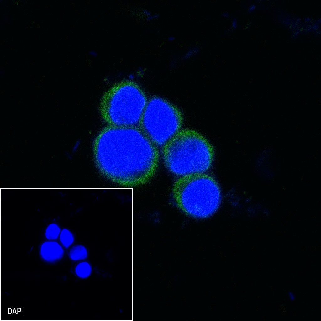
ICC shows positive staining in EL4.IL2 cells. Anti-PD-1 antibody was used at 1/500 dilution (Green) and incubated overnight at 4°C. Goat polyclonal Antibody to rat IgG - H&L (Alexa Fluor® 488) was used as secondary antibody at 1/1000 dilution. The cells were fixed with 100% ice-cold methanol and permeabilized with 0.1% PBS-Triton X-100. Nuclei were counterstained with DAPI (Blue).

Negative control:ICC shows negative staining in NIH/3T3 cells. Anti-PD-1 antibody was used at 1/500 dilution and incubated overnight at 4°C. Goat polyclonal Antibody to rat IgG - H&L (Alexa Fluor® 488) was used as secondary antibody at 1/1000 dilution. The cells were fixed with 100% ice-cold methanol and permeabilized with 0.1% PBS-Triton X-100. Nuclei were counterstained with DAPI (Blue).