Human Surv ELISA Kit
Human Surv ELISA Kit
Price:
Regular price
$368 USD
Regular price
Sale price
$368 USD
Unit price
per
For shipping services or bulk orders, you may request a quotation.
Secure checkout with

View full details
Product Details
Product Details
Product Specification
| protein | Surv | |||||||||||||||||||||||||||||||||||
| Usage |
Self-prepared test equipment required for the experiment: 1 , plate reader ( 450nm ) 2 , high-precision sampler and gun head: 0.5-10uL 、 5-50uL 、 20-200uL 、 200-1000uL 3 、 37℃℃ Incubator 4 Distilled water or deionized water, Sample handling and requirements: Serum: Whole blood samples collected in serum separation tubes were placed at room temperature 2 Hour or 4℃ Overnight, then 1000×g Centrifugation 20 Minutes, take the supernatant, or place the supernatant in -20℃ Or -80℃ Store, but repeated freezing and thawing should be avoided. Plasma: with EDTA Or heparin as an anticoagulant to collect specimens, and collect the specimens after collection 30 Within minutes 2-8℃1000×g Centrifugation 15 Minutes, take the supernatant to detect, or place the supernatant in -20℃ Or -80℃℃ Store, but repeated freezing and thawing should be avoided. Urine Tissue homogenate: with pre-cooled PBS ( 0.01M,pH=7.4 ) Rinse the tissue to remove residual blood (lysed red blood cells in the homogenate can affect the measurement result), and break the tissue after weighing. Combine the fragmented tissue with the corresponding volume PBS (Generally according to 1 : 9 Weight to volume ratio, such as 1g The tissue samples correspond to 9mL Of PBS The specific volume can be appropriately adjusted according to the experimental needs and recorded. Recommended in PBS Add protease inhibitor) into a glass homogenizer and ground thoroughly on ice. For further lysis of tissue cells, the homogenate can be sonicated, or freeze-thawed repeatedly. Finally, the homogenate was mixed in 5000×g Centrifugation 5~10 Minutes, take the supernatant for detection. Cell lysate: pre-cooling for adherent cells PBS Gently washed, followed by trypsinization, 1000×g Centrifugation 5 Cells were collected after minutes; The suspended cells can be collected directly by centrifugation. The collected cells were pre-cooled with PBS Washing 3 Times, every 1×10^6 Added to cells 150-200uL PBS Resuspension (recommended at PBS Adding a protease inhibitor; If the content is very low, it can be appropriately reduced PBS Volume) and the cells were disrupted by repeated freeze-thaw or sonication. The extracts were mixed in 2-8℃ , 1500×g Centrifugation 10 Minutes, take the supernatant for detection. Cell culture supernatant: please 1000×g Centrifugation 20 Minutes, take the supernatant to detect, or place the supernatant in -20℃ Or -80℃ Store, but repeated freezing and thawing should be avoided. Other biological fluids: 1000xg Centrifugation 20 Minutes, take the supernatant to detect. Preparations before testing: 1 , please advance 10 Minutes remove the kit from the refrigerator and equilibrate to room temperature. 2 , Standard gradient working solution preparation: add 1mL Universal diluent into lyophilized standard and let stand 15 Minutes until it is completely dissolved and then gently mix (the concentration is 2000pg/mL ) And then according to the following concentrations: 2000pg/mL 、 1000pg/mL 、 500pg/mL 、 250pg/mL 、 125pg/mL 、 62.5pg/mL 、 31.25pg/mL 、 0pg/mL The dilution was performed. Double dilution method : Take 7 branch EP Tube , added to each tube 500uL Universal diluent ,2000pg/mL Pipette from the standard working solution 500uL To the first EP Mix evenly in a tube 1000pg/mL Standard Working Solution , according to this step, absorb and mix evenly in turn. The last tube is directly used as a blank hole , there is no need to suck liquid from the penultimate tube, as shown in the figure below. 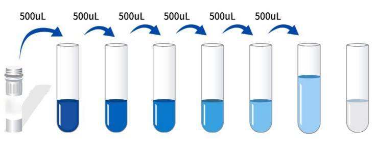 3 Preparation of biotinylated antibody detection working solution: before use 15 Min. The concentrated biotinylated antibody was concentrated in 1000×g Centrifugation 1 Minutes, with a universal diluent 100× The concentrated biotinylated antibody was diluted into 1× Working concentration (ex: 10uL Concentrate +990uL Universal Diluent) , now available for use. 4 Preparation of enzyme conjugate working solution: before use 15 Minutes will 100× Concentrated enzyme conjugate in 1000×g Centrifugation 1 Minutes, with a universal diluent 100× concentrate HRP The enzyme conjugate is diluted into 1× Working concentration (ex: 10uL Concentrate +990uL Universal Diluent) , now available for use. 5 、 1× Wash liquid preparation: Take 10mL 20× Wash liquid to 190mL In distilled water (the concentrated washing liquid taken out of the refrigerator may have crystals, which is a normal phenomenon. It can be left at room temperature and prepared after the crystals are completely dissolved). Operation steps: 1 Equilibration from room temperature 10 After minutes, remove the required slats from the aluminum foil bag, and seal the remaining slats with ziplock bag and put them back 4℃ 。 2 , Adding samples: respectively add samples or different concentration standards according to 100ul Each well is added to the corresponding well, and the blank well is added 100uL Universal diluent. After covering the sealing film 37℃ Incubation 60 Minutes. (Recommendation : Minimum dilution of sample to be tested with universal diluent 1 Times later, add the enzyme labeled plate for testing. So as to reduce the influence of matrix effect on the test results, and finally, the sample concentration needs to be multiplied by the corresponding dilution factor when calculating. It is recommended to set up double wells for all samples and standards to be tested during testing). 3 Add biotinylated antibody: take out the enzyme plate, discard the liquid without washing. Directly add biotinylated antibody working solution to each well 100uL , after covering the sealing film 37℃ Incubation 60 Minutes. 4 Plate washing: discard the liquid and add to each well 300uL 1x Wash liquid, stand 1 Minutes, throw off the washing liquid, pat dry on absorbent paper, and repeat washing the plate 3 Times (the plate can also be washed with a plate washing machine). 5 Adding enzyme conjugate working solution: adding enzyme conjugate working solution to each well 100uL , after covering the sealing film 37℃ Incubation 30 Minutes. 6 Plate washing: discard the liquid according to the steps 4 Washing method, wash plate 5 Times. 7 Substrate addition: substrate is added per well ( TMB ) 90uL Covered with a sealing film, 37℃ Incubation protected from light 15 Minutes. 8 Add stop solution: take out the enzyme label plate and directly add stop solution to each well 50uL , immediately in 450nm Wavelength measurement of each well OD Value. Calculation of experimental results: Result judgment: 1 , calculate the average of the standard and sample replica well OD Value and subtract the blank hole's OD Values as correction values. Taking concentration as the abscissa, OD Value is ordinate , draw the standard curve of the four-parameter logic function on the double logarithmic coordinate paper. 2 If the sample OD If the value is higher than the upper limit of the standard curve, the test should be retested after appropriate dilution and multiplied by the corresponding dilution factor when calculating the sample concentration. 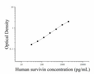 Note: The standard curve data provided is for reference only, and the sample content should be calculated according to the standard curve drawn in the same test. Kit Parameters: 1. Recovery rate: added to selected healthy human serum, plasma and cell culture supernatant 3 People with different concentration levels Surv , calculate the recovery.
2. Sensitivity: 12.52pg/mL 。 3. Linearity: respectively in selected 4 A portion of healthy human serum, plasma and cell culture supernatant were added with high concentration human surv , linearity was assessed by dilution within the standard curve kinetic range.
|
|||||||||||||||||||||||||||||||||||
| Theory | This kit uses double antibody sandwich enzyme-linked immunosorbent assay (ELISA). To the microwells pre-coated with Survivin (Surv) capture antibody, sample, standard product, biotin-labeled detection antibody, and HRP enzyme conjugate are added in sequence, incubated and washed in the middle, and color is developed with substrate TMB. TMB is converted into blue under the catalysis of peroxidase (HRP) and final yellow under the action of acid. There is a positive correlation between the depth of color and Survivin (Surv) in the sample. The absorbance (OD value) was measured with a microplate reader at a wavelength of 450nm, and the sample concentration was calculated. | |||||||||||||||||||||||||||||||||||
| Source | Human | |||||||||||||||||||||||||||||||||||
| Synonym | Human Survivin ELISA Kit | |||||||||||||||||||||||||||||||||||
| Detection Type | Double antibody sandwich method | |||||||||||||||||||||||||||||||||||
| Composition |
|
|||||||||||||||||||||||||||||||||||
| Background | Survivin, also known as the baculovirus inhibitor of 5 containing apoptotic repeats or BIRC5, is a protein encoded by the BIRC5 gene. Survivin is a member of the family of apoptosis inhibitors. The survivin protein has the function of inhibiting caspase activation, which leads to the negative regulation of apoptosis or programmed cell death. This has been shown by disruption of the Survivin-induced pathway, resulting in increased apoptosis and decreased tumor growth. | |||||||||||||||||||||||||||||||||||
| General Notes | 1. Carry out incubation in strict accordance with the specified time and temperature to ensure accurate results. All reagents must reach room temperature 20-25 °C prior to use. Store reagents in refrigeration immediately after use. 2. Incorrect plate washing may lead to inaccurate results. Make sure to drain the liquid from the wells as much as possible before adding the substrate. Do not allow the wells to dry out during incubation. 3. Eliminate the residual liquid and fingerprints at the bottom of the plate, otherwise it will affect the OD value. 4. The substrate color development solution should be colorless or very light in color, and the substrate solution that has turned blue cannot be used. 5. Avoid cross-contamination of reagents and specimens to avoid wrong results. 6. Avoid direct exposure to strong light during storage and incubation. 7. Any reaction reagent cannot come into contact with the bleaching solvent or the strong gas emitted by the bleaching solvent. Any bleaching component will destroy the biological activity of the reaction reagents in the kit. 8. Expired products cannot be used, and components with different item numbers and batch numbers cannot be mixed. 9. Recombinant proteins from sources other than the kit may not match the antibodies in this kit and are not recognized. 10. If the disease may be spread, all samples should be managed well, and the samples and testing devices should be handled according to the prescribed procedures. |
|||||||||||||||||||||||||||||||||||
| Storage Temp. | Unopened kit, stored at 4 °C, shelf life 6 months. | |||||||||||||||||||||||||||||||||||
| Test Range | 31.25-2000pg/mL | |||||||||||||||||||||||||||||||||||
| Applications | Serum, plasma, urine, tissue homogenates, cell lysates, cell culture supernatants and other biological fluids |