
Human GM-CSF Ab ELISA Kit
Human GM-CSF Ab ELISA Kit

Product Details
Product Details
Product Specification
| Usage |
Sample Processing and Requirements 1. 2. 3. 4. Reagent Preparation After removing the kit from the refrigerator, allow it to equilibrate to room temperature before use. Dilution of 20× Wash Buffer: Dilute 1:20 with distilled water, i.e., add 1 part 20× Wash Buffer to 19 parts distilled water. Procedure Calculation of Experimental Results: Using the OD value of the measured standard as the horizontal axis and the concentration of the standard as the vertical axis, draw a standard curve on graph paper or using relevant software. Obtain a linear regression equation. Substitute the OD value of the sample into the equation to calculate the sample concentration. 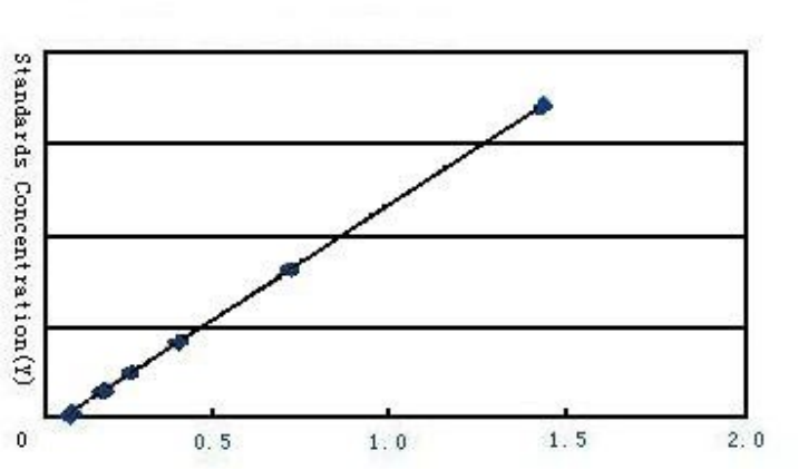 Standard Curve |
||||||||||||||||||||||||||||||
| Sensitivity | The minimum detectable concentration was less than 1.0 pg/mL. | ||||||||||||||||||||||||||||||
| Species Reactivity | Human | ||||||||||||||||||||||||||||||
| Theory | The kit uses an indirect enzyme-linked immunosorbent assay (ELISA). Samples, standards, and HRP-labeled detection antibodies are added sequentially to microwells pre-coated with human granulocyte macrophage colony-stimulating factor (GM-CSF) antibody to capture the antigen. The sample is incubated and washed thoroughly. The color is developed using the substrate TMB, which converts to blue under the catalysis of peroxidase and to yellow under the action of acid. The intensity of the color is positively correlated with the amount of human granulocyte macrophage colony-stimulating factor (GM-CSF) antibody in the sample. The absorbance (OD) is measured at 450 nm using a microplate reader to calculate the sample concentration. | ||||||||||||||||||||||||||||||
| Synonym | Human anti-GM-CSF ELISA Kit | ||||||||||||||||||||||||||||||
| Detection Type | Used for in vitro quantitative detection of the content of human granulocyte macrophage colony-stimulating factor antibody (GM-CSF Ab) in serum, plasma, tissue homogenate and related liquid samples. | ||||||||||||||||||||||||||||||
| Composition |
Remarks: 1. The concentrations of the standard substances are: 320, 160, 80, 40, 20, and 10 pg/mL. 2. After testing a large number of normal specimens, the normal concentration values of the specimens are all within the detection range provided by the kit. During the experiment, 50 μL of sample can be directly sampled. If some sample values exceed the maximum standard concentration, the sample can be appropriately diluted with sample diluent before the experiment.
|
||||||||||||||||||||||||||||||
| General Notes |
1. Strictly follow the specified incubation time and temperature to ensure accurate results. All reagents must reach room temperature (20-25°C) before use. Refrigerate reagents immediately after use. 2. Improper plate washing can lead to inaccurate results. Ensure that the liquid in the wells is as dry as possible before adding substrate. Do not allow the microwells to dry out during incubation. 3. Remove any residual liquid and fingerprints from the bottom of the plate, otherwise it will affect the OD value. 4. The substrate developer solution should be colorless or very light in color. Substrate solution that has turned blue should not be used. 5. Avoid cross-contamination of reagents and specimens to prevent erroneous results. 6. Avoid direct exposure to strong light during storage and incubation. 7. Allow the sealed bag to equilibrate to room temperature before opening to prevent water droplets from condensing on the cold plate strips. 8. No reaction reagents should come into contact with bleaching solvents or the strong fumes emitted by bleaching solvents. Any bleaching component will destroy the biological activity of the reagents in the kit. 9. Do not use expired products. 10. If there is a possibility of disease transmission, all samples should be managed properly and the samples and detection devices should be handled according to the prescribed procedures. |
||||||||||||||||||||||||||||||
| Storage Temp. | Unopened test kit, stored at 2-8°C, has a shelf life of 6 months. | ||||||||||||||||||||||||||||||
| Test Range | 10 pg/mL – 320 pg/mL. |