WB result of Claudin 3 Rabbit mAb
Primary antibody: Claudin 3 Rabbit mAb at 1/5000 dilution
Lane 1: U-87MG whole cell lysate 20 µg
Lane 2: MCF7 whole cell lysate 20 µg
Lane 3: T-47D whole cell lysate 20 µg
Lane 4: HT-29 whole cell lysate 20 µg
Negative control: U-87MG whole cell lysate
Secondary antibody: Goat Anti-rabbit IgG, (H+L), HRP conjugated at 1/10000 dilution
Predicted MW: 23 kDa
Observed MW: 20 kDa
Claudin 3 Recombinant Rabbit mAb (S-819-131)
Claudin 3 Recombinant Rabbit mAb (S-819-131)
Price:
Regular price
$100 USD
Regular price
Sale price
$100 USD
Unit price
per
For shipping services or bulk orders, you may request a quotation.
Secure checkout with

View full details
Product Details
Product Details
Product Specification
| Host | Rabbit |
| Synonyms | Clostridium perfringens enterotoxin receptor 2 (CPE-R 2; CPE-receptor 2), Rat ventral prostate.1 protein homolog (hRVP1), CLDN3, C7orf1, CPETR2 |
| Immunogen | Synthetic Peptide |
| Location | Cell membrane |
| Accession | O15551 |
| Clone Number | S-819-131 |
| Antibody Type | Recombinant mAb |
| Isotype | IgG |
| Application | WB, IHC-P, IP |
| Reactivity | Hu |
| Purification | Protein A |
| Concentration | 2 mg/ml |
| Conjugation | Unconjugated |
| Physical Appearance | Liquid |
| Storage Buffer | PBS, 40% Glycerol, 0.05% BSA, 0.03% Proclin 300 |
| Stability & Storage | 12 months from date of receipt / reconstitution, -20 °C as supplied |
Dilution
| application | dilution | species |
| WB | 1:5000 | null |
| IHC-P | 1:200 | null |
| IP | 1:200 | null |
Background
Claudin 3 protein is a tight junction protein with a molecular weight of approximately 22 kDa. Tight junctions are important membrane-associated complexes between adjacent cells, and Claudin proteins form the main backbone of these structures. Along with other Claudin proteins, Claudin-3 protein stabilizes the structure of intercellular junctions by interacting with each other. It helps maintain the stability of cell gaps and facilitates transmembrane transport/enrichment functions. Claudin-3 protein can also alter the permeability of intercellular junctions, controlling the passage of substances. Claudin-3 protein is often overexpressed in certain cancers, such as ovarian cancer and breast cancer. This increased expression level is associated with the enhancement of tumor cell invasiveness and vitality. Therefore, Claudin-3 may become a target for the treatment of prostate cancer and other malignancies. In cell susceptibility and demyelinating anatomical phenotypes, inhibition of Claudin-3 expression is associated with neurotoxicity.
Picture
Picture
Western Blot
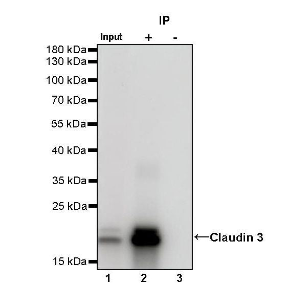
IP

Claudin 3 Rabbit mAb at 1/200 dilution (1 µg) immunoprecipitating Claudin 3 in 0.4 mg MCF7 whole cell lysate.
Western blot was performed on the immunoprecipitate using Claudin 3 Rabbit mAb at 1/1000 dilution.
Secondary antibody (HRP) for IP was used at 1/1000 dilution.
Lane 1: MCF7 whole cell lysate 20 µg (Input)
Lane 2: Claudin 3 Rabbit mAb IP in MCF7 whole cell lysate
Lane 3: Rabbit monoclonal IgG IP in MCF7 whole cell lysate
Predicted MW: 23 kDa
Observed MW: 20 kDa
Immunohistochemistry
IHC shows positive staining in paraffin-embedded human colon. Anti- Claudin 3 antibody was used at 1/200 dilution, followed by a HRP Polymer for Mouse & Rabbit IgG (ready to use). Counterstained with hematoxylin. Heat mediated antigen retrieval with Tris/EDTA buffer pH9.0 was performed before commencing with IHC staining protocol.
IHC shows positive staining in paraffin-embedded human tonsil. Anti- Claudin 3 antibody was used at 1/200 dilution, followed by a HRP Polymer for Mouse & Rabbit IgG (ready to use). Counterstained with hematoxylin. Heat mediated antigen retrieval with Tris/EDTA buffer pH9.0 was performed before commencing with IHC staining protocol.
IHC shows positive staining in paraffin-embedded human colon cancer. Anti- Claudin 3 antibody was used at 1/200 dilution, followed by a HRP Polymer for Mouse & Rabbit IgG (ready to use). Counterstained with hematoxylin. Heat mediated antigen retrieval with Tris/EDTA buffer pH9.0 was performed before commencing with IHC staining protocol.