WB result of S6 Ribosomal Protein Rabbit mAb
Primary antibody: S6 Ribosomal Protein Rabbit mAb at 1/1000 dilution
Lane 1: HeLa whole cell lysate 20 µg
Lane 2: MCF7 whole cell lysate 20 µg
Lane 3: HepG2 whole cell lysate 20 µg
Secondary antibody: Goat Anti-Rabbit IgG, (H+L), HRP conjugated at 1/10000 dilution
Predicted MW: 29 kDa
Observed MW: 29 kDa
S6 Ribosomal Protein Recombinant Rabbit mAb (S-479-1)
S6 Ribosomal Protein Recombinant Rabbit mAb (S-479-1)
Price:
Regular price
$100 USD
Regular price
Sale price
$100 USD
Unit price
per
For shipping services or bulk orders, you may request a quotation.
Secure checkout with

View full details
Product Details
Product Details
Product Specification
| Host | Rabbit |
| Antigen | S6 Ribosomal Protein |
| Synonyms | Small ribosomal subunit protein eS6, 40S ribosomal protein S6, Phosphoprotein NP33, RPS6 |
| Immunogen | Synthetic Peptide |
| Location | Cytoplasm, Nucleus |
| Accession | P62753 |
| Clone Number | S-479-1 |
| Antibody Type | Recombinant mAb |
| Application | WB, IHC-P, IP, ICFCM |
| Reactivity | Hu, Ms, Rt |
| Predicted Reactivity | Av, Bv, Rb, Xe, Fs |
| Purification | Protein A |
| Concentration | 0.5 mg/ml |
| Conjugation | Unconjugated |
| Physical Appearance | Liquid |
| Storage Buffer | PBS, 40% Glycerol, 0.05%BSA, 0.03% Proclin 300 |
| Stability & Storage | 12 months from date of receipt / reconstitution, -20 °C as supplied |
Dilution
| application | dilution | species |
| WB | 1:1000 | |
| IP | 1:50 | |
| IHC | 1:500 | |
| ICFCM | 1:5000 |
Background
Ribosomal protein S6 (rpS6 or eS6) is a component of the 40S ribosomal subunit and is therefore involved in translation. Mouse model studies have shown that phosphorylation of eS6 is involved in the regulation of cell size, cell proliferation, and glucose homeostasis. tudies show that the p70 ribosomal protein S6 kinases (S6K1 and S6K2) and p90 ribosomal protein S6 kinases (RSK) both phosphorylate eS6 and that S6K1 and S6K2 predominate this function. Pathways leading to the induction of human eS6 phosphorylation have been found to enhance IL-8 protein synthesis. This mechanism is dependent on A/U-rich proximal sequences (APS) found in the 3'UTR of IL-8 immediately after the stop codon.
Picture
Picture
Western Blot
WB result of S6 Ribosomal Protein Rabbit mAb
Primary antibody: S6 Ribosomal Protein Rabbit mAb at 1/1000 dilution
Lane 1: NIH/3T3 whole cell lysate 20 µg
Secondary antibody: Goat Anti-Rabbit IgG, (H+L), HRP conjugated at 1/10000 dilution
Predicted MW: 29 kDa
Observed MW: 29 kDa
WB result of S6 Ribosomal Protein Rabbit mAb
Primary antibody: S6 Ribosomal Protein Rabbit mAb at 1/1000 dilution
Lane 1: PC-12 whole cell lysate 20 µg
Secondary antibody: Goat Anti-Rabbit IgG, (H+L), HRP conjugated at 1/10000 dilution
Predicted MW: 29 kDa
Observed MW: 29 kDa
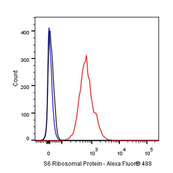
FC
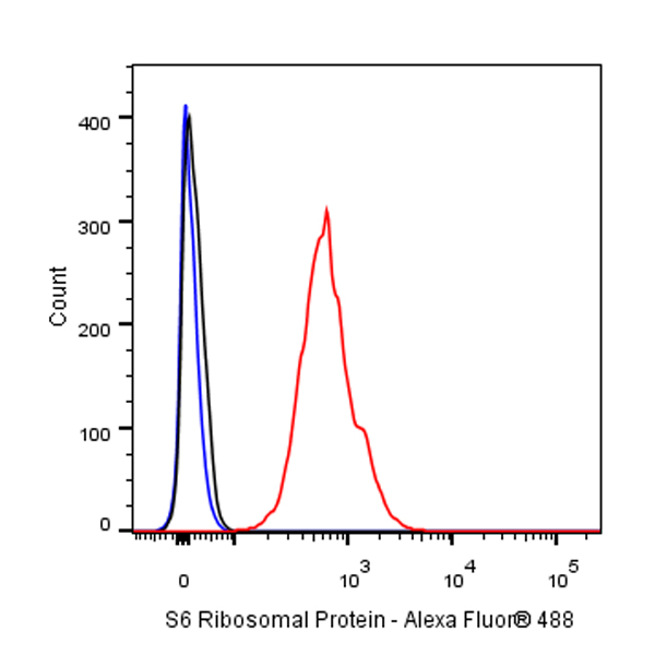
Flow cytometric analysis of 4% PFA fixed 90% methanol permeabilized HeLa (Human cervix adenocarcinoma epithelial cell) cells labeling S6 Ribosomal Protein at 1/5000 dilution (0.01 μg) / (red) compared with a rabbit monoclonal IgG isotype control (black) and an unlabeled control (cells without incubation with primary antibody and secondary antibody) (Blue). Goat Anti - Rabbit IgG Alexa Fluor® 488 was used as the secondary antibody.
IP
S6 Ribosomal Protein Rabbit mAb at 1/50 dilution (1 µg) immunoprecipitating S6 Ribosomal Protein in 0.4 mg HeLa whole cell lysate.
Western blot was performed on the immunoprecipitate using S6 Ribosomal Protein Rabbit mAb at 1/1000 dilution.
Secondary antibody (HRP) for IP was used at 1/400 dilution.
Lane 1: HeLa whole cell lysate 20 µg (Input)
Lane 2: S6 Ribosomal Protein Rabbit mAb IP in HeLa whole cell lysate
Lane 3: Rabbit monoclonal IgG IP in HeLa whole cell lysate
Predicted MW: 29 kDa
Observed MW: 29 kDa
Immunohistochemistry
IHC shows positive staining in paraffin-embedded human tonsil. Anti-S6 Ribosomal Protein antibody was used at 1/500 dilution, followed by a HRP Polymer for Mouse & Rabbit IgG (ready to use). Counterstained with hematoxylin. Heat mediated antigen retrieval with Tris/EDTA buffer pH9.0 was performed before commencing with IHC staining protocol.
IHC shows positive staining in paraffin-embedded human cerebral cortex. Anti-S6 Ribosomal Protein antibody was used at 1/500 dilution, followed by a HRP Polymer for Mouse & Rabbit IgG (ready to use). Counterstained with hematoxylin. Heat mediated antigen retrieval with Tris/EDTA buffer pH9.0 was performed before commencing with IHC staining protocol.
IHC shows positive staining in paraffin-embedded human diffuse large B-cell lymphoma. Anti-S6 Ribosomal Protein antibody was used at 1/500 dilution, followed by a HRP Polymer for Mouse & Rabbit IgG (ready to use). Counterstained with hematoxylin. Heat mediated antigen retrieval with Tris/EDTA buffer pH9.0 was performed before commencing with IHC staining protocol.
IHC shows positive staining in paraffin-embedded mouse cerebellum. Anti-S6 Ribosomal Protein antibody was used at 1/500 dilution, followed by a HRP Polymer for Mouse & Rabbit IgG (ready to use). Counterstained with hematoxylin. Heat mediated antigen retrieval with Tris/EDTA buffer pH9.0 was performed before commencing with IHC staining protocol.
IHC shows positive staining in paraffin-embedded rat cerebellum. Anti-S6 Ribosomal Protein antibody was used at 1/500 dilution, followed by a HRP Polymer for Mouse & Rabbit IgG (ready to use). Counterstained with hematoxylin. Heat mediated antigen retrieval with Tris/EDTA buffer pH9.0 was performed before commencing with IHC staining protocol.