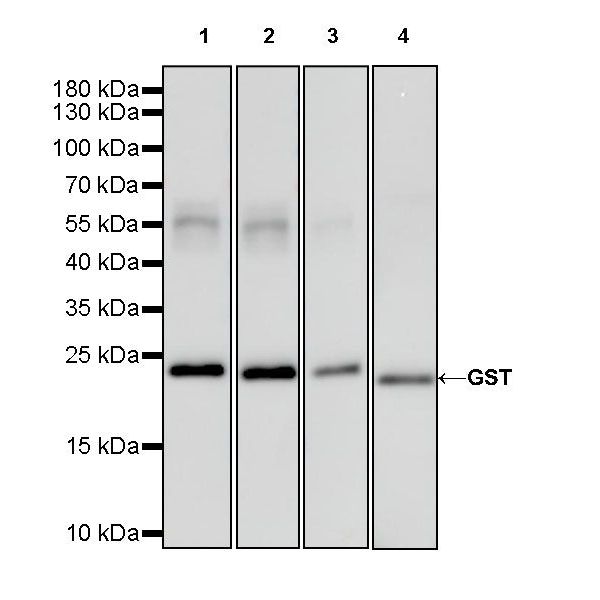
WB result of GST Rabbit mAb
Lysate: Recombinant Schistosoma Japonicum GST Protein 20ng
Primary antibody:
Lane 1: GST Rabbit mAb at 1/5000 dilution
Lane 2: GST Rabbit mAb at 1/10000 dilution
Lane 3: GST Rabbit mAb at 1/50000 dilution
Lane 4: GST Rabbit mAb at 1/100000 dilution
Secondary antibody: Goat Anti-Rabbit IgG, (H+L), HRP conjugated at 1/10000 dilution
Predicted MW: 25 kDa
Observed MW: 24 kDa
S-RMab® GST Tag Recombinant Rabbit mAb (S-372-19)
S-RMab® GST Tag Recombinant Rabbit mAb (S-372-19)
Price:
Regular price
$70 USD
Regular price
Sale price
$70 USD
Unit price
per
For shipping services or bulk orders, you may request a quotation.
Secure checkout with

View full details
Product Details
Product Details
Product Specification
| Host | Rabbit |
| Synonyms | Glutathione S-transferase class-mu 26 kDa isozyme, GST 26, Sj26 antigen, SjGST |
| Immunogen | Recombinant Protein |
| Accession | P08515 |
| Clone Number | S-372-19 |
| Antibody Type | Rabbit mAb |
| Application | WB, ICC, ICFCM |
| Reactivity | Species Independent |
| Purification | Protein A |
| Concentration | 0.5 mg/ml |
| Conjugation | Unconjugated |
| Physical Appearance | Liquid |
| Storage Buffer | PBS, 40% Glycerol, 0.05% BSA, 0.03% Proclin 300 |
| Stability & Storage | 12 months from date of receipt / reconstitution, -20 °C as supplied. |
Dilution
| application | dilution | species |
| WB | 1:5000-1:50000 | |
| ICC | 1:500 | |
| ICFCM | 1:250 |
Background
Glutathione S-transferases (GSTs), previously known as ligandins, are a family of eukaryotic and prokaryotic phase II metabolic isozymes best known for their ability to catalyze the conjugation of the reduced form of glutathione (GSH) to xenobiotic substrates for the purpose of detoxification [PMID: 15717864]. A GST-tag is often used to separate and purify proteins that contain the GST-fusion protein. The tag is 220 amino acids (roughly 26 kDa) in size, which, compared to tags such as the Myc-tag or the FLAG-tag, is quite large. It can be fused to either the N-terminus or C-terminus of a protein.
Picture
Picture
Western Blot
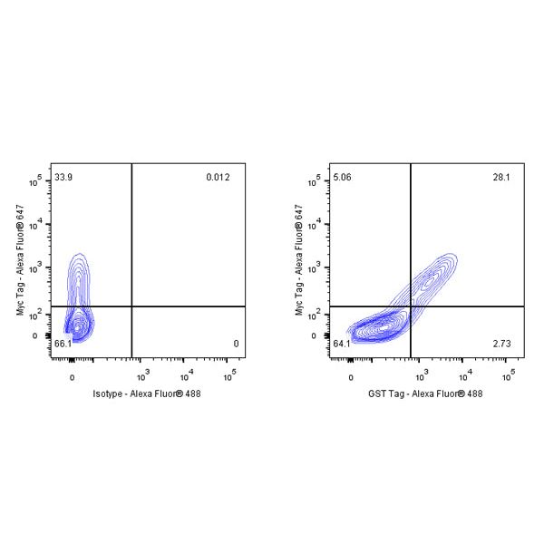
FC
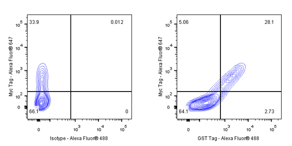
Flow cytometric analysis of 4% PFA fixed 0.1% Tween-20 permeabilized Histone H3, GST Tag, HA Tag, Myc tag transfected 293T (Human embryonic kidney epithelial cell) labelling GST Tag antibody at 1/250 (0.1 μg) dilution/ (right panel) compared with a Rabbit IgG Isotype Control / (left panel). Goat Anti-Rabbit IgG Alexa Fluor® 488 was used as the secondary antibody. Then cells were stained with Myc - Alexa Fluor® 647 separately.
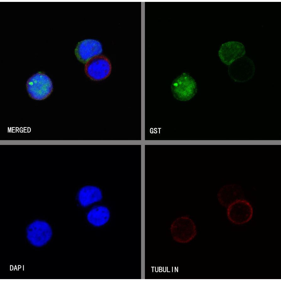
Immunocytochemistry
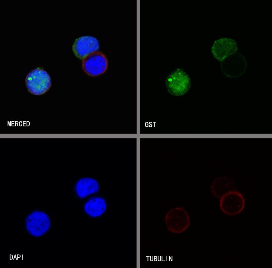
ICC shows positive staining in GST-Histone H3 transfected 293T cells. Anti-GST Tag antibody was used at 1/500 dilution (Green) and incubated overnight at 4°C. Goat polyclonal Antibody to Rabbit IgG - H&L (Alexa Fluor® 488) was used as secondary antibody at 1/1000 dilution. The cells were fixed with 100% ice-cold methanol and permeabilized with 0.1% PBS-Triton X-100. Nuclei were counterstained with DAPI (Blue). Counterstain with tubulin (Red).
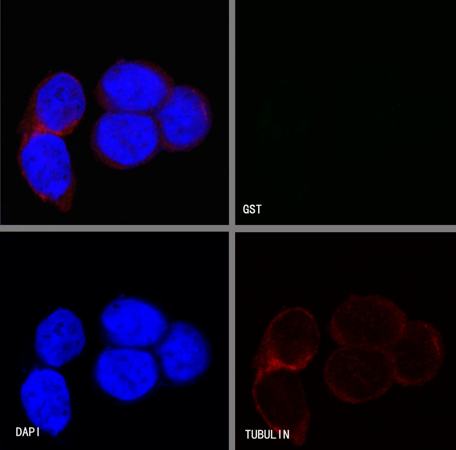
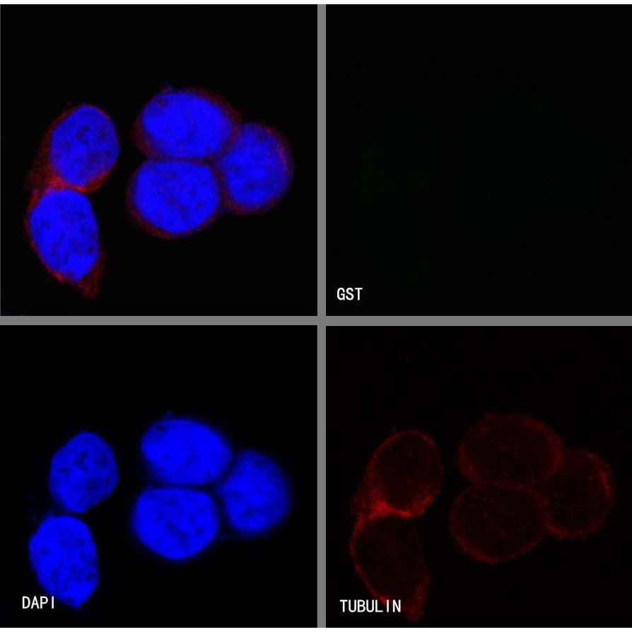
Negative control: ICC shows negative staining in vector-transfected 293T cells. Anti-GST Tag antibody was used at 1/500 dilution and incubated overnight at 4°C. Goat polyclonal Antibody to Rabbit IgG - H&L (Alexa Fluor® 488) was used as secondary antibody at 1/1000 dilution. The cells were fixed with 100% ice-cold methanol and permeabilized with 0.1% PBS-Triton X-100. Nuclei were counterstained with DAPI (Blue). Counterstain with tubulin (Red).