WB result of p21 Waf1/Cip1 Mouse mAb Primary antibody: p21 Waf1/Cip1 Mouse mAb at 1/1000 dilution Lane 1: HeLa whole cell lysate 20 µg Lane 2: MCF7 whole cell lysate 20 µg Low expression control: HeLa whole cell lysate Secondary antibody: Goat Anti-Mouse IgG, (H+L), HRP conjugated at 1/10000 dilution Predicted MW: 18 kDa Observed MW: 18 kDa
p21 Waf1/Cip1 Mouse mAb (469-2)
p21 Waf1/Cip1 Mouse mAb (469-2)
Price:
Regular price
$100 USD
Regular price
Sale price
$100 USD
Unit price
per
For shipping services or bulk orders, you may request a quotation.
Secure checkout with

View full details
Product Details
Product Details
Product Specification
| Host | Mouse |
| Antigen | p21 Waf1/Cip1 |
| Synonyms | WAFT, Cyclin-dependent kinase inhibitor 1, CDK-interacting protein 1, MDA-6, p21, CDKN1A, CAP20, CDKN1, CIP1, MDA6, PIC1, SDI1 |
| Immunogen | Synthetic Peptide |
| Location | Nucleus |
| Accession | P38936 |
| Clone Number | 469-2 |
| Antibody Type | Mouse mAb |
| Isotype | IgG2a |
| Application | WB, IHC-P, ICC, ICFCM |
| Reactivity | Hu |
| Purification | Protein A |
| Concentration | 2 mg/ml |
| Conjugation | Unconjugated |
| Physical Appearance | Liquid |
| Storage Buffer | PBS, 40% Glycerol, 0.05% BSA, 0.03% Proclin 300 |
| Stability & Storage | 12 months from date of receipt / reconstitution, -20 °C as supplied |
Dilution
| application | dilution | species |
| WB | 1:1000 | |
| IHC | 1:500 | |
| ICC | 1:500 | |
| ICFCM | 1:50 |
Background
p21Cip1 (alternatively p21Waf1), also known as cyclin-dependent kinase inhibitor 1 or CDK-interacting protein 1, is a cyclin-dependent kinase inhibitor (CKI) that is capable of inhibiting all cyclin/CDK complexes [PMID: 8259214], though is primarily associated with inhibition of CDK2 [PMID: 8259214, PMID: 8242751]. p21 represents a major target of p53 activity and thus is associated with linking DNA damage to cell cycle arrest [PMID: 9822382]. his protein is encoded by the CDKN1A gene located on chromosome 6 (6p21.2) in humans.
Picture
Picture
Western Blot
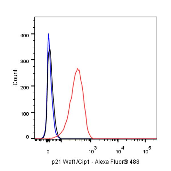
FC
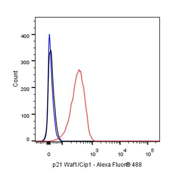
Flow cytometric analysis of 4% PFA fixed 90% methanol permeabilized HeLa (Human cervix adenocarcinoma epithelial cell) cells labeling p21 Waf1/Cip1 at 1/50 dilution (1 μg) / (red) compared with a mouse monoclonal IgG isotype control (black) and an unlabeled control (cells without incubation with primary antibody and secondary antibody) (Blue). Goat Anti - Mouse IgG Alexa Fluor® 488 was used as the secondary antibody.
Immunohistochemistry
IHC shows positive staining in paraffin-embedded human tonsil. Anti-p21 Waf1/Cip1 antibody was used at 1/500 dilution, followed by a HRP Polymer for Mouse & Rabbit IgG (ready to use). Counterstained with hematoxylin. Heat mediated antigen retrieval with Tris/EDTA buffer pH9.0 was performed before commencing with IHC staining protocol.
IHC shows positive staining in paraffin-embedded human colon cancer. Anti-p21 Waf1/Cip1 antibody was used at 1/500 dilution, followed by a HRP Polymer for Mouse & Rabbit IgG (ready to use). Counterstained with hematoxylin. Heat mediated antigen retrieval with Tris/EDTA buffer pH9.0 was performed before commencing with IHC staining protocol.
IHC shows positive staining in paraffin-embedded human cervical squamous cell carcinoma. Anti-p21 Waf1/Cip1 antibody was used at 1/500 dilution, followed by a HRP Polymer for Mouse & Rabbit IgG (ready to use). Counterstained with hematoxylin. Heat mediated antigen retrieval with Tris/EDTA buffer pH9.0 was performed before commencing with IHC staining protocol.
IHC shows positive staining in paraffin-embedded human endometrial cancer. Anti-p21 Waf1/Cip1 antibody was used at 1/500 dilution, followed by a HRP Polymer for Mouse & Rabbit IgG (ready to use). Counterstained with hematoxylin. Heat mediated antigen retrieval with Tris/EDTA buffer pH9.0 was performed before commencing with IHC staining protocol.
Immunocytochemistry
ICC shows positive staining in MCF7 cells. Anti- p21 Waf1/Cip1 antibody was used at 1/500 dilution (Green) and incubated overnight at 4°C. Goat polyclonal Antibody to Mouse IgG - H&L (Alexa Fluor® 488) was used as secondary antibody at 1/1000 dilution. The cells were fixed with 4% PFA and permeabilized with 0.1% PBS-Triton X-100. Nuclei were counterstained with DAPI (Blue).
ICC shows positive staining in HeLa (low expression) cells. Anti-p21 Waf1/Cip1 antibody was used at 1/500 dilution (Green) and incubated overnight at 4°C. Goat polyclonal Antibody to Mouse IgG - H&L (Alexa Fluor® 488) was used as secondary antibody at 1/1000 dilution. The cells were fixed with 4% PFA and permeabilized with 0.1% PBS-Triton X-100. Nuclei were counterstained with DAPI (Blue).