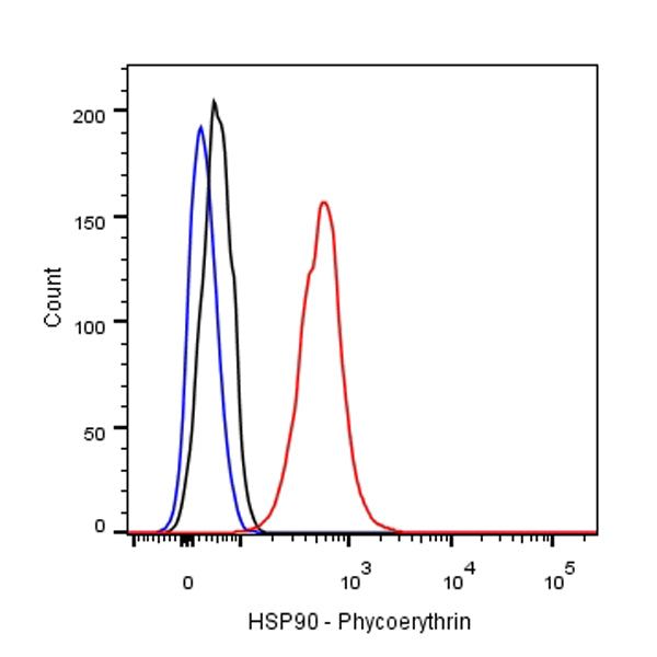
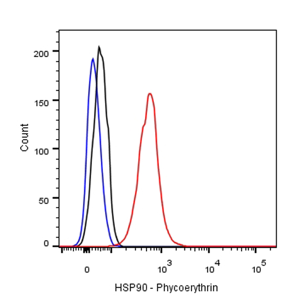
Flow cytometric analysis of HeLa cells labelling HSP60 (Alexa Fluor 647 Conjugate) antibody at 1/2000 (0.1 μg) dilution/ (red) compared with a Rabbit monoclonal IgG (Black) isotype control and an unlabelled control (cells without incubation with primary antibody and secondary antibody) (Blue).
HSP60 Recombinant Rabbit mAb (Alexa Fluor® 647 Conjugate) (SDT-R012)
HSP60 Recombinant Rabbit mAb (Alexa Fluor® 647 Conjugate) (SDT-R012)
Price:
Regular price
$45 USD
Regular price
Sale price
$45 USD
Unit price
per
For shipping services or bulk orders, you may request a quotation.
Secure checkout with

View full details
Product Details
Product Details
Product Specification
| Host | Rabbit |
| Antigen | HSP60 |
| Synonyms | 60 kDa chaperonin, Chaperonin 60, CPN60, Heat shock protein 60, HuCHA60, Mitochondrial matrix protein P1, P60 lymphocyte protein |
| Immunogen | N/A |
| Location | Mitochondrion Matrix |
| Accession | P10809 |
| Clone Number | SDT-R012 |
| Antibody Type | Recombinant mAb |
| Application | ICC, ICFCM |
| Reactivity | Hu, Ms, Rt |
| Purification | Protein A |
| Concentration | 2 mg/ml |
| Conjugation | Alexa Fluor® 647 |
| Physical Appearance | Liquid |
| Storage Buffer | PBS, 0.1% BSA, 0.01% Proclin 300 |
| Stability & Storage | 12 months from date of receipt / reconstitution, 2 to 8 °C as supplied. |
Dilution
| application | dilution | species |
| ICC | 1:500 |
Background
HSP60, also known as chaperonins (Cpn), is a family of heat shock proteins originally sorted by their 60kDa molecular mass. They prevent misfolding of proteins during stressful situations such as high heat, by assisting protein folding. HSP60 belongs to a large class of molecules that assist protein folding, called molecular chaperones.
Picture
Picture
FC
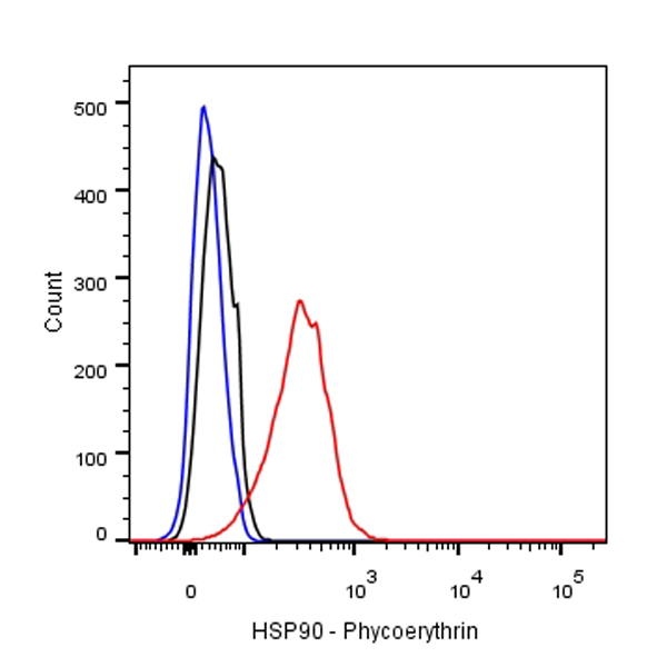
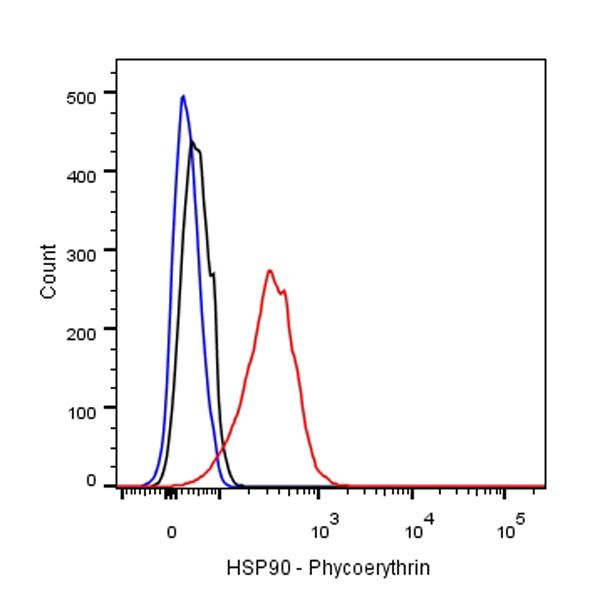
Flow cytometric analysis of C6 cells labelling HSP60 (Alexa Fluor 647 Conjugate) antibody at 1/2000 (0.1 μg) dilution/ (red) compared with a Rabbit monoclonal IgG (Black) isotype control and an unlabelled control (cells without incubation with primary antibody and secondary antibody) (Blue).
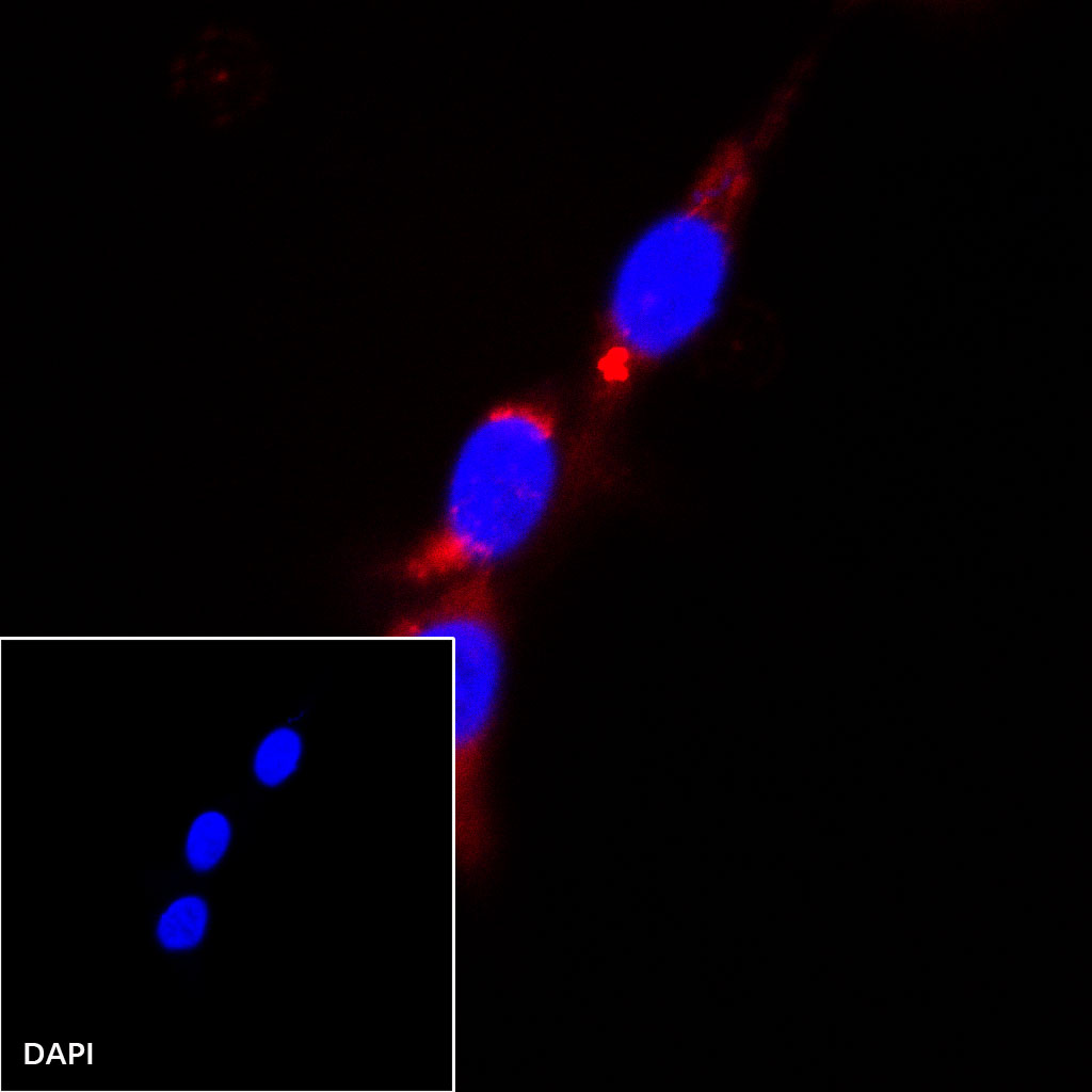
Immunocytochemistry
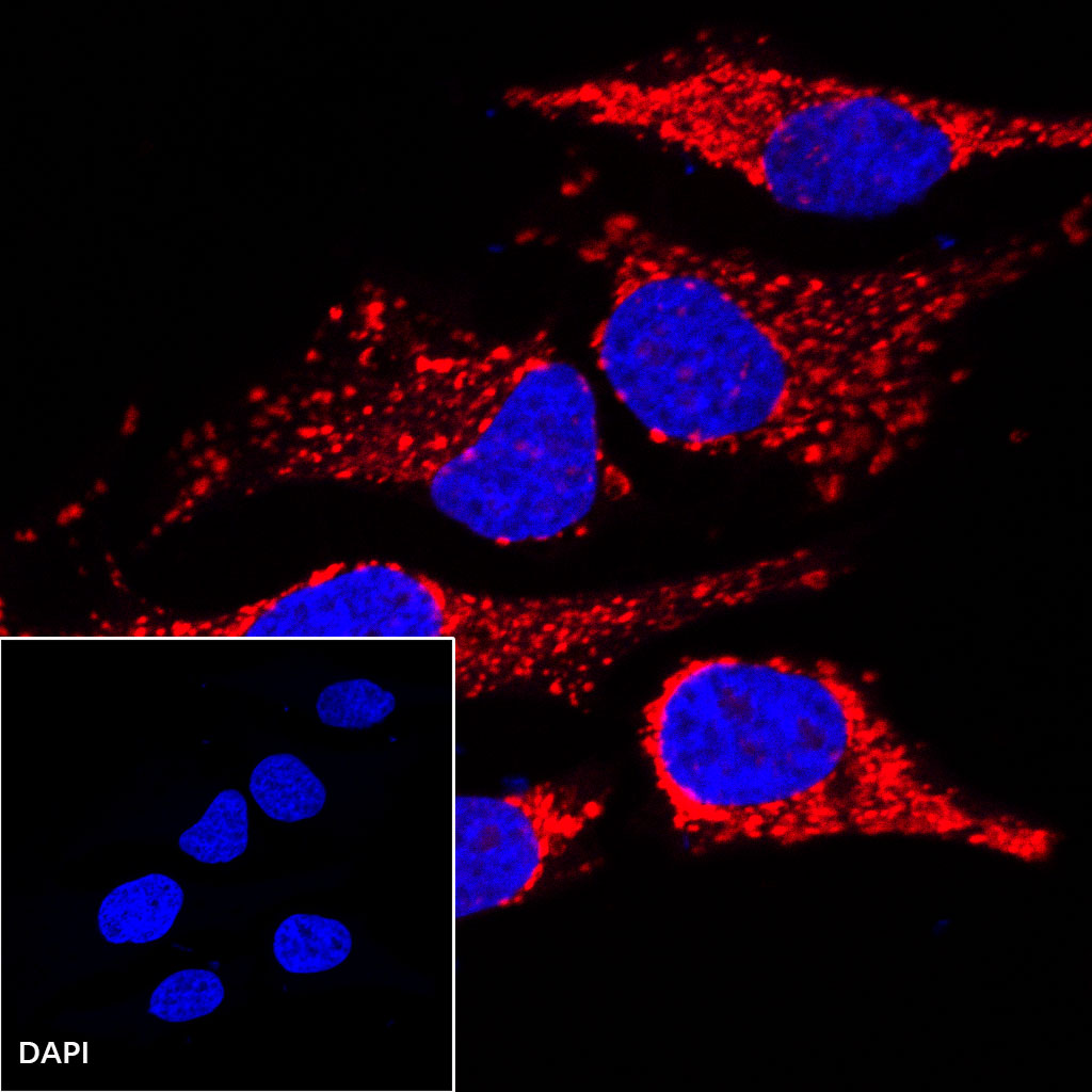
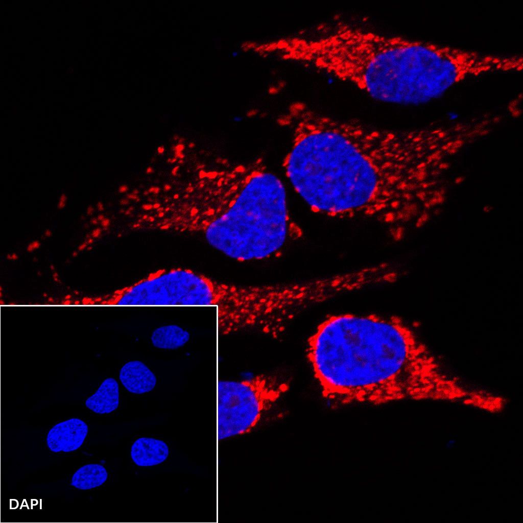
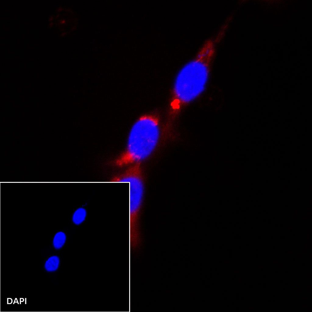
ICC shows positive staining in HeLa cells. Anti-HSP60 (Alexa Fluor® 647 Conjugate) antibody was used at 1/500 dilution (magenta) and incubated overnight at 4°C. The cells were fixed with 100% ice-cold methanol and permeabilized with 0.1% PBS-Triton X-100. Nuclei were counterstained with DAPI (Blue).
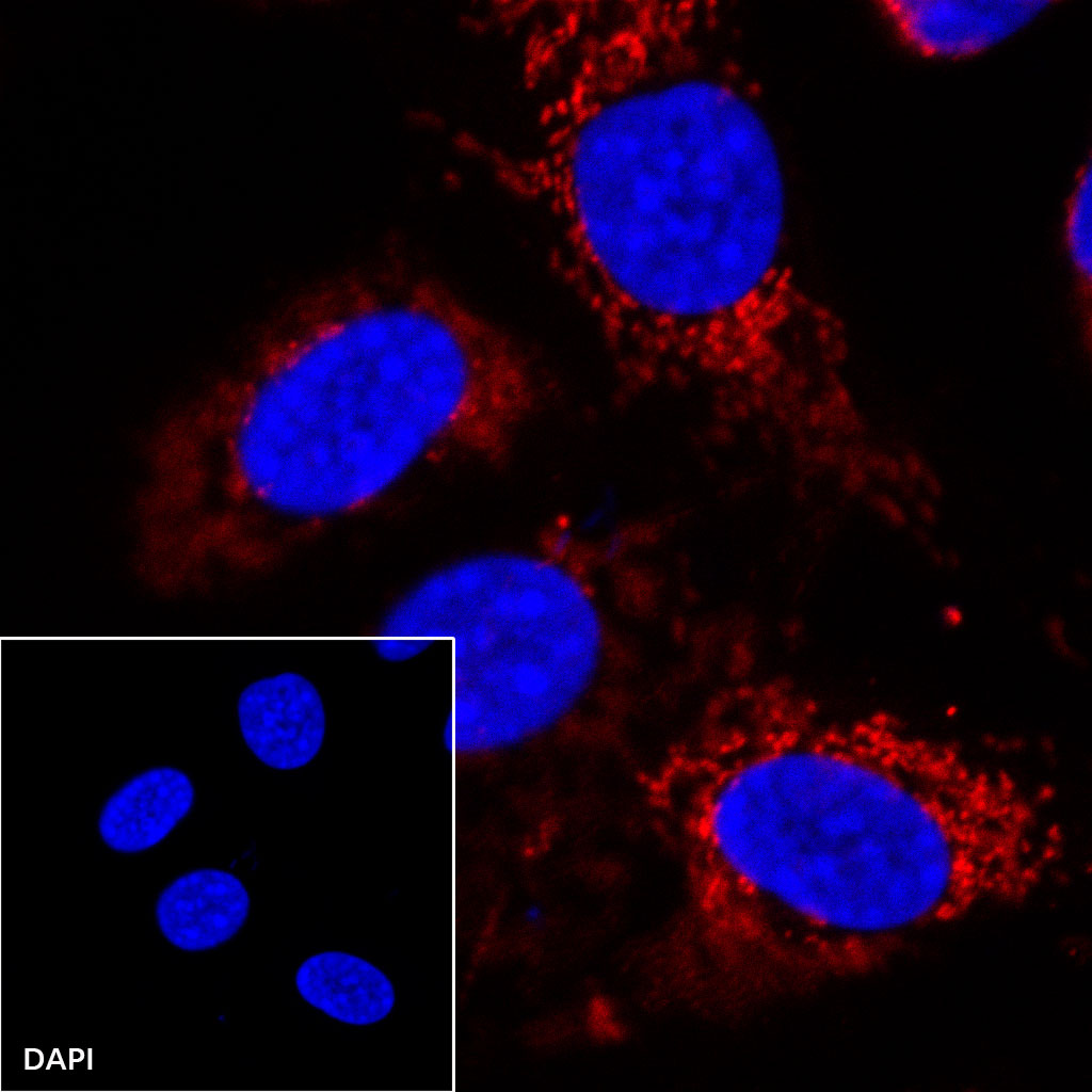
ICC shows positive staining in NIH/3T3 cells. Anti-HSP60 (Alexa Fluor® 647 Conjugate) antibody was used at 1/500 dilution (magenta) and incubated overnight at 4°C. The cells were fixed with 4% PFA and permeabilized with 0.1% PBS-Triton X-100. Nuclei were counterstained with DAPI (Blue).
ICC shows positive staining in C6 cells. Anti-HSP60 (Alexa Fluor® 647 Conjugate) antibody was used at 1/500 dilution (magenta) and incubated overnight at 4°C. The cells were fixed with 100% ice-cold methanol and permeabilized with 0.1% PBS-Triton X-100. Nuclei were counterstained with DAPI (Blue).