WB result of Histone H3 (acetyl K27) Rabbit pAb
Primary antibody: Histone H3 (acetyl K27) Rabbit pAb at 1/1000 dilution
Lane 1: HeLa whole cell lysate 20 µg
Lane 2: HeLa treated with TSA (500 ng/ml, 4h) whole cell lysate 20 µg
Secondary antibody: Goat Anti-Rabbit IgG, (H+L), HRP conjugated at 1/10000 dilution
Predicted MW: 15 kDa
Observed MW: 14 kDa
Exposure time: 90 s
Histone H3 (acetyl K27) Rabbit pAb
Histone H3 (acetyl K27) Rabbit pAb
Price:
Regular price
$35 USD
Regular price
Sale price
$35 USD
Unit price
per
For shipping services or bulk orders, you may request a quotation.
Secure checkout with

View full details
Product Details
Product Details
Product Specification
| Host | Rabbit |
| Antigen | Histone H3 (acetyl K27) |
| Synonyms | H3K27ac; Acetyl-Histone H3 (Lys27) |
| Immunogen | Synthetic Peptide |
| Location | Nucleus |
| Accession | P68431 |
| Antibody Type | Polyclonal antibody |
| Isotype | IgG |
| Application | WB, IHC-P, ICC, ChIP |
| Reactivity | Hu, Ms, Rt |
| Purification | Protein A |
| Concentration | 0.5 mg/ml |
| Conjugation | Unconjugated |
| Physical Appearance | Liquid |
| Storage Buffer | PBS, 40% Glycerol, 0.05%BSA, 0.03% Proclin 300 |
| Stability & Storage | 12 months from date of receipt / reconstitution, -20 °C as supplied |
Dilution
| application | dilution | species |
| Dot Blot | 1:1000 | |
| WB | 1:1000 | |
| IHC-P | 1:500 | |
| ICC | 1:500 | |
| ChIP | 1:20~1:50 |
Background
H3K27ac is an epigenetic modification to the DNA packaging protein histone H3. It is a mark that indicates acetylation of the lysine residue at N-terminal position 27 of the histone H3 protein. H3K27ac is associated with the higher activation of transcription and therefore defined as an active enhancer mark. H3K27ac is found at both proximal and distal regions of transcription start site (TSS).
Picture
Picture
Western Blot
WB result of Histone H3 (acetyl K27) Rabbit pAb
Primary antibody: Histone H3 (acetyl K27) Rabbit pAb at 1/1000 dilution
Lane 1: NIH/3T3 whole cell lysate 20 µg
Lane 2: NIH/3T3 treated with TSA (500 ng/ml, 4h) whole cell lysate 20 µg
Secondary antibody: Goat Anti-Rabbit IgG, (H+L), HRP conjugated at 1/10000 dilution
Predicted MW: 15 kDa
Observed MW: 14 kDa
Exposure time: 90 s
Dot Blot
Dot blot result of Histone H3 (acetyl K27) Rabbit pAb
Lane 1: Histone H3 (acetyl K27) peptide
Lane 2: Histone H3 unmodified peptide
Primary antibody: Histone H3 (acetyl K27) Rabbit pAb at 1/1000 dilution
Secondary antibody: Goat Anti-Rabbit IgG, (H+L), HRP conjugated at 1/10000 dilution
Exposure time: 30 s
Immunohistochemistry
IHC shows positive staining in paraffin-embedded human kidney. Anti-Histone H3 (acetyl K27) antibody was used at 1/500 dilution, followed by a HRP Polymer for Mouse & Rabbit IgG (ready to use). Counterstained with hematoxylin. Heat mediated antigen retrieval with Tris/EDTA buffer pH9.0 was performed before commencing with IHC staining protocol.
IHC shows positive staining in paraffin-embedded human testis. Anti-Histone H3 (acetyl K27) antibody was used at 1/500 dilution, followed by a HRP Polymer for Mouse & Rabbit IgG (ready to use). Counterstained with hematoxylin. Heat mediated antigen retrieval with Tris/EDTA buffer pH9.0 was performed before commencing with IHC staining protocol.
IHC shows positive staining in paraffin-embedded human colon cancer. Anti-Histone H3 (acetyl K27) antibody was used at 1/500 dilution, followed by a HRP Polymer for Mouse & Rabbit IgG (ready to use). Counterstained with hematoxylin. Heat mediated antigen retrieval with Tris/EDTA buffer pH9.0 was performed before commencing with IHC staining protocol.
IHC shows positive staining in paraffin-embedded human hepatocellular carcinoma. Anti-Histone H3 (acetyl K27) antibody was used at 1/500 dilution, followed by a HRP Polymer for Mouse & Rabbit IgG (ready to use). Counterstained with hematoxylin. Heat mediated antigen retrieval with Tris/EDTA buffer pH9.0 was performed before commencing with IHC staining protocol.
IHC shows positive staining in paraffin-embedded mouse lung. Anti-Histone H3 (acetyl K27) antibody was used at 1/500 dilution, followed by a HRP Polymer for Mouse & Rabbit IgG (ready to use). Counterstained with hematoxylin. Heat mediated antigen retrieval with Tris/EDTA buffer pH9.0 was performed before commencing with IHC staining protocol.
IHC shows positive staining in paraffin-embedded rat kidney. Anti-Histone H3 (acetyl K27) antibody was used at 1/500 dilution, followed by a HRP Polymer for Mouse & Rabbit IgG (ready to use). Counterstained with hematoxylin. Heat mediated antigen retrieval with Tris/EDTA buffer pH9.0 was performed before commencing with IHC staining protocol.
Immunocytochemistry
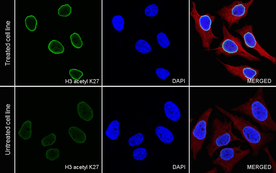
ICC analysis of HeLa cells treated with TSA(500ng/ml, 4h) (top panel) and HeLa cells untreated with TSA(500ng/ml, 4h)(below panel). Anti-Histone H3 (acetyl K27) antibody was used at 1/500 dilution (Green) and incubated overnight at 4°C. Goat polyclonal Antibody to Rabbit IgG - H&L (Alexa Fluor® 488) was used as secondary antibody at 1/1000 dilution. The cells were fixed with 4% PFA and permeabilized with 0.1% PBS-Triton X-100. Nuclei were counterstained with DAPI (Blue). Counterstain with tubulin (Red).
ChIP
Chromatin immunoprecipitation (ChIP) was performed on HeLa cells cross - linked with 1% formaldehyde for 10 min, then chromatin was fragmented by sonication. Parallel reactions used Histone H3 (acetyl K27) Rabbit pAb and Rabbit mAb IgG Isotype Control (SDT-R173) at 1:50 for immunoprecipitation. Post - immunoprecipitation, both samples were washed, eluted, and cross - links reversed. Purified DNA was analyzed by qPCR.
qPCR (%input: immunoprecipitated DNA/input DNA) showed the enrichment of RPL30, GAPDH, MYOD1, AFM, SAT-α and SAT-2 in Histone H3 (acetyl K27) Rabbit pAb - immunoprecipitated sample.