WB result of Granzyme B Rabbit mAb
Primary antibody: Granzyme B Rabbit mAb at 1/1000 dilution
Lane 1: Jurkat whole cell lysate 20 µg
Lane 2: NK-92 whole cell lysate 20 µg
Negative control: Jurkat whole cell lysate
Secondary antibody: Goat Anti-Rabbit IgG, (H+L), HRP conjugated at 1/10000 dilution
Predicted MW: 28 kDa
Observed MW: 30, 65 kDa
Granzyme B Recombinant Rabbit mAb (S-357-54)
Granzyme B Recombinant Rabbit mAb (S-357-54)
Price:
Regular price
$100 USD
Regular price
Sale price
$100 USD
Unit price
per
For shipping services or bulk orders, you may request a quotation.
Secure checkout with

View full details
Product Details
Product Details
Product Specification
| Host | Rabbit |
| Antigen | Granzyme B |
| Synonyms | C11, CTLA-1, Cathepsin G-like 1 (CTSGL1), Cytotoxic T-lymphocyte proteinase 2 (Lymphocyte protease), Fragmentin-2, Granzyme-2, Human lymphocyte protein (HLP), SECT, T-cell serine protease 1-3E |
| Immunogen | Recombinant Protein |
| Location | Secreted |
| Accession | P10144 |
| Clone Number | S-357-54 |
| Antibody Type | Recombinant mAb |
| Isotype | IgG |
| Application | WB, IHC-P, ICC, ICFCM |
| Reactivity | Hu |
| Purification | Protein A |
| Concentration | 0.5 mg/ml |
| Conjugation | Unconjugated |
| Physical Appearance | Liquid |
| Storage Buffer | PBS, 40% Glycerol, 0.05%BSA, 0.03% Proclin 300 |
| Stability & Storage | 12 months from date of receipt / reconstitution, -20 °C as supplied |
Dilution
| application | dilution | species |
| WB | 1:1000 | null |
| IHC | 1:250 | null |
| ICFCM | 1:50 | null |
| ICC | 1:50 | null |
Background
Granzyme B (GrB) is one of the serine protease granzymes most commonly found in the granules of natural killer cells (NK cells) and cytotoxic T cells. It is secreted by these cells along with the pore forming protein perforin to mediate apoptosis in target cells. Granzyme B has also been found to be produced by a wide range of non-cytotoxic cells ranging from basophils and mast cells to smooth muscle cells. The secondary functions of granzyme B are also numerous. Granzyme B has shown to be involved in inducing inflammation by stimulating cytokine release and is also involved in extracellular matrix remodelling. Elevated levels of granzyme B are also implicated in a number of autoimmune diseases, several skin diseases, and type 1 diabetes.
Picture
Picture
Western Blot
FC
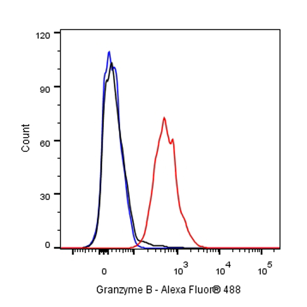
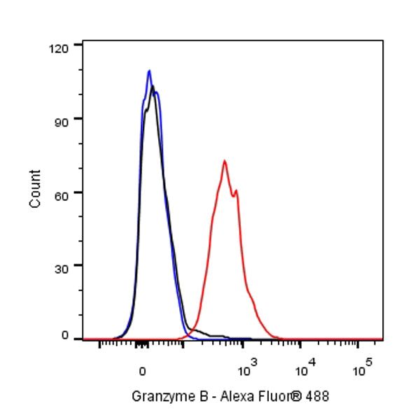
Flow cytometric analysis of 4% PFA fixed 90% methanol permeabilized NK92 (Human Natural Killer cells) cells labelling Granzyme B antibody at 1/50 dilution (1 μg)/ (Red) compared with a Rabbit monoclonal IgG (Black) isotype control and an unlabelled control (cells without incubation with primary antibody and secondary antibody) (Blue). Goat Anti - Rabbit IgG Alexa Fluor® 488 was used as the secondary antibody.
Immunohistochemistry
IHC shows positive staining in paraffin-embedded human tonsil. Anti-Granzyme B antibody was used at 1/250 dilution, followed by a HRP Polymer for Mouse & Rabbit IgG (ready to use). Counterstained with hematoxylin. Heat mediated antigen retrieval with Tris/EDTA buffer pH9.0 was performed before commencing with IHC staining protocol.
IHC shows positive staining in paraffin-embedded human spleen. Anti-Granzyme B antibody was used at 1/250 dilution, followed by a HRP Polymer for Mouse & Rabbit IgG (ready to use). Counterstained with hematoxylin. Heat mediated antigen retrieval with Tris/EDTA buffer pH9.0 was performed before commencing with IHC staining protocol.
IHC shows positive staining in paraffin-embedded human colon. Anti-Granzyme B antibody was used at 1/250 dilution, followed by a HRP Polymer for Mouse & Rabbit IgG (ready to use). Counterstained with hematoxylin. Heat mediated antigen retrieval with Tris/EDTA buffer pH9.0 was performed before commencing with IHC staining protocol.
IHC shows positive staining in paraffin-embedded human breast cancer. Anti-Granzyme B antibody was used at 1/250 dilution, followed by a HRP Polymer for Mouse & Rabbit IgG (ready to use). Counterstained with hematoxylin. Heat mediated antigen retrieval with Tris/EDTA buffer pH9.0 was performed before commencing with IHC staining protocol.
IHC shows positive staining in paraffin-embedded human cervical squamous cell carcinoma. Anti-Granzyme B antibody was used at 1/250 dilution, followed by a HRP Polymer for Mouse & Rabbit IgG (ready to use). Counterstained with hematoxylin. Heat mediated antigen retrieval with Tris/EDTA buffer pH9.0 was performed before commencing with IHC staining protocol.
Immunocytochemistry
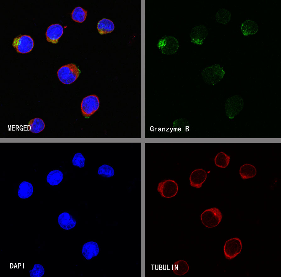
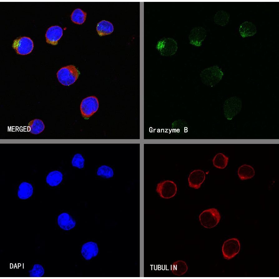
ICC shows positive staining in NK92 cells. Anti-Granzyme B antibody was used at 1/50 dilution (Green) and incubated overnight at 4°C. Goat polyclonal Antibody to Rabbit IgG - H&L (Alexa Fluor® 488) was used as secondary antibody at 1/1000 dilution. The cells were fixed with 4%PFA and permeabilized with 0.1% PBS-Triton X-100. Nuclei were counterstained with DAPI (Blue).Counterstain with tubulin (Red).
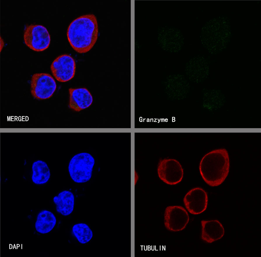
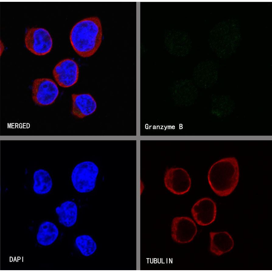
Negative control:ICC shows negative staining in Jurkat cells. Anti-Granzyme B antibody was used at 1/50 dilution and incubated overnight at 4°C. Goat polyclonal Antibody to Rabbit IgG - H&L (Alexa Fluor® 488) was used as secondary antibody at 1/1000 dilution. The cells were fixed with 4%PFA and permeabilized with 0.1% PBS-Triton X-100. Nuclei were counterstained with DAPI (Blue).Counterstain with tubulin (Red).