Flow cytometric analysis of THP-1 (Human monocytic leukemia monocyte, left) / Jurkat (Human T cell leukemia T lymphocyte, Right) cells labelling CD28 antibody at 1/1500 dilution (0.1 μg) / (Red) compared with a Mouse monoclonal IgG (Black) isotype control and an unlabelled control (cells without incubation with primary antibody and secondary antibody) (Blue). Goat Anti - Mouse IgG Alexa Fluor® 488 was used as the secondary antibody.
Negative control: THP-1
CD28 Mouse mAb (S-714-76)
CD28 Mouse mAb (S-714-76)
Price:
Regular price
$100 USD
Regular price
Sale price
$100 USD
Unit price
per
For shipping services or bulk orders, you may request a quotation.
Secure checkout with

View full details
Product Details
Product Details
Product Specification
| Host | Mouse |
| Antigen | CD28 |
| Synonyms | T-cell-specific surface glycoprotein CD28, TP44 |
| Immunogen | Recombinant Protein |
| Location | Cell membrane |
| Accession | P10747 |
| Clone Number | S-714-76 |
| Antibody Type | Mouse mAb |
| Isotype | IgG1,k |
| Application | ICC, FCM |
| Reactivity | Hu |
| Purification | Protein G |
| Concentration | 1.5 mg/ml |
| Conjugation | Unconjugated |
| Physical Appearance | Liquid |
| Storage Buffer | PBS, 40% Glycerol, 0.05%BSA, 0.03% Proclin 300 |
| Stability & Storage | 12 months from date of receipt / reconstitution, -20 °C as supplied |
Dilution
| application | dilution | species |
| FCM | 1:1500 | null |
| ICC | 1:500 | null |
Background
CD28 (Cluster of Differentiation 28) is one of the proteins expressed on T cells that provide co-stimulatory signals required for T cell activation and survival. T cell stimulation through CD28 in addition to the T-cell receptor (TCR) can provide a potent signal for the production of various interleukins (IL-6 in particular). CD28 is the receptor for CD80 (B7.1) and CD86 (B7.2) proteins. When activated by Toll-like receptor ligands, the CD80 expression is upregulated in antigen-presenting cells (APCs). The CD86 expression on antigen-presenting cells is constitutive (expression is independent of environmental factors). It is generally reported, that CD28 is expressed on 50% of CD8+ T cells and more than 80% CD4+ T cells in human, but during the course of activation some T cells lose this molecule. In general, CD28 is a primary costimulatory molecule for T cell activation.
Picture
Picture
FC
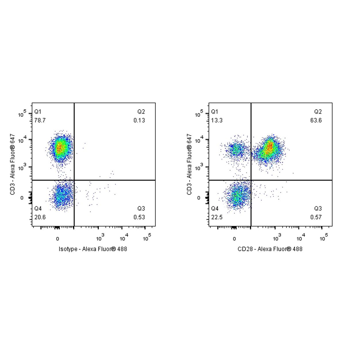
Flow cytometric analysis of human PBMC (human peripheral blood mononuclear cell) labelling CD28 antibody at 1/1500 (0.1 μg) dilution (Right) compared with a Mouse monoclonal IgG isotype control (Left). Goat Anti - Mouse IgG Alexa Fluor® 488 was used as the secondary antibody. Then cells were stained with CD3 - Alexa Fluor® 647 separately. Gated on total viable cells.
Immunocytochemistry
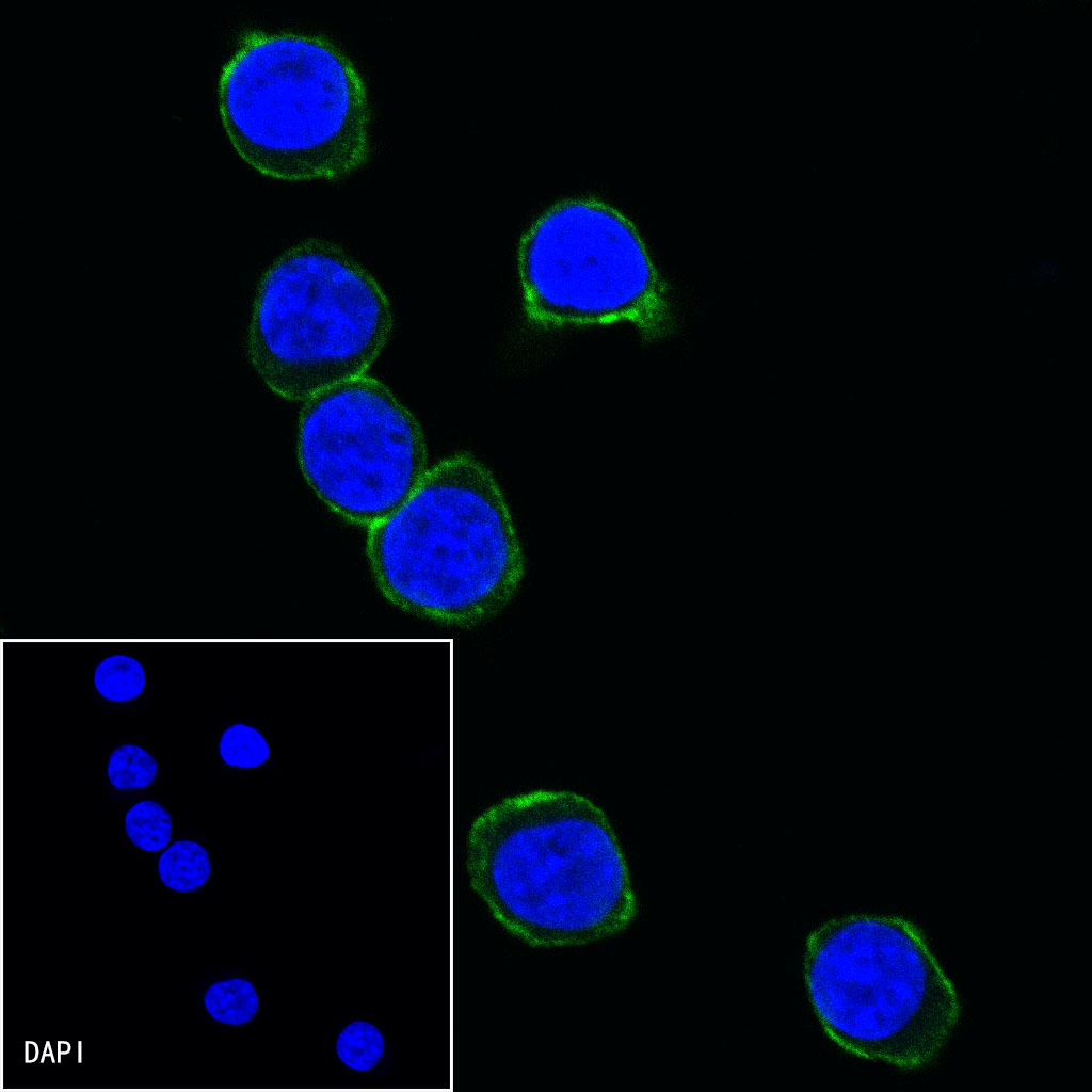
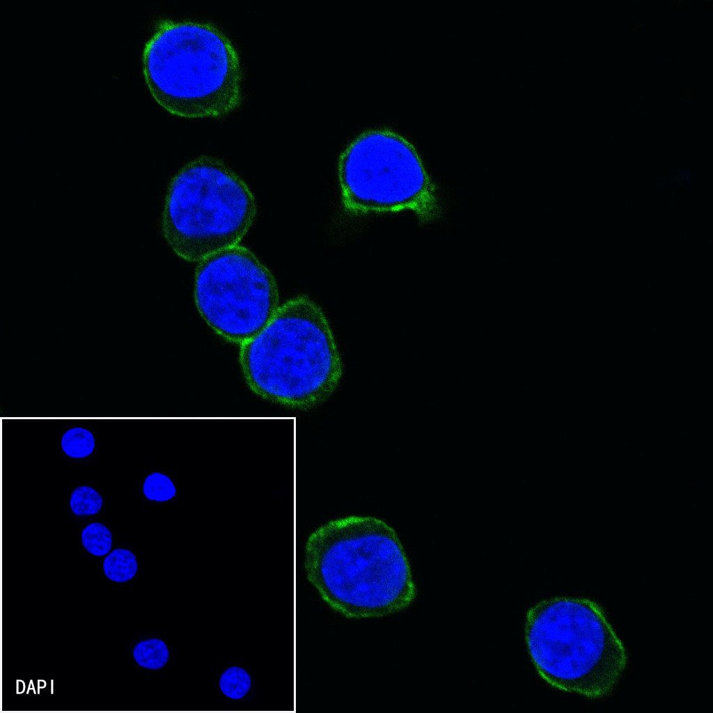
ICC shows positive staining in Jurkat cells. Anti-CD28 antibody was used at 1/500 dilution (Green) and incubated overnight at 4°C. Goat polyclonal Antibody to mouse IgG - H&L (Alexa Fluor® 488) was used as secondary antibody at 1/1000 dilution. The cells were fixed with 4% PFA and permeabilized with 0.1% PBS-Triton X-100. Nuclei were counterstained with DAPI (Blue).

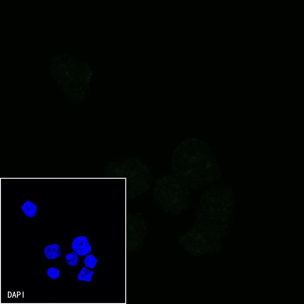
Negative control:ICC shows negative staining in THP-1 cells. Anti-CD28 antibody was used at 1/500 dilution and incubated overnight at 4°C. Goat polyclonal Antibody to mouse IgG - H&L (Alexa Fluor® 488) was used as secondary antibody at 1/1000 dilution. The cells were fixed with 4% PFA and permeabilized with 0.1% PBS-Triton X-100. Nuclei were counterstained with DAPI (Blue).