WB result of CD239 Rabbit mAb
Primary antibody: CD239 Rabbit mAb at 1/1000 dilution
Lane 1: SH-SY5Y whole cell lysate 20 µg
Lane 2: A431 whole cell lysate 20 µg
Negative control: SH-SY5Y whole cell lysate
Secondary antibody: Goat Anti-Rabbit IgG, (H+L), HRP conjugated at 1/10000 dilution
Predicted MW: 67 kDa
Observed MW: 85 kDa
(This blot was developed with high sensitivity substrate)
CD239 Recombinant Rabbit mAb (S-R284)
CD239 Recombinant Rabbit mAb (S-R284)
Price:
Regular price
$100 USD
Regular price
Sale price
$100 USD
Unit price
per
For shipping services or bulk orders, you may request a quotation.
Secure checkout with

View full details
Product Details
Product Details
Product Specification
| Host | Rabbit |
| Antigen | CD239 |
| Synonyms | Basal cell adhesion molecule, Auberger B antigen, B-CAM cell surface glycoprotein, F8/G253 antigen, Lutheran antigen, Lutheran blood group glycoprotein, BCAM, LU, MSK19 |
| Location | Membrane |
| Accession | P50895 |
| Clone Number | S-R284 |
| Antibody Type | Recombinant mAb |
| Isotype | IgG |
| Application | WB, IHC-P, IP |
| Reactivity | Hu |
| Purification | Protein A |
| Concentration | 0.5 mg/ml |
| Conjugation | Unconjugated |
| Physical Appearance | Liquid |
| Storage Buffer | PBS, 40% Glycerol, 0.05%BSA, 0.03% Proclin 300 |
| Stability & Storage | 12 months from date of receipt / reconstitution, -20 °C as supplied |
Dilution
| application | dilution | species |
| WB | 1:1000 | null |
| IHC | 1:500 | null |
| IP | 1:50 | null |
Background
Basal cell adhesion molecule, also known as Lutheran antigen, is a plasma membrane glycoprotein that in humans is encoded by the BCAM gene. BCAM has also recently been designated CD239 (cluster of differentiation 239). It is a member of the immunoglobulin superfamily and a receptor for the extracellular matrix protein, laminin. The protein contains five, N-terminus, extracellular immunoglobulin domains, a single transmembrane domain, and a short, C-terminal cytoplasmic tail. This protein may play a role in epithelial cell cancer and in vaso-occlusion of red blood cells in sickle cell disease. BCAM has been shown to interact with Laminin, alpha 5. BCAM has also been shown to promote the metastasis of ovarian cancer.
Picture
Picture
Western Blot
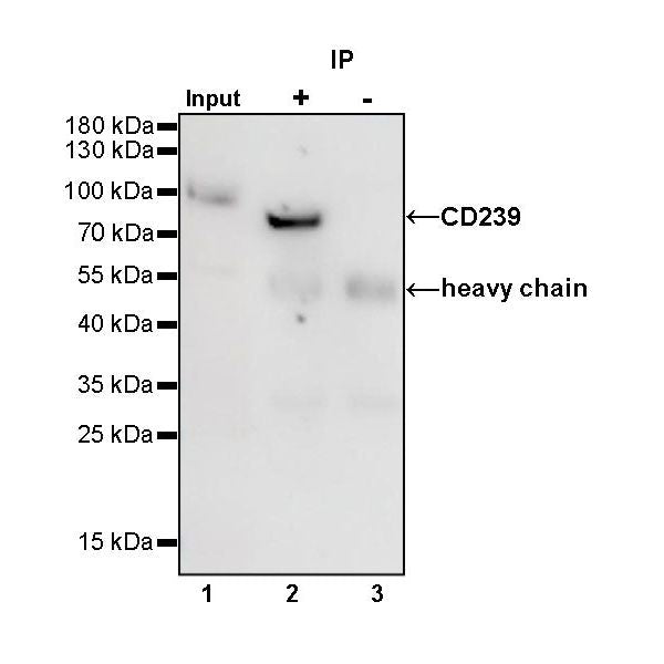
IP
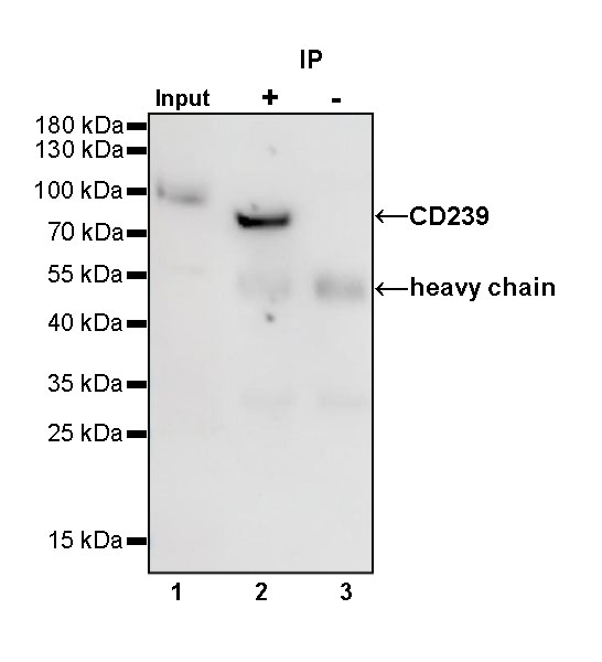
CD239 Rabbit mAb at 1/50 dilution (1 µg) immunoprecipitating CD239 in 0.4 mg A431 whole cell lysate.
Western blot was performed on the immunoprecipitate using CD239 Rabbit mAb at 1/1000 dilution.
Secondary antibody (HRP) for IP was used at 1/400 dilution.
Lane 1: A431 whole cell lysate 20 µg (Input)
Lane 2: CD239 Rabbit mAb IP in A431 whole cell lysate
Lane 3: Rabbit monoclonal IgG IP in A431 whole cell lysate
Predicted MW: 67 kDa
Observed MW: 85 kDa
(This blot was developed with high sensitivity substrate)
Immunohistochemistry
IHC shows positive staining in paraffin-embedded human kidney. Anti-CD239 antibody was used at 1/500 dilution, followed by a HRP Polymer for Mouse & Rabbit IgG (ready to use). Counterstained with hematoxylin. Heat mediated antigen retrieval with Tris/EDTA buffer pH9.0 was performed before commencing with IHC staining protocol.
IHC shows positive staining in paraffin-embedded human prostate. Anti-CD239 antibody was used at 1/500 dilution, followed by a HRP Polymer for Mouse & Rabbit IgG (ready to use). Counterstained with hematoxylin. Heat mediated antigen retrieval with Tris/EDTA buffer pH9.0 was performed before commencing with IHC staining protocol.
IHC shows positive staining in paraffin-embedded human cerebral cortex. Anti-CD239 antibody was used at 1/500 dilution, followed by a HRP Polymer for Mouse & Rabbit IgG (ready to use). Counterstained with hematoxylin. Heat mediated antigen retrieval with Tris/EDTA buffer pH9.0 was performed before commencing with IHC staining protocol.
IHC shows positive staining in paraffin-embedded human ovarian cancer. Anti-CD239 antibody was used at 1/500 dilution, followed by a HRP Polymer for Mouse & Rabbit IgG (ready to use). Counterstained with hematoxylin. Heat mediated antigen retrieval with Tris/EDTA buffer pH9.0 was performed before commencing with IHC staining protocol.