Flow cytometric analysis of Ramos (Human Burkitt's lymphoma B lymphocyte, left) / Jurkat (Human T cell leukemia T lymphocyte, right) cells labelling CD2 antibody at 1/2000 dilution (0.1 μg) / (red) compared with a Mouse monoclonal IgG (Black) isotype control and an unlabelled control (cells without incubation with primary antibody and secondary antibody) (Blue). Goat Anti - Mouse IgG Alexa Fluor® 488 was used as the secondary antibody.
CD2 Mouse mAb (S-633-25)
CD2 Mouse mAb (S-633-25)
Price:
Regular price
$100 USD
Regular price
Sale price
$100 USD
Unit price
per
For shipping services or bulk orders, you may request a quotation.
Secure checkout with

View full details
Product Details
Product Details
Product Specification
| Host | Mouse |
| Antigen | CD2 |
| Synonyms | T-cell surface antigen CD2, Erythrocyte receptor, LFA-2, LFA-3 receptor, Rosette receptor, T-cell surface antigen T11/Leu-5, SRBC |
| Immunogen | Recombinant Protein |
| Location | Cell membrane |
| Accession | P06729 |
| Clone Number | S-633-25 |
| Antibody Type | Mouse mAb |
| Isotype | IgG1,k |
| Application | ICC, FCM |
| Reactivity | Hu |
| Purification | Protein G |
| Concentration | 2 mg/ml |
| Conjugation | Unconjugated |
| Physical Appearance | Liquid |
| Storage Buffer | PBS, 40% Glycerol, 0.05%BSA, 0.03% Proclin 300 |
| Stability & Storage | 12 months from date of receipt / reconstitution, -20 °C as supplied |
Dilution
| application | dilution | species |
| FCM | 1:2000 | null |
| ICC | 1:500 | null |
Background
CD2 (cluster of differentiation 2) is a cell adhesion molecule found on the surface of T cells and natural killer (NK) cells. It has also been called T-cell surface antigen T11/Leu-5, LFA-2, LFA-3 receptor, erythrocyte receptor and rosette receptor. It interacts with other adhesion molecules, such as lymphocyte function-associated antigen-3 (LFA-3/CD58) in humans, or CD48 in rodents, which are expressed on the surfaces of other cells. CD2 is a specific marker for T cells and NK cells, and can therefore be used in immunohistochemistry to identify the presence of such cells in tissue sections. The great majority of T cell lymphomas and leukaemias also express CD2, making it possible to use the presence of the antigen to distinguish these conditions from B cell neoplasms.
Picture
Picture
FC
Immunocytochemistry

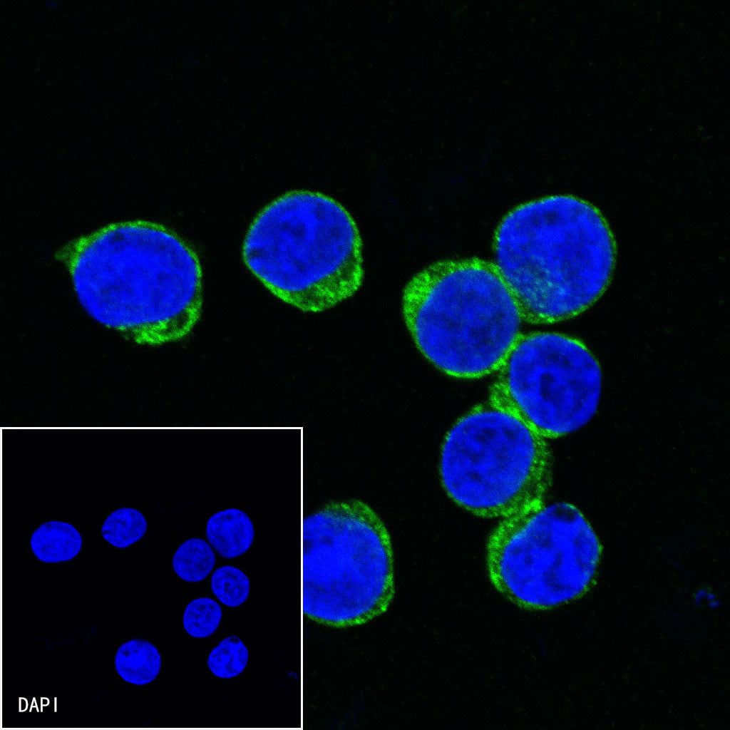
ICC shows positive staining in Jurkat cells. Anti-CD2 antibody was used at 1/500 dilution (Green) and incubated overnight at 4°C. Goat polyclonal Antibody to Mouse IgG - H&L (Alexa Fluor® 488) was used as secondary antibody at 1/1000 dilution. The cells were fixed with 4%PFA and permeabilized with 0.1% PBS-Triton X-100. Nuclei were counterstained with DAPI (Blue).
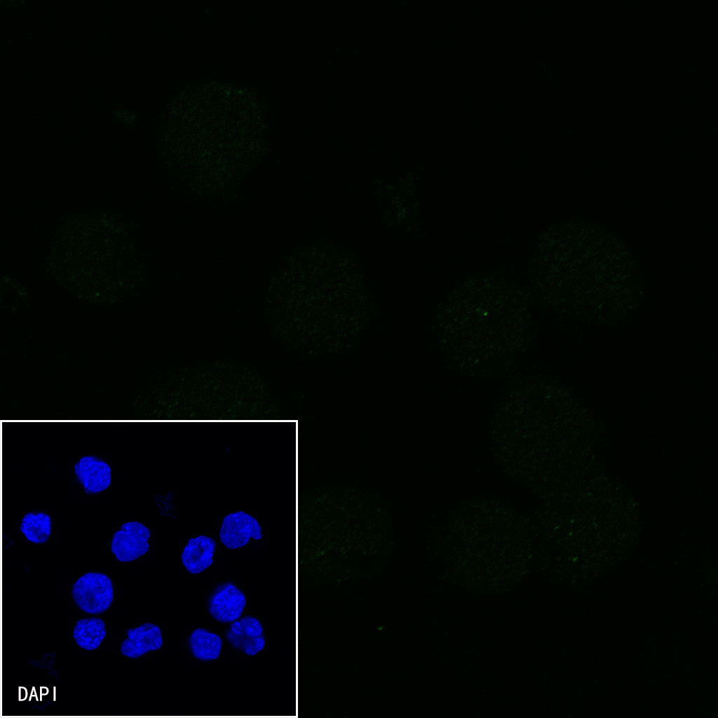
Negative control: ICC shows negative staining in Daudi cells. Anti-CD2 antibody was used at 1/500 dilution and incubated overnight at 4°C. Goat polyclonal Antibody to Mouse IgG - H&L (Alexa Fluor® 488) was used as secondary antibody at 1/1000 dilution. The cells were fixed with 4%PFA and permeabilized with 0.1% PBS-Triton X-100. Nuclei were counterstained with DAPI (Blue).